Intersex
•Download as PPT, PDF•
12 likes•6,099 views

The document discusses ambiguous genitalia or intersex conditions. It defines ambiguous genitalia and provides classifications based on karyotype and etiology. Key causes include congenital adrenal hyperplasia, androgen insensitivity syndrome, and true gonadal intersex. The document outlines approaches to diagnosis, including family history, physical exam, imaging, hormonal testing, and biopsy. Determining the karyotype, presence of uterus, and hormone levels helps to differentiate between conditions like hermaphroditism versus disorders of sex development.

Recommended
More Related Content
What's hot
What's hot (20)
Viewers also liked
Viewers also liked (17)
Similar to Intersex
Similar to Intersex (20)
More from Soumya Ranjan Parida
More from Soumya Ranjan Parida (20)
Recently uploaded
Recently uploaded (20)
Intersex
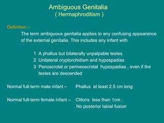
- 1. Ambiguous Genitalia ( Hermaphroditism ) Definition – The term ambiguous genitalia applies to any confusing appearance of the external genitalia. This includes any infant with 1 A phallus but bilaterally unpalpable testes 2 Unilateral cryptorchidism and hypospadias 3 Penoscrotal or perineoscrotal hypospadias , even if the testes are descended Normal full-term male infant – Phallus at least 2.5 cm long Normal full-term female infant – Clitoris less than 1cm , No posterior labial fusion
- 2. INTERSEX • Soumya Ranjan Parida • Basic B.Sc. Nursing 4th year • Sum Nursing College
- 3. Normal sexual differentiation Female phenotype development • It requires 46,XX ch complement, WNT-4 signaling molecule, DAX-1 genetic factor. • It is independent of fetal gonads • Ovaries are formed by 13-16 wks of gestation • Mullerian ducts – fallopian tubes, uterus and upper portion of vagina • Urogenital tubercle, swelling and folds – clitoris, labia majora and labia minora. Male phenotype development- • It requires 46,XY ch complement, SRY gene, SOX-9, SF-1 and WT1 genatic factors • 6-7 wks – AMH production by sertoli cells • 8 wks – Testosterone by Leydig Cells • 8-12 wks – hCG by placenta • > 12 wks – LH by pituitary • Virilization of wolffian duct –epididymis, vas, seminal vesical • DHT – penis and scrotum
- 4. Etiological classification of intersex conditions 46,XX-Intersex ( 46,XX-Virilized ) Androgen Exposure. Fetal source 21- Hydroxylase deficiency 11β- Hydroxylase deficiency 3β-HSD װ deficiency Aromatase deficiency Glucocorticoid receptor gene mutation Maternal source Virilizing ovarian tumor Virilizing adrenal tumor Androgenic drugs 46,XY-Intersex ( 46,XY-Undervirilized ) Defects in testicular differentiation Denys-Drash syndrome WAGR syndrome Camptomelic syndrome XY pure gonadal dysgenesis Mutation in SRY gene XY gonadal agenesis Deficiency of testicular hormones Leydig cell aplasia Mutation in LH receptor Lipoid adrenal hyperplasia 3β-HSD װ deficiency 17- Hydroxylase deficiency Persistent mullerian duct syndrome Antimullerian hormone Receptor defects for anti-mullerian hormone Defects in Androgen action 5α-Reductase װ mutations Androgen receptor defects Complete AIS Partial AIS Smith-Lemli-Opitz syndrome Defect in conversion of 7- dehydrocholesterol to cholesterol True Gonadal Intersex X X XY XX / XY chimeras
- 5. Pathways of steroid biosynthesis Cholesterol Cholesterol Pregnenolone 17OH Pregnenolone DHEA Progesterone DOC Corticosterone 18-OH Corticosterone Aldosterone 17OH Progesterone 11- Deoxycortisol Cortisol Cortisone Androstenedione Estrone DHT Testosterone Esradiol StAR Cho desmolase 3β-HSD 3β-HSD 3β-HSD 21-Hydroxylase 21-Hydroxylase Aromatase Aromatase 17β-HSD 17β-HSD 11β-Hydroxylase 18-Hydroxylase 18-oxidase 17α-Hydroxylase 17α-Hydroxylase 17,20-Lyase 17,20-Lyase 11β-Hydroxylase 11β-HSD 5α-reductase
- 6. 46,XX Intersex ( 46,XX with virilization ) ( female pseudohermaphroditism ) Genotype is XX, gonads are ovaries but external genitalia are virilized Uterus, tubes and ovaries are developed Clitoral hypertrophy and labioscrotal fusion a) Congenital adrenal hyperplasia - 21-hydrxylase, 11-hydrxylase, type װ3 β-HSD - Salt looser have more virilization than non salt loosers b) Aromatase deficiency - Hypergonadotropic hypogonadism - Enlargement of clitoris and posterior labial fusion - Large ovarian cysts - Low estrogen,high serum andrgens, high GnRH
- 7. c) Glucocorticoid receptor gene mutation- - Autosomal dominant - Elevated cortisol level - Homozygus mutation in exons 5 of receptor d) Virilizing maternal tumors – - Adrenal adenoma, androblastoma, luteomas, krukenberg tumor - Mother will manifest enlargement of clitoris, acne, deepening of voice, decreased laction, hirsutism - Elevated levels of testosterone, DHEA, androstenedione - Infant will show enlargement of clitoris and labial fusion e) Androgenic drugs- - Testosterone, 17 methyl testosterone, progesterone 46,XX Intersex ( 46,XX with virilization ) ( female pseudohermaphroditism )
- 8. 46,XY Intersex (46,XY with undervirilization ) ( Male pseudohermaphroditism ) Genotype is male, but external genitalia are ambiguous or completely female Defects in testicular differentiation – Deletion in short arm of Y chromosome , male differentiation does not occur..Phenotype is female. Mullerian ducts are well developed. Azoospermia and short stature. (a) Denys –Drash syndrome – - Focal and diffuse mesengial sclerosis - Male pseudohermaphroditism - Bilateral wilms tumor (b) WAGR syndrome – - Wilms tumor - Aniridia - Genitourinary malformations - Retardation
- 9. (c) Camtomelic syndrome – - Short limb dysplasia, anterior bowing of tibia and fibula. - External internal genitalia are female. - Gene responsible is SOX-9. - It regulates the type 2 collagen gene development. (d) XY Pure gonadal dysgenesis (SWYER syndrome) – - X-linked recessive, mutation in SYR gene - Normal stature - Female phenotype - Vagina, uterus ,fallopian tubes are present - Gonads consist of undifferentiated streaks - Breast development and menarche fail to occur at puberty (e) XY Pure gonadal agenesis – - External genitalia female - No uterus vagina and gonadal tissue found - Hypoplasia of labia, labioscrotal fusion, perianal urethra - No sexual development occur at puberty 46,XY Intersex (46,XY with undervirilization ) ( Male pseudohermaphroditism )
- 10. Defects in testicular hormones – a) Leydig cell aplasia - Autosomal dominant - Phenotype female - Testes, vas, epididymis are present - Uterus and fallopian tubes are absent - No secondary sexual characteristics at puberty - Testosterone decreased, no hCG response, increased LH, low FSH b) Lipoid adrenal hyperplasia - Mutation in StAR - Enlarged adrenal gland due to accumulation of chlosterol and cholesteraol esters - All steroids are low where as corticotropin and plasma renin level are high - Phenotype is female in genetic male and female - Genetic males produce AMH but no steroids - Present in infancy as acute adrenal crisis with salt wasting 46,XY Intersex (46,XY with undervirilization ) ( Male pseudohermaphroditism )
- 11. 46,XY Intersex (46,XY with undervirilization ) ( Male pseudohermaphroditism ) c) 3β HSD deficiency – - Point mutation in type װ3 β HSD in gonads and adrena0ls - Hypospadias with or without bifid scrotum and cryptorchidism - Salt loosing manifestations shortly after birth - Normal pubertal development in some patients due to type 1 3β HSD in peripheral tissue d) 17 hydroxylase & 17,20 lyase deficiency – - Genetic males usually present with undervirilization from labioscrotal fusion to perineal hypospadias and cryptorchidism - DOC level is increased leading to hypokalemia and hypertension - Renin- aldosterone axis is suppressed - Cortisol levels are low, corticotropin and corticosterone are high - Virilization does not occur at puberty
- 12. 46,XY Intersex (46,XY with undervirilization ) ( Male pseudohermaphroditism e) 17 ketosteroid reductase deficiency – - Autosomal recessive - 46 xy males with complete femal phenotype - Mullerian ducts are absent - Diagnosed at puberty due to failure to menstruate and virilization f) Persistent mullerian duct syndrome – - Cryptorchidism in 80% cases - Testicular functions are normal - Low AMH level - Some patients may acquire testicular tumor
- 13. 46,XY Intersex (46,XY with undervirilization ) ( Male pseudohermaphroditism Defects in androgen action – a) 5α Reductase deficiency - Autosomal recessive - Biosynthesis and peripheral conversion actions of testosterone are normal - Small phallus,bifid scrotum, urogenital sinus with perineal hypospadias and blind vaginal pouch - Growth of facial hair and of prostate are DHT dependent - Normal testosterone level, low DHT, testosterone : DHT >17 - High ratio of urinary etiocholanolone to androsterone and 5β to 5 α metabolites b) Androgen insensitivity syndrome – - X-linked recessive, normal 46 XY chromosome - Testicular tissue and testosterone are normal - Genetic males appear female at birth - At puberty, normal development of breast, habitus is female but menstruation does not occur - Normal levels of testosterone and DHT, high gonadotropin level - Azoospermia and infertility are common
- 14. True hermaphroditism • Both ovarian and testicular tissue present either in the same or in the opposite gonads • Kayotypes – 46,XX ( 70% ); – 46,XY ( 10% ); – 46,XX / 46,XY ( 20% ). • Most frequent gonads are bilateral ovotestes • Ovarian tissue is normal but testicular tissue is dysgenetic • Patient who are highly virilized, have good testicular function, and have no uterus are usually reared as males • If uterus exists, virilization mild, testicular function minimal, reared as females • 5α reductase deficiency – reared as males • Androgen receptor defect – reared as females • 46,XX / 46,XY – reared as females • XY males with receptor defect and micropenis are treated by three monthly IM injections 25-50 mg of testosterone enanthate.
- 15. Clinical approach to the diagnosis of ambiguous genitalia Family history – - Testicular feminization – X-linked recessive - CAH – Autosomal recessive - Gestational history – H/O testosterone and progesterone Growth failure – - Girls with Turner – short - Boys with klenifelter – Tall - CAH – short Gonads – - Palpable gonad at inguinal ring is always testes - Ovary seldom herniats at inguinal ring - Rectal examination – useful for evaluation of vaginal pouch, uterus or prostate Imaging techniques – - Bone age advanced – CAH - Bone age delayed – gonadal dysgenesis, hypopituitarism - Retrograde genitourethrogram – urogenital sinus - Pelvic USG,CT, MRI – Internal genitalia, undescended gonads, adrenal anomaly
- 16. Clinical approach to the dignosis of ambiguous genitalia Peripheral blood karyotype – a) If sex chromatin is positive or karyotype is 46,XX the patient may be true hermaphrodite or female pseudohermaphrodite b) If sex chromatin is negative or karyotype is 46,XY the patient may be male pseudohermaphrodite and true hermaphrodite is rare Hormonal investigations – - Basal testosterone, estrogen and gonadotropin levels - Testosterone / DHT ratio - 24 hr urinary 17 ketosteroids and pregnanetriol - Adrenal steroids – 17-OHP , DHEA, cortisol
- 17. Ambiguous genitalia 46,XX Karyotype Uterus present 17 hydroxyprogesterone Normal NormalIncreased Ovotestes on USG Biopsy: ovarian follicle And testicular tissue HERMAPHRODITISM Bisexual gonads CAH Maternal virilization/ Exogenous androgen exposure Maternal virilizing disorders Medications: progestins Tumors:adrenal /ovarian Luteoma of pregnancy Aromatase deficiency
- 18. Ambiguous genitalia Karyotype 46,XY Uterus present No uterus Testosterone and DHT assay Normal or elevated T and DHT Partial AIS Idiopathic Gonadal biopsy: True hermaphrodite Partial gonadal dysgenesis Normal T –low DHT 5α reductase deficiency 5β to 5 α ratios Low T and DHT LH/FSH } Partial gonadal dysgenesis Gonadal biopsy } Leydig cell hypoplasia Precursor steroids T biosynthetic defect