SlideShare a Scribd company logo
1 of 46
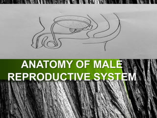
ANATOMY OF MALE
REPRODUCTIVE SYSTEM
PARTS OF MALE REPRODUCTIVE SYSTEM
The external structures of male reproductive system include:
 The penis
 The scrotum
 The testicles &Epididymis
The internal organs of the male reproductive system, also called
accessory organs, include the following:
 Vas deferens
 Ejaculatory ducts
 Urethra
 Seminal vesicles
 Prostate gland
 Bulbourethral glands
The penis
The penis is an external organ of the male
reproductive system.
STRUCTURE OF THE PENIS
The penis can be anatomically divided into
three parts:
Root – the most proximal, fixed part of the
penis.
• It is located in the superficial perineal
pouch of the pelvic floor, and is not
visible externally.
• The root contains three erectile tissues
(two crura and bulb of the penis), and
two muscles (ischiocavernosus and
bulbospongiosus).
• Body – the free part of the penis, located between
the root and glans. It is suspended from the pubic
symphysis. It is composed of three cylinders of
erectile tissue – two corpora cavernosa, and the
corpus spongiosum.
• Glans – the most distal part of the of penis. It is
conical in shape, and is formed by the distal
expansion of the corpus spongiosum. This contains
the opening of the urethra, termed the external
urethral orifice.
Erectile Tissues
• The erectile tissues fill with blood during sexual arousal,
producing an erection. The root and body of the penis
are spanned by three masses of erectile tissue.
• In the root, these tissues are known as the left and
right crura, and the bulb of the penis. The bulb is
situated in the midline of the penile root, and is
traversed by the urethra. The left and right crura are
located laterally; attached to the ipsilateral ischial
ramus, and covered by the paired ischiocavernosal
muscles.
• The erectile tissues continue into the body
of the penis. The left and right crura
continue anteriorly into the dorsal part of
the penis – they form the two corpora
cavernosa. They are separated by the
septum of the penis, although often
incompletely.
• The bulb forms the corpus
spongiosum, which lies
ventrally. The male urethra
runs through the corpus
spongiosum – to prevent it
becoming occluded during
erection the corpus
spongiosum fills to a reduced
pressure.
• Distally, the corpus
spongiosum expands to form
the glans penis.
The erectile tissues of the penis.
Muscles
• There are four muscles located in the root of the
penis:
• Bulbospongiosus (x2) – associated with the bulb of
the penis. It contracts to empty the spongy urethra
of any residual semen and urine.
• Ischiocavernosus (x2) – surrounds the left and right
crura of the penis. It contracts to force blood from
the cavernous spaces in the crura into the corpus
cavernosa – this helps maintain erection.
Ligaments
• The root of the penis is supported by two ligaments,
which attach it to the surrounding structures:
• Suspensory ligament – a condensation of deep
fascia. It connects the erectile bodies of the penis to
the pubic symphysis.
• Fundiform ligament – a condensation of abdominal
subcutaneous tissue. It runs down from the linea
alba, surrounding the penis like a sling, and attaching
to the pubic symphysis.
• Skin
• The skin of the penis is more heavily pigmented than
that of the rest of the body. It is connected to the
underlying fascias by loose connective tissue.
• The prepuce (foreskin) is a double layer of skin and
fascia, located at the neck of the glans. It covers the
glans to a variable extent. The prepuce is connected
to the surface of the glans by the frenulum, a median
fold of skin on the ventral surface of the penis.
Neurovascular Supply
Vasculature
• The penis receives arterial supply from three sources:
• Dorsal arteries of the penis
• Deep arteries of the penis
• Bulbourethral artery
• These arteries are all branches of the internal pudendal
artery. This vessel arises from the anterior division of
the internal iliac artery.
Venous drainage:
Venous blood is drained from the penis by paired veins. The
cavernous spaces are drained by the deep dorsal vein of
the penis – this empties into the prostatic venous plexus
. The superficial dorsal veins drain the superficial structures
of the penis, such as the skin and cutaneous tissues.
Nerve Innervation:
• The penis is supplied by S2-S4 spinal cord segments and
spinal ganglia.
• Sensory and sympathetic innervation to the skin and glans
penis is supplied by the dorsal nerve of the penis, a branch
of the pudendal nerve.
Clinical Relevance
Paraphimosis
• Paraphimosis is an acute condition that occurs when
a tight prepuce is left retracted under the glans: this
may cause oedema of the soft prepuce and further
strangulation occurs.
Scrotum
• The scrotum is a fibromuscular cutaneous sac,
located between the penis and anus. It is dual-
chambered, forming an expansion of the
perineum.
• Embryologically, the scrotum is derived from
the paired genital swellings. During
development, the genital swellings fuse in the
midline – in the adult this fusion is marked by
the scrotal raphe. The scrotum is biologically
homologous to the labia majora.
The scrotum, muscle layer and contents
Neurovascular Supply
• Vessels
• The scrotum receives arterial supply from the
anterior and posterior scrotal arteries. The anterior
scrotal artery arises from the external pudendal
artery, while the posterior is derived from the
internal pudendal artery.
• The scrotal veins follow the major arteries, draining
into the external pudendal veins.
• Nerves
• Cutaneous innervation to the scrotum is supplied via
several nerves, according to the topography:
• Anterior and anterolateral aspect – Anterior scrotal
nerves derived from the genital branch of
genitofemoral nerve and ilioinguinal nerve
• Posterior aspect – Posterior scrotal nerves derived
from the perineal branches of the pudendal nerve
and posterior femoral cutaneous nerve.
• Lymphatics
• The lymphatic fluid from the scrotum drains to the
nearby superficial inguinal nodes.
CLINICAL RELEVANCE:
Haematoma of the Scrotum
• A haematoma may develop in the scrotum as a result
of scrotal surgery or trauma in the genital region.
• This results in swelling (oedema) and discolouration
of the scrotal skin.
The Testes and Epididymis
The testes and epididymis are paired structures, located
within the scrotum. The testes are the site of sperm
production and hormone synthesis, while the epididymis
has a role in the storage of sperm.
ANATOMICAL POSITION:
The testes are located within the scrotum, with the
epididymis situated on the posterolateral aspect of each
testicle.
Originally, the testes are located on the posterior
abdominal wall. During embryonic development they
descend down the abdomen, and through the inguinal
canal to reach the scrotum
The testes and epididymis, surrounded by the tunica
vaginalis.
Anatomical Structure
• The testes have an ellipsoid shape. They consist of a
series of lobules, each containing seminiferous
tubules supported by interstitial tissue. The
seminiferous tubules are lined by Sertoli cells that aid
the maturation process of the spermatozoa.
• Spermatozoa are produced in the seminiferous
tubules. The developing sperm travels through the
tubules, collecting in the rete testes.
• Inside the scrotum, the testes are covered almost
entirely by the tunica vaginalis, a closed sac of parietal
peritoneal origin that contains a small amount of
viscous fluid
• The testicular parenchyma is protected by the tunica
albuginea, a fibrous capsule that encloses the testes.
• The epididymis consists of a single heavily coiled duct. It
can be divided into three parts; head, body and tail.
• Head – The most proximal part of the epididymis. It is
formed by the efferent tubules of the testes, which
transport sperm from the testes to the epididymis.
• Body – Formed by the heavily coiled duct of the
epididymis.
• Tail – The most distal part of the epididymis. It marks the
origin of the vas deferens, which transports sperm to the
prostatic portion of the urethra for ejaculation.
•
Structure of the testes and epididymis
Vascular Supply
ARTERIAL SUPPLY: paired testicular arteries, which
arise directly from the abdominal aorta.
• Testes are also supplied by branches of the
cremasteric artery (from the inferior epigastric
artery) and the artery of the vas deferens (from the
inferior vesical artery).
VENOUS DRAINAGE: Paired testicular veins. They are
formed from the pampiniform plexus in the scrotum
– a network of veins wrapped around the testicular
artery.
INNERVATION:
The testes and epididymis receive innervation from the
testicular plexus – a network of nerves derived from
the renal and aortic plexi. They receive autonomic
and sensory fibres.
LYMPHATICS:
The lymphatic drainage is to the lumbar and para-
aortic nodes, along the lumbar vertebrae.
• This is in contrast to the scrotum, which drains into
the nearby superficial inguinal nodes.
Clinical Relevance
• Inguinal hernia – where the contents of the abdominal
cavity protrude into the scrotum, via the inguinal canal.
• Hydrocoele – a collection of serous fluid within the tunica
vaginalis. The congenital form is most commonly due to a
failure of the processus vaginalis to close. Adult hydrocele
is often associated with inflammation or trauma and
rarely, testicular tumors.
• Haematocoele – a collection of blood in the tunica
vaginalis. It can be distinguished from a hydrocoele
by transillumination (where a light is applied to the
testicular swelling).
• Varicocoele – gross dilation of the veins draining the
testes. The left testicle is more commonly affected,
as the left testicular vein is longer and drains into the
left renal vein at a perpendicular angle.
• Epididymitis – inflammation of the epididymis,
usually caused by bacterial or viral infection
INTERNAL ORGANS/ACCESSORY ORGANS
• Vas deferens — The vas deferens is a long, muscular tube
that travels from the epididymis into the pelvic cavity, to just
behind the bladder. The vas deferens transports mature
sperm to the urethra in preparation for ejaculation.
• Ejaculatory ducts — These are formed by the fusion of the
vas deferens and the seminal vesicles. The ejaculatory ducts
empty into the urethra.
• Urethra — The urethra is the tube that carries urine from the
bladder to outside of the body. In males, it has the additional
function of expelling (ejaculating) semen when the man
reaches orgasm. When the penis is erect during sex, the flow
of urine is blocked from the urethra, allowing only semen to
be ejaculated at orgasm.
Inferior view of the structures in the male
reproductive system.
The Seminal Vesicles
The seminal vesicles (also
known as the vesicular or
seminal glands) are a pair
of glands found in the
male pelvis, which
function to produce many
of the constituent
ingredients of semen.
They ultimately provide
around 70% of the total
volume of semen. Anatomical position of the seminal
vesicles in relation to the vas deferens
and prostate.
Anatomical Position and Structure
• The seminal glands are a pair of 5cm long tubular
glands. They are located between the
bladder fundus and the rectum (separated from
the latter by the rectovesicle pouch and the
rectoprostatic fascia).
• Their most important anatomical relation is with
the vas deferens, which combine with the duct of
the seminal vesicles to form the ejaculatory duct,
which subsequently drains into the prostatic
urethra.
• Internally the gland has a honeycombed, lobulated
structure with a mucosa lined by pseudostratified
columnar epithelium. These columnar cells are highly
influenced by testosterone, growing taller with higher
levels, and are responsible for the production of seminal
secretions.
Embryology
• The Seminal glands, along with the Ejaculatory ducts,
Epididymis and Ductus (vas) deferens, are derived from
the mesonephric ducts, the precursor structure of male
internal genitalia.
VASCULATURE
• The arteries to the seminal gland are derived from
the inferior vesicle, internal pudendal and middle
rectal arteries, all of which stem from the internal
iliac artery.
Innervation:The innervation of the gland, like much of
the male internal genitalia, is mainly sympathetic in
origin.
Lymphatic Drainage
• The lymphatic drainage of the gland is the external
and internal iliac lymph nodes.
The Prostate Gland
• The prostate is the largest accessory gland in the
male reproductive system.
• It secretes proteolytic enzymes into the semen,
which act to break down clotting factors in the
ejaculate. This allows the semen to remain in a fluid
state, moving throughout the female reproductive
tract for potential fertilisation.
Anatomical Position
• The prostate is positioned inferiorly to the neck of
the bladder and superiorly to the external urethral
sphincter, with the levator ani muscle lying
inferolaterally to the gland.
• The proteolytic enzymes leave the prostate via
the prostatic ducts. These open into the prostatic
portion of the urethra, through 10-12 openings at
each side of the seminal colliculus (or
verumontanum); secreting the enzymes into the
semen immediately before ejaculation.
Anatomical Structure
• The prostate is commonly described as being the size
of a walnut.
• Roughly two-thirds of the prostate is glandular in
structure and the remaining third is fibromuscular.
The gland itself is surrounded by a thin fibrous
capsule of the prostate.
• This is not a real capsule; it rather resembles the thin
connective tissue known as adventitia in the large
blood vessels.
• More important clinically
is the histological division
of the prostate into three
zones
• Central zone – surrounds
the ejaculatory ducts,
comprising approximately
25% of normal prostate
volume.The ducts of the
glands from the central
zone are obliquely
emptying in the prostatic
urethra, thus being rather
immune to urine reflux. The anatomical position and zones of the
prostate
• Transitional zone – located centrally and surrounds the
urethra, comprising approximately 5-10% of normal
prostate volume.
• The glands of the transitional zone are those that typically
undergo benign hyperplasia (BPH)
• Peripheral zone – makes up the main body of the gland
(approximately 65%) and is located posteriorly.The ducts of
the glands from the peripheral zone are vertically emptying
in the prostatic urethra; that may explain the tendency of
these glands to permit urine reflux.
• The fibromuscular stroma (or fourth zone for some) is
situated anteriorly in the gland. It merges with the tissue of
the urogenital diaphragm.
Vasculature
The arterial supply to the prostate comes from the prostatic
arteries, which are mainly derived from the internal iliac
arteries. Some branches may also arise from the internal
pudendal and middle rectal arteries.
• Venous drainage of the prostate is via the prostatic venous
plexus, draining into the internal iliac veins.
• Innervation
• The prostate receives sympathetic, parasympathetic and
sensory innervation from the inferior hypogastric plexus.
The smooth muscle of the prostate gland is innervated by
sympathetic fibres, which activate during ejaculation.
Clinical Relevance -
• Prostatic Carcinoma: Prostatic carcinoma represents the
most commonly diagnosed cancer in men, especially in
countries with high sociodemographic index. The malignant
cells commonly originate from the peripheral zone,
although carcinomas may arise (more rarely) from the
central and transition zones too. It is still debatable that the
latter tumors may present with lower malignant potential.
• However the proximity of the peripheral zone to the
neurovascular bundle that surrounds the prostate may
facilitate spread along perineural and lymphatic pathways,
thus increasing the metastatic potential of these tumors.
• Benign Prostatic Hyperplasia (BPH)
• Benign prostatic hyperplasia is the increase in size of
the prostate, without the presence of malignancy. It
is much more common with advancing age, although
initial histological evidence of hyperplasia may be
evident from much earlier ages (<40 yrs old).
• PROSTATIS :Inflammation of the prostate
The Bulbourethral Glands
• The bulbourethral glands (also
known as Cowper’s glands) are a
pair of pea shaped exocrine
glands located posterolateral to
the membranous urethra. They
contribute to the final volume of
semen by producing a lubricating
mucus secretion.
• Anatomical Position and
Structure:
• the bulbourethral glands can be
found in the deep perineal pouch
of the male. Anatomical position of the bulbourethral
gland.
• They are situated posterolaterally to the membranous
urethra and superiorly to the bulb of the penis. Anatomical
position of the bulbourethral gland.
• The ducts of the gland penetrate the perineal membrane
alongside the membranous urethra and open into the
proximal portion of the spongy urethra.
• The glands themselves can be described as compound
tubulo-alveolar glands lined by columnar epithelium.
Embryology
• Embryologically the bulbourethral glands are derived from
the urogenital sinus, along with the bladder, prostate and
urethra. Their development is greatly influenced by DHT
(dihydrotestosterone).
VASCULATURE
• The arterial supply of the bulbourethral glands is derived
from the arteries to the bulb of the penis.
• Innervation
• In a mammal study (in pigs), neurons projecting to the
bulbourethral glands were found in pelvic ganglia (PG),
sympathetic chain ganglia (L2–S3), the caudal mesenteric
ganglion and dorsal root ganglia (L1–L3, S1–S3);
• They reach the bulbourethral glands via the the
hypogastric nerve and the pelvic nerve or pelvic branch of
the pudendal nerve.
Lymphatics
• Like the seminal vesicles the
bulbourethral glands drain into the
internal and external iliac lymph nodes
Male Reproductive Anatomy Guide

More Related Content

What's hot

The Male Reproductive System
The Male Reproductive System The Male Reproductive System
The Male Reproductive System maratima
 
Anatomy & Histology of ovary and Oogenesis
Anatomy & Histology of ovary and Oogenesis Anatomy & Histology of ovary and Oogenesis
Anatomy & Histology of ovary and Oogenesis Dr. Devi Shankar
 
Histology of male reproductive system
Histology of male reproductive systemHistology of male reproductive system
Histology of male reproductive systemDr Laxman Khanal
 
09.24.08: Skin and Glands
09.24.08: Skin and Glands 09.24.08: Skin and Glands
09.24.08: Skin and Glands Open.Michigan
 
Female Reproductive system
Female Reproductive systemFemale Reproductive system
Female Reproductive systemJegan Nadar
 
3rd week of development and derivatives of germ
3rd week of development and derivatives of germ3rd week of development and derivatives of germ
3rd week of development and derivatives of germAbdul Ansari
 
Spermatogenesis-converted.pdf
Spermatogenesis-converted.pdfSpermatogenesis-converted.pdf
Spermatogenesis-converted.pdfPooja Rani
 
Female reproductive system
Female reproductive systemFemale reproductive system
Female reproductive systemChintan Chavda
 
Anatomy and Physiology of Male reproductive system
Anatomy and Physiology of Male reproductive systemAnatomy and Physiology of Male reproductive system
Anatomy and Physiology of Male reproductive systemDR .PALLAVI PATHANIA
 
Histology of special senses
Histology of special sensesHistology of special senses
Histology of special sensesmgmcri1234
 
Histology of small intestine
Histology of small intestine Histology of small intestine
Histology of small intestine Shizra Imtiaz
 
Female reproductive system
Female reproductive systemFemale reproductive system
Female reproductive systemUnknown Jaffar
 
Female reproductive system
Female reproductive systemFemale reproductive system
Female reproductive systemUE
 
Glandular epith
Glandular epithGlandular epith
Glandular epithAhmedooz
 
Male reproductive system
Male reproductive systemMale reproductive system
Male reproductive systemwaheed Bahir
 
Spermatogenesis (Male Reproductive System-2)
Spermatogenesis (Male Reproductive System-2)Spermatogenesis (Male Reproductive System-2)
Spermatogenesis (Male Reproductive System-2)Dr. Sarita Sharma
 

What's hot (20)

Male reproductive system
Male reproductive systemMale reproductive system
Male reproductive system
 
The Male Reproductive System
The Male Reproductive System The Male Reproductive System
The Male Reproductive System
 
Anatomy & Histology of ovary and Oogenesis
Anatomy & Histology of ovary and Oogenesis Anatomy & Histology of ovary and Oogenesis
Anatomy & Histology of ovary and Oogenesis
 
Histology of male reproductive system
Histology of male reproductive systemHistology of male reproductive system
Histology of male reproductive system
 
09.24.08: Skin and Glands
09.24.08: Skin and Glands 09.24.08: Skin and Glands
09.24.08: Skin and Glands
 
Female Reproductive system
Female Reproductive systemFemale Reproductive system
Female Reproductive system
 
3rd week of development and derivatives of germ
3rd week of development and derivatives of germ3rd week of development and derivatives of germ
3rd week of development and derivatives of germ
 
Spermatogenesis-converted.pdf
Spermatogenesis-converted.pdfSpermatogenesis-converted.pdf
Spermatogenesis-converted.pdf
 
Female reproductive system
Female reproductive systemFemale reproductive system
Female reproductive system
 
Anatomy and Physiology of Male reproductive system
Anatomy and Physiology of Male reproductive systemAnatomy and Physiology of Male reproductive system
Anatomy and Physiology of Male reproductive system
 
Histology of special senses
Histology of special sensesHistology of special senses
Histology of special senses
 
Oogenesis
OogenesisOogenesis
Oogenesis
 
Histology of small intestine
Histology of small intestine Histology of small intestine
Histology of small intestine
 
Female reproductive system
Female reproductive systemFemale reproductive system
Female reproductive system
 
Physiology of reproductive system
Physiology of reproductive systemPhysiology of reproductive system
Physiology of reproductive system
 
Female reproductive system
Female reproductive systemFemale reproductive system
Female reproductive system
 
Glandular epith
Glandular epithGlandular epith
Glandular epith
 
Male reproductive system
Male reproductive systemMale reproductive system
Male reproductive system
 
Female reproductive system
Female reproductive system Female reproductive system
Female reproductive system
 
Spermatogenesis (Male Reproductive System-2)
Spermatogenesis (Male Reproductive System-2)Spermatogenesis (Male Reproductive System-2)
Spermatogenesis (Male Reproductive System-2)
 

Similar to Male Reproductive Anatomy Guide

Male Reproductive System Anju - Copy.pptx
Male Reproductive System Anju - Copy.pptxMale Reproductive System Anju - Copy.pptx
Male Reproductive System Anju - Copy.pptxAnju Kumawat
 
malereproductivesystemnew-190314170104.pdf
malereproductivesystemnew-190314170104.pdfmalereproductivesystemnew-190314170104.pdf
malereproductivesystemnew-190314170104.pdfDennis Agyemfra
 
Anatomy of the Male External genitalia.pptx
Anatomy of the Male External genitalia.pptxAnatomy of the Male External genitalia.pptx
Anatomy of the Male External genitalia.pptxDr Ndayisaba Corneille
 
male reproductive system.pptx
male reproductive system.pptxmale reproductive system.pptx
male reproductive system.pptxAnumSajid12
 
Male reproductive system - Anatomy
Male reproductive system - AnatomyMale reproductive system - Anatomy
Male reproductive system - AnatomyMohammad Ihmeidan
 
Genitourinary system surgical antomy.pptx
Genitourinary system surgical antomy.pptxGenitourinary system surgical antomy.pptx
Genitourinary system surgical antomy.pptxPradeep Pande
 
malereproductivesystem
malereproductivesystemmalereproductivesystem
malereproductivesystemRathiNivedhana
 
2. Male reproductive (Acessory glands, Urethra and penis)
2. Male reproductive (Acessory glands, Urethra  and penis)2. Male reproductive (Acessory glands, Urethra  and penis)
2. Male reproductive (Acessory glands, Urethra and penis)Dr. Mohammad Mahmoud
 
Female reproductive system
Female reproductive systemFemale reproductive system
Female reproductive systemSubhashini N
 
male external genital organs.pptx
male external genital organs.pptxmale external genital organs.pptx
male external genital organs.pptxDr Mohammad Amaan
 
Lect 2 anterior abdominal wall
Lect 2  anterior abdominal wallLect 2  anterior abdominal wall
Lect 2 anterior abdominal wallMohaned Lehya
 
Anatomy & embryology of urinary bladder
Anatomy & embryology of urinary bladderAnatomy & embryology of urinary bladder
Anatomy & embryology of urinary bladderDeepesh Kalra
 
male reproductive organs
male reproductive organsmale reproductive organs
male reproductive organsMohanad Mohanad
 
Male Reproductive system (without Audio).pptx
Male Reproductive system (without Audio).pptxMale Reproductive system (without Audio).pptx
Male Reproductive system (without Audio).pptxAsifBahadurAli
 
LECT. 13 MALE REPRODUCTIVE SYSTEM.ppt
LECT. 13 MALE REPRODUCTIVE SYSTEM.pptLECT. 13 MALE REPRODUCTIVE SYSTEM.ppt
LECT. 13 MALE REPRODUCTIVE SYSTEM.pptHajiDrammeh
 

Similar to Male Reproductive Anatomy Guide (20)

Male reproductive system
Male reproductive systemMale reproductive system
Male reproductive system
 
Male Reproductive System Anju - Copy.pptx
Male Reproductive System Anju - Copy.pptxMale Reproductive System Anju - Copy.pptx
Male Reproductive System Anju - Copy.pptx
 
malereproductivesystemnew-190314170104.pdf
malereproductivesystemnew-190314170104.pdfmalereproductivesystemnew-190314170104.pdf
malereproductivesystemnew-190314170104.pdf
 
Anatomy of the Male External genitalia.pptx
Anatomy of the Male External genitalia.pptxAnatomy of the Male External genitalia.pptx
Anatomy of the Male External genitalia.pptx
 
male reproductive system.pptx
male reproductive system.pptxmale reproductive system.pptx
male reproductive system.pptx
 
Male reproductive system
Male reproductive systemMale reproductive system
Male reproductive system
 
Male reproductive system - Anatomy
Male reproductive system - AnatomyMale reproductive system - Anatomy
Male reproductive system - Anatomy
 
Anatomy penis.pptx
Anatomy penis.pptxAnatomy penis.pptx
Anatomy penis.pptx
 
Genitourinary system surgical antomy.pptx
Genitourinary system surgical antomy.pptxGenitourinary system surgical antomy.pptx
Genitourinary system surgical antomy.pptx
 
Male reproductive system anatomy
Male reproductive system anatomyMale reproductive system anatomy
Male reproductive system anatomy
 
malereproductivesystem
malereproductivesystemmalereproductivesystem
malereproductivesystem
 
2. Male reproductive (Acessory glands, Urethra and penis)
2. Male reproductive (Acessory glands, Urethra  and penis)2. Male reproductive (Acessory glands, Urethra  and penis)
2. Male reproductive (Acessory glands, Urethra and penis)
 
Female reproductive system
Female reproductive systemFemale reproductive system
Female reproductive system
 
male external genital organs.pptx
male external genital organs.pptxmale external genital organs.pptx
male external genital organs.pptx
 
Lect 2 anterior abdominal wall
Lect 2  anterior abdominal wallLect 2  anterior abdominal wall
Lect 2 anterior abdominal wall
 
Anatomy & embryology of urinary bladder
Anatomy & embryology of urinary bladderAnatomy & embryology of urinary bladder
Anatomy & embryology of urinary bladder
 
male reproductive organs
male reproductive organsmale reproductive organs
male reproductive organs
 
Male external genitalia
Male external genitaliaMale external genitalia
Male external genitalia
 
Male Reproductive system (without Audio).pptx
Male Reproductive system (without Audio).pptxMale Reproductive system (without Audio).pptx
Male Reproductive system (without Audio).pptx
 
LECT. 13 MALE REPRODUCTIVE SYSTEM.ppt
LECT. 13 MALE REPRODUCTIVE SYSTEM.pptLECT. 13 MALE REPRODUCTIVE SYSTEM.ppt
LECT. 13 MALE REPRODUCTIVE SYSTEM.ppt
 

More from Subhashini N

Benifits of e learning
Benifits of e learningBenifits of e learning
Benifits of e learningSubhashini N
 
Sample size calculation for cohort studies
Sample size calculation for cohort studies Sample size calculation for cohort studies
Sample size calculation for cohort studies Subhashini N
 
Types of NANDA-I Nursing Diagnosis
 Types of NANDA-I Nursing Diagnosis Types of NANDA-I Nursing Diagnosis
Types of NANDA-I Nursing DiagnosisSubhashini N
 
ANATOMY OF MAMMARY GLAND
ANATOMY OF MAMMARY GLANDANATOMY OF MAMMARY GLAND
ANATOMY OF MAMMARY GLANDSubhashini N
 
Individualized instruction teaching method
Individualized instruction teaching methodIndividualized instruction teaching method
Individualized instruction teaching methodSubhashini N
 
Normal ecg interpretation
Normal ecg interpretationNormal ecg interpretation
Normal ecg interpretationSubhashini N
 

More from Subhashini N (7)

Benifits of e learning
Benifits of e learningBenifits of e learning
Benifits of e learning
 
Sample size calculation for cohort studies
Sample size calculation for cohort studies Sample size calculation for cohort studies
Sample size calculation for cohort studies
 
Types of NANDA-I Nursing Diagnosis
 Types of NANDA-I Nursing Diagnosis Types of NANDA-I Nursing Diagnosis
Types of NANDA-I Nursing Diagnosis
 
ANATOMY OF MAMMARY GLAND
ANATOMY OF MAMMARY GLANDANATOMY OF MAMMARY GLAND
ANATOMY OF MAMMARY GLAND
 
Lung cancer
Lung cancerLung cancer
Lung cancer
 
Individualized instruction teaching method
Individualized instruction teaching methodIndividualized instruction teaching method
Individualized instruction teaching method
 
Normal ecg interpretation
Normal ecg interpretationNormal ecg interpretation
Normal ecg interpretation
 

Recently uploaded

Call Girl Bangalore Nandini 7001305949 Independent Escort Service Bangalore
Call Girl Bangalore Nandini 7001305949 Independent Escort Service BangaloreCall Girl Bangalore Nandini 7001305949 Independent Escort Service Bangalore
Call Girl Bangalore Nandini 7001305949 Independent Escort Service Bangalorenarwatsonia7
 
Low Rate Call Girls Pune Esha 9907093804 Short 1500 Night 6000 Best call girl...
Low Rate Call Girls Pune Esha 9907093804 Short 1500 Night 6000 Best call girl...Low Rate Call Girls Pune Esha 9907093804 Short 1500 Night 6000 Best call girl...
Low Rate Call Girls Pune Esha 9907093804 Short 1500 Night 6000 Best call girl...Miss joya
 
Low Rate Call Girls Mumbai Suman 9910780858 Independent Escort Service Mumbai
Low Rate Call Girls Mumbai Suman 9910780858 Independent Escort Service MumbaiLow Rate Call Girls Mumbai Suman 9910780858 Independent Escort Service Mumbai
Low Rate Call Girls Mumbai Suman 9910780858 Independent Escort Service Mumbaisonalikaur4
 
Call Girls Jp Nagar Just Call 7001305949 Top Class Call Girl Service Available
Call Girls Jp Nagar Just Call 7001305949 Top Class Call Girl Service AvailableCall Girls Jp Nagar Just Call 7001305949 Top Class Call Girl Service Available
Call Girls Jp Nagar Just Call 7001305949 Top Class Call Girl Service Availablenarwatsonia7
 
call girls in munirka DELHI 🔝 >༒9540349809 🔝 genuine Escort Service 🔝✔️✔️
call girls in munirka  DELHI 🔝 >༒9540349809 🔝 genuine Escort Service 🔝✔️✔️call girls in munirka  DELHI 🔝 >༒9540349809 🔝 genuine Escort Service 🔝✔️✔️
call girls in munirka DELHI 🔝 >༒9540349809 🔝 genuine Escort Service 🔝✔️✔️saminamagar
 
Call Girls Service in Bommanahalli - 7001305949 with real photos and phone nu...
Call Girls Service in Bommanahalli - 7001305949 with real photos and phone nu...Call Girls Service in Bommanahalli - 7001305949 with real photos and phone nu...
Call Girls Service in Bommanahalli - 7001305949 with real photos and phone nu...narwatsonia7
 
call girls in Connaught Place DELHI 🔝 >༒9540349809 🔝 genuine Escort Service ...
call girls in Connaught Place  DELHI 🔝 >༒9540349809 🔝 genuine Escort Service ...call girls in Connaught Place  DELHI 🔝 >༒9540349809 🔝 genuine Escort Service ...
call girls in Connaught Place DELHI 🔝 >༒9540349809 🔝 genuine Escort Service ...saminamagar
 
Call Girls ITPL Just Call 7001305949 Top Class Call Girl Service Available
Call Girls ITPL Just Call 7001305949 Top Class Call Girl Service AvailableCall Girls ITPL Just Call 7001305949 Top Class Call Girl Service Available
Call Girls ITPL Just Call 7001305949 Top Class Call Girl Service Availablenarwatsonia7
 
VIP Call Girls Mumbai Arpita 9910780858 Independent Escort Service Mumbai
VIP Call Girls Mumbai Arpita 9910780858 Independent Escort Service MumbaiVIP Call Girls Mumbai Arpita 9910780858 Independent Escort Service Mumbai
VIP Call Girls Mumbai Arpita 9910780858 Independent Escort Service Mumbaisonalikaur4
 
Call Girls Hsr Layout Just Call 7001305949 Top Class Call Girl Service Available
Call Girls Hsr Layout Just Call 7001305949 Top Class Call Girl Service AvailableCall Girls Hsr Layout Just Call 7001305949 Top Class Call Girl Service Available
Call Girls Hsr Layout Just Call 7001305949 Top Class Call Girl Service Availablenarwatsonia7
 
Bangalore Call Girls Marathahalli 📞 9907093804 High Profile Service 100% Safe
Bangalore Call Girls Marathahalli 📞 9907093804 High Profile Service 100% SafeBangalore Call Girls Marathahalli 📞 9907093804 High Profile Service 100% Safe
Bangalore Call Girls Marathahalli 📞 9907093804 High Profile Service 100% Safenarwatsonia7
 
Call Girl Lucknow Mallika 7001305949 Independent Escort Service Lucknow
Call Girl Lucknow Mallika 7001305949 Independent Escort Service LucknowCall Girl Lucknow Mallika 7001305949 Independent Escort Service Lucknow
Call Girl Lucknow Mallika 7001305949 Independent Escort Service Lucknownarwatsonia7
 
Book Call Girls in Kasavanahalli - 7001305949 with real photos and phone numbers
Book Call Girls in Kasavanahalli - 7001305949 with real photos and phone numbersBook Call Girls in Kasavanahalli - 7001305949 with real photos and phone numbers
Book Call Girls in Kasavanahalli - 7001305949 with real photos and phone numbersnarwatsonia7
 
Russian Call Girls Chickpet - 7001305949 Booking and charges genuine rate for...
Russian Call Girls Chickpet - 7001305949 Booking and charges genuine rate for...Russian Call Girls Chickpet - 7001305949 Booking and charges genuine rate for...
Russian Call Girls Chickpet - 7001305949 Booking and charges genuine rate for...narwatsonia7
 
Call Girls Hosur Just Call 7001305949 Top Class Call Girl Service Available
Call Girls Hosur Just Call 7001305949 Top Class Call Girl Service AvailableCall Girls Hosur Just Call 7001305949 Top Class Call Girl Service Available
Call Girls Hosur Just Call 7001305949 Top Class Call Girl Service Availablenarwatsonia7
 
VIP Call Girls Lucknow Nandini 7001305949 Independent Escort Service Lucknow
VIP Call Girls Lucknow Nandini 7001305949 Independent Escort Service LucknowVIP Call Girls Lucknow Nandini 7001305949 Independent Escort Service Lucknow
VIP Call Girls Lucknow Nandini 7001305949 Independent Escort Service Lucknownarwatsonia7
 
Housewife Call Girls Bangalore - Call 7001305949 Rs-3500 with A/C Room Cash o...
Housewife Call Girls Bangalore - Call 7001305949 Rs-3500 with A/C Room Cash o...Housewife Call Girls Bangalore - Call 7001305949 Rs-3500 with A/C Room Cash o...
Housewife Call Girls Bangalore - Call 7001305949 Rs-3500 with A/C Room Cash o...narwatsonia7
 
Kolkata Call Girls Services 9907093804 @24x7 High Class Babes Here Call Now
Kolkata Call Girls Services 9907093804 @24x7 High Class Babes Here Call NowKolkata Call Girls Services 9907093804 @24x7 High Class Babes Here Call Now
Kolkata Call Girls Services 9907093804 @24x7 High Class Babes Here Call NowNehru place Escorts
 
Ahmedabad Call Girls CG Road 🔝9907093804 Short 1500 💋 Night 6000
Ahmedabad Call Girls CG Road 🔝9907093804  Short 1500  💋 Night 6000Ahmedabad Call Girls CG Road 🔝9907093804  Short 1500  💋 Night 6000
Ahmedabad Call Girls CG Road 🔝9907093804 Short 1500 💋 Night 6000aliya bhat
 
Book Call Girls in Yelahanka - For 7001305949 Cheap & Best with original Photos
Book Call Girls in Yelahanka - For 7001305949 Cheap & Best with original PhotosBook Call Girls in Yelahanka - For 7001305949 Cheap & Best with original Photos
Book Call Girls in Yelahanka - For 7001305949 Cheap & Best with original Photosnarwatsonia7
 

Recently uploaded (20)

Call Girl Bangalore Nandini 7001305949 Independent Escort Service Bangalore
Call Girl Bangalore Nandini 7001305949 Independent Escort Service BangaloreCall Girl Bangalore Nandini 7001305949 Independent Escort Service Bangalore
Call Girl Bangalore Nandini 7001305949 Independent Escort Service Bangalore
 
Low Rate Call Girls Pune Esha 9907093804 Short 1500 Night 6000 Best call girl...
Low Rate Call Girls Pune Esha 9907093804 Short 1500 Night 6000 Best call girl...Low Rate Call Girls Pune Esha 9907093804 Short 1500 Night 6000 Best call girl...
Low Rate Call Girls Pune Esha 9907093804 Short 1500 Night 6000 Best call girl...
 
Low Rate Call Girls Mumbai Suman 9910780858 Independent Escort Service Mumbai
Low Rate Call Girls Mumbai Suman 9910780858 Independent Escort Service MumbaiLow Rate Call Girls Mumbai Suman 9910780858 Independent Escort Service Mumbai
Low Rate Call Girls Mumbai Suman 9910780858 Independent Escort Service Mumbai
 
Call Girls Jp Nagar Just Call 7001305949 Top Class Call Girl Service Available
Call Girls Jp Nagar Just Call 7001305949 Top Class Call Girl Service AvailableCall Girls Jp Nagar Just Call 7001305949 Top Class Call Girl Service Available
Call Girls Jp Nagar Just Call 7001305949 Top Class Call Girl Service Available
 
call girls in munirka DELHI 🔝 >༒9540349809 🔝 genuine Escort Service 🔝✔️✔️
call girls in munirka  DELHI 🔝 >༒9540349809 🔝 genuine Escort Service 🔝✔️✔️call girls in munirka  DELHI 🔝 >༒9540349809 🔝 genuine Escort Service 🔝✔️✔️
call girls in munirka DELHI 🔝 >༒9540349809 🔝 genuine Escort Service 🔝✔️✔️
 
Call Girls Service in Bommanahalli - 7001305949 with real photos and phone nu...
Call Girls Service in Bommanahalli - 7001305949 with real photos and phone nu...Call Girls Service in Bommanahalli - 7001305949 with real photos and phone nu...
Call Girls Service in Bommanahalli - 7001305949 with real photos and phone nu...
 
call girls in Connaught Place DELHI 🔝 >༒9540349809 🔝 genuine Escort Service ...
call girls in Connaught Place  DELHI 🔝 >༒9540349809 🔝 genuine Escort Service ...call girls in Connaught Place  DELHI 🔝 >༒9540349809 🔝 genuine Escort Service ...
call girls in Connaught Place DELHI 🔝 >༒9540349809 🔝 genuine Escort Service ...
 
Call Girls ITPL Just Call 7001305949 Top Class Call Girl Service Available
Call Girls ITPL Just Call 7001305949 Top Class Call Girl Service AvailableCall Girls ITPL Just Call 7001305949 Top Class Call Girl Service Available
Call Girls ITPL Just Call 7001305949 Top Class Call Girl Service Available
 
VIP Call Girls Mumbai Arpita 9910780858 Independent Escort Service Mumbai
VIP Call Girls Mumbai Arpita 9910780858 Independent Escort Service MumbaiVIP Call Girls Mumbai Arpita 9910780858 Independent Escort Service Mumbai
VIP Call Girls Mumbai Arpita 9910780858 Independent Escort Service Mumbai
 
Call Girls Hsr Layout Just Call 7001305949 Top Class Call Girl Service Available
Call Girls Hsr Layout Just Call 7001305949 Top Class Call Girl Service AvailableCall Girls Hsr Layout Just Call 7001305949 Top Class Call Girl Service Available
Call Girls Hsr Layout Just Call 7001305949 Top Class Call Girl Service Available
 
Bangalore Call Girls Marathahalli 📞 9907093804 High Profile Service 100% Safe
Bangalore Call Girls Marathahalli 📞 9907093804 High Profile Service 100% SafeBangalore Call Girls Marathahalli 📞 9907093804 High Profile Service 100% Safe
Bangalore Call Girls Marathahalli 📞 9907093804 High Profile Service 100% Safe
 
Call Girl Lucknow Mallika 7001305949 Independent Escort Service Lucknow
Call Girl Lucknow Mallika 7001305949 Independent Escort Service LucknowCall Girl Lucknow Mallika 7001305949 Independent Escort Service Lucknow
Call Girl Lucknow Mallika 7001305949 Independent Escort Service Lucknow
 
Book Call Girls in Kasavanahalli - 7001305949 with real photos and phone numbers
Book Call Girls in Kasavanahalli - 7001305949 with real photos and phone numbersBook Call Girls in Kasavanahalli - 7001305949 with real photos and phone numbers
Book Call Girls in Kasavanahalli - 7001305949 with real photos and phone numbers
 
Russian Call Girls Chickpet - 7001305949 Booking and charges genuine rate for...
Russian Call Girls Chickpet - 7001305949 Booking and charges genuine rate for...Russian Call Girls Chickpet - 7001305949 Booking and charges genuine rate for...
Russian Call Girls Chickpet - 7001305949 Booking and charges genuine rate for...
 
Call Girls Hosur Just Call 7001305949 Top Class Call Girl Service Available
Call Girls Hosur Just Call 7001305949 Top Class Call Girl Service AvailableCall Girls Hosur Just Call 7001305949 Top Class Call Girl Service Available
Call Girls Hosur Just Call 7001305949 Top Class Call Girl Service Available
 
VIP Call Girls Lucknow Nandini 7001305949 Independent Escort Service Lucknow
VIP Call Girls Lucknow Nandini 7001305949 Independent Escort Service LucknowVIP Call Girls Lucknow Nandini 7001305949 Independent Escort Service Lucknow
VIP Call Girls Lucknow Nandini 7001305949 Independent Escort Service Lucknow
 
Housewife Call Girls Bangalore - Call 7001305949 Rs-3500 with A/C Room Cash o...
Housewife Call Girls Bangalore - Call 7001305949 Rs-3500 with A/C Room Cash o...Housewife Call Girls Bangalore - Call 7001305949 Rs-3500 with A/C Room Cash o...
Housewife Call Girls Bangalore - Call 7001305949 Rs-3500 with A/C Room Cash o...
 
Kolkata Call Girls Services 9907093804 @24x7 High Class Babes Here Call Now
Kolkata Call Girls Services 9907093804 @24x7 High Class Babes Here Call NowKolkata Call Girls Services 9907093804 @24x7 High Class Babes Here Call Now
Kolkata Call Girls Services 9907093804 @24x7 High Class Babes Here Call Now
 
Ahmedabad Call Girls CG Road 🔝9907093804 Short 1500 💋 Night 6000
Ahmedabad Call Girls CG Road 🔝9907093804  Short 1500  💋 Night 6000Ahmedabad Call Girls CG Road 🔝9907093804  Short 1500  💋 Night 6000
Ahmedabad Call Girls CG Road 🔝9907093804 Short 1500 💋 Night 6000
 
Book Call Girls in Yelahanka - For 7001305949 Cheap & Best with original Photos
Book Call Girls in Yelahanka - For 7001305949 Cheap & Best with original PhotosBook Call Girls in Yelahanka - For 7001305949 Cheap & Best with original Photos
Book Call Girls in Yelahanka - For 7001305949 Cheap & Best with original Photos
 

Male Reproductive Anatomy Guide

  • 2. PARTS OF MALE REPRODUCTIVE SYSTEM The external structures of male reproductive system include:  The penis  The scrotum  The testicles &Epididymis The internal organs of the male reproductive system, also called accessory organs, include the following:  Vas deferens  Ejaculatory ducts  Urethra  Seminal vesicles  Prostate gland  Bulbourethral glands
  • 3. The penis The penis is an external organ of the male reproductive system. STRUCTURE OF THE PENIS The penis can be anatomically divided into three parts: Root – the most proximal, fixed part of the penis. • It is located in the superficial perineal pouch of the pelvic floor, and is not visible externally. • The root contains three erectile tissues (two crura and bulb of the penis), and two muscles (ischiocavernosus and bulbospongiosus).
  • 4. • Body – the free part of the penis, located between the root and glans. It is suspended from the pubic symphysis. It is composed of three cylinders of erectile tissue – two corpora cavernosa, and the corpus spongiosum. • Glans – the most distal part of the of penis. It is conical in shape, and is formed by the distal expansion of the corpus spongiosum. This contains the opening of the urethra, termed the external urethral orifice.
  • 5. Erectile Tissues • The erectile tissues fill with blood during sexual arousal, producing an erection. The root and body of the penis are spanned by three masses of erectile tissue. • In the root, these tissues are known as the left and right crura, and the bulb of the penis. The bulb is situated in the midline of the penile root, and is traversed by the urethra. The left and right crura are located laterally; attached to the ipsilateral ischial ramus, and covered by the paired ischiocavernosal muscles.
  • 6. • The erectile tissues continue into the body of the penis. The left and right crura continue anteriorly into the dorsal part of the penis – they form the two corpora cavernosa. They are separated by the septum of the penis, although often incompletely.
  • 7. • The bulb forms the corpus spongiosum, which lies ventrally. The male urethra runs through the corpus spongiosum – to prevent it becoming occluded during erection the corpus spongiosum fills to a reduced pressure. • Distally, the corpus spongiosum expands to form the glans penis. The erectile tissues of the penis.
  • 8. Muscles • There are four muscles located in the root of the penis: • Bulbospongiosus (x2) – associated with the bulb of the penis. It contracts to empty the spongy urethra of any residual semen and urine. • Ischiocavernosus (x2) – surrounds the left and right crura of the penis. It contracts to force blood from the cavernous spaces in the crura into the corpus cavernosa – this helps maintain erection.
  • 9. Ligaments • The root of the penis is supported by two ligaments, which attach it to the surrounding structures: • Suspensory ligament – a condensation of deep fascia. It connects the erectile bodies of the penis to the pubic symphysis. • Fundiform ligament – a condensation of abdominal subcutaneous tissue. It runs down from the linea alba, surrounding the penis like a sling, and attaching to the pubic symphysis.
  • 10. • Skin • The skin of the penis is more heavily pigmented than that of the rest of the body. It is connected to the underlying fascias by loose connective tissue. • The prepuce (foreskin) is a double layer of skin and fascia, located at the neck of the glans. It covers the glans to a variable extent. The prepuce is connected to the surface of the glans by the frenulum, a median fold of skin on the ventral surface of the penis.
  • 11. Neurovascular Supply Vasculature • The penis receives arterial supply from three sources: • Dorsal arteries of the penis • Deep arteries of the penis • Bulbourethral artery • These arteries are all branches of the internal pudendal artery. This vessel arises from the anterior division of the internal iliac artery.
  • 12. Venous drainage: Venous blood is drained from the penis by paired veins. The cavernous spaces are drained by the deep dorsal vein of the penis – this empties into the prostatic venous plexus . The superficial dorsal veins drain the superficial structures of the penis, such as the skin and cutaneous tissues. Nerve Innervation: • The penis is supplied by S2-S4 spinal cord segments and spinal ganglia. • Sensory and sympathetic innervation to the skin and glans penis is supplied by the dorsal nerve of the penis, a branch of the pudendal nerve.
  • 13. Clinical Relevance Paraphimosis • Paraphimosis is an acute condition that occurs when a tight prepuce is left retracted under the glans: this may cause oedema of the soft prepuce and further strangulation occurs.
  • 14. Scrotum • The scrotum is a fibromuscular cutaneous sac, located between the penis and anus. It is dual- chambered, forming an expansion of the perineum. • Embryologically, the scrotum is derived from the paired genital swellings. During development, the genital swellings fuse in the midline – in the adult this fusion is marked by the scrotal raphe. The scrotum is biologically homologous to the labia majora.
  • 15. The scrotum, muscle layer and contents
  • 16. Neurovascular Supply • Vessels • The scrotum receives arterial supply from the anterior and posterior scrotal arteries. The anterior scrotal artery arises from the external pudendal artery, while the posterior is derived from the internal pudendal artery. • The scrotal veins follow the major arteries, draining into the external pudendal veins. • Nerves
  • 17. • Cutaneous innervation to the scrotum is supplied via several nerves, according to the topography: • Anterior and anterolateral aspect – Anterior scrotal nerves derived from the genital branch of genitofemoral nerve and ilioinguinal nerve • Posterior aspect – Posterior scrotal nerves derived from the perineal branches of the pudendal nerve and posterior femoral cutaneous nerve.
  • 18. • Lymphatics • The lymphatic fluid from the scrotum drains to the nearby superficial inguinal nodes. CLINICAL RELEVANCE: Haematoma of the Scrotum • A haematoma may develop in the scrotum as a result of scrotal surgery or trauma in the genital region. • This results in swelling (oedema) and discolouration of the scrotal skin.
  • 19. The Testes and Epididymis The testes and epididymis are paired structures, located within the scrotum. The testes are the site of sperm production and hormone synthesis, while the epididymis has a role in the storage of sperm. ANATOMICAL POSITION: The testes are located within the scrotum, with the epididymis situated on the posterolateral aspect of each testicle. Originally, the testes are located on the posterior abdominal wall. During embryonic development they descend down the abdomen, and through the inguinal canal to reach the scrotum
  • 20. The testes and epididymis, surrounded by the tunica vaginalis.
  • 21. Anatomical Structure • The testes have an ellipsoid shape. They consist of a series of lobules, each containing seminiferous tubules supported by interstitial tissue. The seminiferous tubules are lined by Sertoli cells that aid the maturation process of the spermatozoa. • Spermatozoa are produced in the seminiferous tubules. The developing sperm travels through the tubules, collecting in the rete testes. • Inside the scrotum, the testes are covered almost entirely by the tunica vaginalis, a closed sac of parietal peritoneal origin that contains a small amount of viscous fluid
  • 22. • The testicular parenchyma is protected by the tunica albuginea, a fibrous capsule that encloses the testes. • The epididymis consists of a single heavily coiled duct. It can be divided into three parts; head, body and tail. • Head – The most proximal part of the epididymis. It is formed by the efferent tubules of the testes, which transport sperm from the testes to the epididymis. • Body – Formed by the heavily coiled duct of the epididymis. • Tail – The most distal part of the epididymis. It marks the origin of the vas deferens, which transports sperm to the prostatic portion of the urethra for ejaculation. •
  • 23. Structure of the testes and epididymis
  • 24. Vascular Supply ARTERIAL SUPPLY: paired testicular arteries, which arise directly from the abdominal aorta. • Testes are also supplied by branches of the cremasteric artery (from the inferior epigastric artery) and the artery of the vas deferens (from the inferior vesical artery). VENOUS DRAINAGE: Paired testicular veins. They are formed from the pampiniform plexus in the scrotum – a network of veins wrapped around the testicular artery.
  • 25. INNERVATION: The testes and epididymis receive innervation from the testicular plexus – a network of nerves derived from the renal and aortic plexi. They receive autonomic and sensory fibres. LYMPHATICS: The lymphatic drainage is to the lumbar and para- aortic nodes, along the lumbar vertebrae. • This is in contrast to the scrotum, which drains into the nearby superficial inguinal nodes.
  • 26. Clinical Relevance • Inguinal hernia – where the contents of the abdominal cavity protrude into the scrotum, via the inguinal canal. • Hydrocoele – a collection of serous fluid within the tunica vaginalis. The congenital form is most commonly due to a failure of the processus vaginalis to close. Adult hydrocele is often associated with inflammation or trauma and rarely, testicular tumors. • Haematocoele – a collection of blood in the tunica vaginalis. It can be distinguished from a hydrocoele by transillumination (where a light is applied to the testicular swelling).
  • 27. • Varicocoele – gross dilation of the veins draining the testes. The left testicle is more commonly affected, as the left testicular vein is longer and drains into the left renal vein at a perpendicular angle. • Epididymitis – inflammation of the epididymis, usually caused by bacterial or viral infection
  • 28. INTERNAL ORGANS/ACCESSORY ORGANS • Vas deferens — The vas deferens is a long, muscular tube that travels from the epididymis into the pelvic cavity, to just behind the bladder. The vas deferens transports mature sperm to the urethra in preparation for ejaculation. • Ejaculatory ducts — These are formed by the fusion of the vas deferens and the seminal vesicles. The ejaculatory ducts empty into the urethra. • Urethra — The urethra is the tube that carries urine from the bladder to outside of the body. In males, it has the additional function of expelling (ejaculating) semen when the man reaches orgasm. When the penis is erect during sex, the flow of urine is blocked from the urethra, allowing only semen to be ejaculated at orgasm.
  • 29. Inferior view of the structures in the male reproductive system.
  • 30. The Seminal Vesicles The seminal vesicles (also known as the vesicular or seminal glands) are a pair of glands found in the male pelvis, which function to produce many of the constituent ingredients of semen. They ultimately provide around 70% of the total volume of semen. Anatomical position of the seminal vesicles in relation to the vas deferens and prostate.
  • 31. Anatomical Position and Structure • The seminal glands are a pair of 5cm long tubular glands. They are located between the bladder fundus and the rectum (separated from the latter by the rectovesicle pouch and the rectoprostatic fascia). • Their most important anatomical relation is with the vas deferens, which combine with the duct of the seminal vesicles to form the ejaculatory duct, which subsequently drains into the prostatic urethra.
  • 32. • Internally the gland has a honeycombed, lobulated structure with a mucosa lined by pseudostratified columnar epithelium. These columnar cells are highly influenced by testosterone, growing taller with higher levels, and are responsible for the production of seminal secretions. Embryology • The Seminal glands, along with the Ejaculatory ducts, Epididymis and Ductus (vas) deferens, are derived from the mesonephric ducts, the precursor structure of male internal genitalia.
  • 33. VASCULATURE • The arteries to the seminal gland are derived from the inferior vesicle, internal pudendal and middle rectal arteries, all of which stem from the internal iliac artery. Innervation:The innervation of the gland, like much of the male internal genitalia, is mainly sympathetic in origin. Lymphatic Drainage • The lymphatic drainage of the gland is the external and internal iliac lymph nodes.
  • 34. The Prostate Gland • The prostate is the largest accessory gland in the male reproductive system. • It secretes proteolytic enzymes into the semen, which act to break down clotting factors in the ejaculate. This allows the semen to remain in a fluid state, moving throughout the female reproductive tract for potential fertilisation.
  • 35. Anatomical Position • The prostate is positioned inferiorly to the neck of the bladder and superiorly to the external urethral sphincter, with the levator ani muscle lying inferolaterally to the gland. • The proteolytic enzymes leave the prostate via the prostatic ducts. These open into the prostatic portion of the urethra, through 10-12 openings at each side of the seminal colliculus (or verumontanum); secreting the enzymes into the semen immediately before ejaculation.
  • 36. Anatomical Structure • The prostate is commonly described as being the size of a walnut. • Roughly two-thirds of the prostate is glandular in structure and the remaining third is fibromuscular. The gland itself is surrounded by a thin fibrous capsule of the prostate. • This is not a real capsule; it rather resembles the thin connective tissue known as adventitia in the large blood vessels.
  • 37. • More important clinically is the histological division of the prostate into three zones • Central zone – surrounds the ejaculatory ducts, comprising approximately 25% of normal prostate volume.The ducts of the glands from the central zone are obliquely emptying in the prostatic urethra, thus being rather immune to urine reflux. The anatomical position and zones of the prostate
  • 38. • Transitional zone – located centrally and surrounds the urethra, comprising approximately 5-10% of normal prostate volume. • The glands of the transitional zone are those that typically undergo benign hyperplasia (BPH) • Peripheral zone – makes up the main body of the gland (approximately 65%) and is located posteriorly.The ducts of the glands from the peripheral zone are vertically emptying in the prostatic urethra; that may explain the tendency of these glands to permit urine reflux. • The fibromuscular stroma (or fourth zone for some) is situated anteriorly in the gland. It merges with the tissue of the urogenital diaphragm.
  • 39. Vasculature The arterial supply to the prostate comes from the prostatic arteries, which are mainly derived from the internal iliac arteries. Some branches may also arise from the internal pudendal and middle rectal arteries. • Venous drainage of the prostate is via the prostatic venous plexus, draining into the internal iliac veins. • Innervation • The prostate receives sympathetic, parasympathetic and sensory innervation from the inferior hypogastric plexus. The smooth muscle of the prostate gland is innervated by sympathetic fibres, which activate during ejaculation.
  • 40. Clinical Relevance - • Prostatic Carcinoma: Prostatic carcinoma represents the most commonly diagnosed cancer in men, especially in countries with high sociodemographic index. The malignant cells commonly originate from the peripheral zone, although carcinomas may arise (more rarely) from the central and transition zones too. It is still debatable that the latter tumors may present with lower malignant potential. • However the proximity of the peripheral zone to the neurovascular bundle that surrounds the prostate may facilitate spread along perineural and lymphatic pathways, thus increasing the metastatic potential of these tumors.
  • 41. • Benign Prostatic Hyperplasia (BPH) • Benign prostatic hyperplasia is the increase in size of the prostate, without the presence of malignancy. It is much more common with advancing age, although initial histological evidence of hyperplasia may be evident from much earlier ages (<40 yrs old). • PROSTATIS :Inflammation of the prostate
  • 42. The Bulbourethral Glands • The bulbourethral glands (also known as Cowper’s glands) are a pair of pea shaped exocrine glands located posterolateral to the membranous urethra. They contribute to the final volume of semen by producing a lubricating mucus secretion. • Anatomical Position and Structure: • the bulbourethral glands can be found in the deep perineal pouch of the male. Anatomical position of the bulbourethral gland.
  • 43. • They are situated posterolaterally to the membranous urethra and superiorly to the bulb of the penis. Anatomical position of the bulbourethral gland. • The ducts of the gland penetrate the perineal membrane alongside the membranous urethra and open into the proximal portion of the spongy urethra. • The glands themselves can be described as compound tubulo-alveolar glands lined by columnar epithelium. Embryology • Embryologically the bulbourethral glands are derived from the urogenital sinus, along with the bladder, prostate and urethra. Their development is greatly influenced by DHT (dihydrotestosterone).
  • 44. VASCULATURE • The arterial supply of the bulbourethral glands is derived from the arteries to the bulb of the penis. • Innervation • In a mammal study (in pigs), neurons projecting to the bulbourethral glands were found in pelvic ganglia (PG), sympathetic chain ganglia (L2–S3), the caudal mesenteric ganglion and dorsal root ganglia (L1–L3, S1–S3); • They reach the bulbourethral glands via the the hypogastric nerve and the pelvic nerve or pelvic branch of the pudendal nerve.
  • 45. Lymphatics • Like the seminal vesicles the bulbourethral glands drain into the internal and external iliac lymph nodes