Medical Parasitology Laboratory
•Download as PPTX, PDF•
12 likes•5,575 views

Medical parasitology : study of parasites that infect human, diseases caused by them, clinical picture, their diagnosis, treatment and prevention as well as controls. It involves drug development, epidemiological studies and study of zoonoses. To know various terms related to parasitology. To know about general parasites and parasitic infections. To get knowledge about laboratory diagnosis and its importance. To gain idea about general epidemiological aspects of parasites that affect human. Apply basic methods of specimen collection , preservation and processing in lab. To prevent ourselves from these infections and apply control measures.

Recommended
More Related Content
What's hot
What's hot (20)
Similar to Medical Parasitology Laboratory
Similar to Medical Parasitology Laboratory (20)
More from Tapeshwar Yadav
More from Tapeshwar Yadav (20)
Recently uploaded
Recently uploaded (20)
Medical Parasitology Laboratory
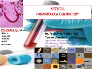
- 1. MEDICAL PARASITOLOGYLABORATORY PresentedTo : Mr.TapeshwarYadav (Sir) (teacher/mentor) Department of Laboratory Medicine, Nobel College of Medical Sciences, Kathmandu, Nepal Presented By : Group H -Moona -Yuyasha - Renuka - Rohit - Akshana
- 2. Preface It is a golden opportunity for us to work on this presentation titled as “PARASITOLOGY” to enhance our speaking and presenting skills under the faculty of BSc.MLT in Nobel College.The basic objective of presentation is to gather knowledge about various topics emphasizing on our course contents. This presentation includes explanation on different laboratory diagnosis, treatment, control of parasitic diseases.
- 3. Acknowledgement For any project, essential requirement is correct guidance and references. We are very thankful to Mr.TapeshwarYadav sir who provided us this opportunity and motivation to gain knowledge through this presentation. We have got knowledge about the way to search information, filter them and utilize various resources for gathering information. Some of the sources are: google scholar, slide share, wikipedia,etc.
- 4. CONTENTS OBJECTIVES SCOPE AND IMPORTANCE DEFINITIONS RELATIONSHIP OF PARASITES CLASSIFICATION OF PARASITES MODE OFTRANSMISSION SAMPLE COLLECTION DIFFERENTTESTS GENERAL STAINS ANDTHEIR PURPOSES PREVENTION AND CONTROL
- 5. Scope And Importance To provide knowledge on parasitic infections, their diagnosis , prevent and control them Know the lifecycle of specific parasites and identify important parasitic agents affecting human health Be able to prepare reagents necessary for parasitological laboratory test Apply necessary procedures for diagnosis of parasites in laboratory
- 6. OBJECTIVES To know various terms related to parasitology. To know about general parasites and parasitic infections. To get knowledge about laboratory diagnosis and its importance. To gain idea about general epidemiological aspects of parasites that affect human. Apply basic methods of specimen collection , preservation and processing in lab. To prevent ourselves from these infections and apply control measures.
- 7. Definitions Parasitology: Scientific study of parasites, their hosts and relationship among them. Father of parasitology : Francesco Redi Medical parasitology : study of parasites that infect human, diseases caused by them, clinical picture, their diagnosis, treatment and prevention as well as controls. It involves drug development, epidemiological studies and study of zoonoses. Parasites : organism which depends upon host eg: Plasmodium vivax
- 8. Vector : living organism that carries a disease-causing organism to new hosts Host : organism on which parasite is depended. eg: human beings 2 types: Definitive host :Host in which the parasite reaches maturity and reproduces sexually. Intermediate host: Host in which a parasite undergoes development but not sexual.
- 9. Some relationships Symbiosis: living together where either one or both are benefitted while none is harmed Commensalism: one symbionts is benefitted other is unaffected Mutualism: both symbionts are benefitted Parasitism: one symbiont is benefitted whereas the next is damaged
- 10. PARASITE DISEASE Plasmodium vivax Malaria Solid transmitted helminths Roundworm (Ascaris) Pnemonitis, intestinal obstruction Whipworm (Trichuris) Blood diarrhoea, rectal prolapse Hookworm (Ancylostoma and Necator ) Coughing, wheezing, abdominal pain and anaemia Schistosoma Renal tract and intestinal disease Filariae Lymphatic filariasis and elephantiasis Trypanosoma cruzi Chage diseases (cardiovascular) Leishmania Cutaneous, mucocutaneous and visceral leishmaniasis
- 12. FILARIASIS
- 18. Classification of parasites Protozoa: Unicellular organism. Eg: Plasmodium spp., the protozoan parasite which causes malaria Helminthes: Multicellular organisms and intestinal worms (helminths) such as Schistosoma spp., Wuchereria bancrofti, Necator americanus Taenia spp. (tapeworm)
- 19. PROTOZOA Unicellular eukaryotic microorganism Lack capability for photosynthesis exc. Euglena Heterotrophic mode of nutrition Have indefinite shape and size Able to move independently by flagellum or cilia (9+2) arrangement Moist habitat Study: protozoology
- 21. Protozoan diseases Protozoa Diseases Giardia lamblia Leishmania tropica Leishmania donovani Entamoeba histolytica Plasmodium vivax Cryptosporidium parvum Trichomonas vaginalis Giardiasis Cutaneous leishmaniasis Kala-azar(visceral leishmaniasis) Amebic dysentery Malaria Cryptosporidiasis Vaginal trichomoniasis
- 22. Helminths Parasitic intestinal worms Bilateral symmetry Lacks digestive system Reduced nervous system Complex reproductive system( produce large no. of eggs) Means of locomotion is reduced or complete lacking
- 25. Nomenclature of parasites Binominial nomenclature Protozoa: 1. Sacromastigophora: Entamoeba histolytica, Giardia lamblia 2. Apicomplexa: Plasmodium vivax, P. malaria, P. falciparum 3. Ciliophora: Balantidium coli 4. Microspora: Enterocytozoon bieneusi, E. hellam, Encephalitozoon cuniculi Platyhelminthes : 1. Cestoda: Diphyllobothrium latum, Taenia solium 2. Trematoda: Schiotosoma mansoni, S. japonicm,etc 3. Nemathelminthes: Ascaris lumbricoides
- 26. PARASITIC INFECTION An infection caused or transmitted by parasites Source of infection Contaminated water Eggs containing food ingestion Arthropods bites Penetration of intact skin or mucuos membrane
- 27. Mode of transmission MODE OFTRANSMISSION DISEASES FAECAL-ORAL ROUTE , INGESTION Ascaris, Balantidiasis, Giardia,Taenia, Cryptosporidiasis,Cyclospora, Fasciola, VECTOR BORNE Kissing and hugging: Trypanosoma Mosquito: Plasmodium, Wuchereria Sand fly: Leishmania Tsetse fly: Trypanosoma SEXUAL CONTACT Entameoba, Giardia,Trichomonas INHALATION Acanthameoba, Enterobius, Naegleria CONTACT OF SKIN(PENETRATION) Ancylostoma, Necator, Schistosoma CONTACT OF EYES(PENETRATION) Acanthamoeba
- 29. General Classification of parasites Ecto parasite: lives outside the host body Endo parasite: lives inside host body (blood, tissues, body cavities, digestive tract) Temporary parasite: visit host for short time Permanent parasite: throughout whole lifespan Facultative parasite: on parasites when opportunity arise Obligatory parasite: only on living host body Wandering parasite: at place where it cant live Occasional parasite: attacks on unusual host
- 30. Plasmodiumspp.
- 33. Tests Stool physical consistency Color and odor Quantity Mucus Chemical Ph Reducing substances Microscopic Leukocytes RBCs Ova and parasites Meatfibres, musclefibres fat Urine Physical Color and odor Turbidity Specific gravity pH Chemical Protein Glucose Ketones, nitrites Urobilinogen Bilirubin Blood microscopic RBCs,WBCs Epithelial cells microorganis ms Blood Thin smear Thick smear
- 34. INSTRUMENTS USED IN LAB
- 35. Sample Collection 1. The amount should be sufficient (4.0ml) 2. It should be collected in a non-absorbtive container 3. Stool sample should be examined as soon as possible 4. Sample should not be left exposed to air which leads to drying of stool 5. Stool mixed with urine should never be accepted for investigation (urine destroys trophozoite forms of parasites 6. While making smears, sample should be taken from well inside formed stool and from mucus or blood stained portions of loose stool
- 36. PHYSICAL COLOR Normal color - presence of stercobilinogen - light or dark brown Abnormal stool colour seen in different disease condition - Black : bleeding from upper gastrointestinal tract tumors - Brighter : bleeding from lower gastrointestinal tract - Blood and mucus : amoebic dysentery - Clay coloured :post hepatic jaundice obstruction to the flow of bile to intestine(biliary obstructions) - Yellow or yellowish green – diarrhoea - Maroon or pink –due to tumors, haemorhoids,tissue or inflammatory processs from lower gastrointestinaltract
- 37. ODOR The normal sample may smell offensive but not excessively foul, other can be described foul sour Foul odor caused by the undigested protein and by excessive intake of carbohydrate Offensive : amoebic dysentery Nil : bacillary dysentery QUANTITY Normally, there is 100 to 200 gm / day Many disorders cause large bulky stools even in people who Don’t eat a lot. Some gastrointestinal order also cause poor food breakdown and absorption which leads to large, bulky stools
- 38. CONSISTENCY Normal stool is well formed . Abnormal stool consistency seen in different physical and pathological condition . Stools may be loosely formed stools, Watery stools, Thin stools, Dry and hard stools, Putty like stools, Small round hard stool(habitual constipation), Pasty stools, diarrhoeal stool are watery, Steatorrhea stool are large in amount, forthy, foul smelling Constipated stools are firm and may be spherical masses Ribbon like stool
- 39. MUCUS Mucus is not present in normal stool . Seen in amoeba of Giardia lamblia . when urea gets high than the normal mucus can be seen in stool. It is also present in carcinoma condition. Pure mucous ie. translucent gelatinous material clinging to the surface of stool. This may be seen in severe constipation, mucous colitis , excessive staining of stool, emotionally unstable patient Mucus and diarrhoea with microscopically patient with RBCs and WBCs is seen in bacilliary dysentary, ulcerative colitis, intestinal tuberculosis, ameobiasis, enteritis, acute diverticulis, ulcerating malignancy of the colon
- 41. CHEMICAL Stool pH: This depends upon the dietary intake. Normally, slightly acidic or alkaline pH (7.0 to7.5) Alkaline pH : Colitis Villous adenoma Diarrhoea Antibiotic therapy Acidic pH: Fat and carbohydrate mal-absorption Disaccharidase deficiency
- 42. Reducing substances Glucose, fructose, galactose,etc are reducing substance Lactose intolerance may occur after a prolonged episode of viral gastroenteritis Fructose not absorbed can cause a positive test for reducing sugar in stool Reducing subs. are reported as: Negative –this is the normal result and means that the body is digesting and absorbing sugars properly. Positive- this means these are substances in the stool that can act as reducing agents i.e. there are forms of sugar in the stool that have not been absorbed by the body
- 43. MICROSCOPIC It includes Presence of leukocytes Presence of RBCs Presence of ova and parasites Presence of meat fibers and muscle fibers Presence of fat
- 44. 1. Presence of leukocytes Normally there are no WBCs Increase no of WBCs in stool Bacillary dysentery Chronic ulcerative colitis Shigellosis Salmonella E.coli diarrhoea Fistula of anus or rectum Localized abscess WBCs may appear in typhoid Absence of WBCs seen in some of the diarrhoeal condition ‐ Cholera ‐ Viral diarrhoea ‐ Drug induced diarrhoea ‐ Amoebic colitis ‐ Non-invarsive diarrhoea ‐ Parasitic infestation
- 45. 2. Presence of RBCs in the stool Blood in the stool can be Bright red: from the bleeding in the lower GI tract Maroon in color Black & tarry: from bleeding from the upper GI tract Occult (not visible to naked eye) Causes of blood in stool Hemorrhoids Cancer Dysentry
- 46. 3. Ova and parasites Normally there are no parasites or eggs in the stool sample Multiple stool sample are needed to rule out parasitic infestation at least three consecutive days. An abnormal results means parasites or eggs are present in stool such infections include - Roundworms: Ascaris lumbricoides - Hook worms: Necator americanus - Pinworms : Enterobius vermicularis - Tapeworms: Taenia solium, Diphyllebothrium latum - Protozoa : Entamoeba histolytica, Giardia lamblia
- 47. 4.Presence of meat and muscle fibers Their presence show some defects in the digestion. Increased amount of meat fibers are found in : Mal-absorption syndrome Pancreatic functional defect like cystic fibrosis 5. Presence of fat The fat in the stool shows the possibility of : Mal-absorption syndrome Deficiency of pancreatic digestive enzyme Deficiency of bile
- 48. Serodiagnosis Serological test becomes positive only in invasive amoebisis. Various serological tests done include : Indirect Hemagglutination (IHA)Test: Serum with antibody titer of 1:256 or more by IHA Is diagnostic of amoebic hepatitis. Latex agglutination test Enzyme-linked immunosorbent assay (ELISA): Commercially available tests that use ELISA to detect Entamoeba antigens (E. histolytica) Greater sensitivity than microscopic test
- 49. STOOL CULTURE It is a sensitive method in diagnosinf chronic and asymptomatic intestinal amoebiasis. Media used for stool culture: Boeck and Drbohlav media NIH polygenic media Craig’s medium Nelson’s medium Robinson’s medium
- 50. Faecal Occult Blood Test Generally, stool of normal person doesn’t contain blood. It may be present due to various pathological and physiological condition. In some cases, very scanty blood may be present and can’t be detected by physical appearance. This test is used to detect trace amount of blood in stool for which various chemicals are used.
- 53. VOLUME − Normal range (600-2000ml) − Polyuria : more than 2500ml (within 24 hours) − Oliguria : less than 500ml urine day − Anuria : complete suppression of urine formation COLOUR AND ODOR - light yellow to pale yellow or white clear - concentration of urochrome pigment. − Diet, medicines and various chemical disease can affect the colour − Urine becomes more ammonia-like due to bacterial activity.
- 54. ReactionandpH − Ranges from 4.5 -8.0, average :6.0( i.e.slightly acidic) − High protein intake : acidic urine − High vegetative diets and bacterial infections : alkaline − In case of UTI urine is acidic in case of infection with E.coli Specific Gravity Normal range : 1.003- 1.035 gm/cm^3 Measurement of density of urine high specific gravity: Diabetes mellitus, adrenal abnormalities or excessive water loss, diarrhoea or kidney inflammation low specific gravity: Diabetes insipidus, excessive water intake, chronic renal failure
- 55. APPEARANCE − Normal urine is usually clear − Cloudy: presence of amorphous phosphate − Turbid : presence of WBC / epithelial cells and bacteria − Milky : presence of fat SEDIMENT FORMATION In case of WBC, amorphous phosphate epithelial cells, sediments formation occurs at the bottom of container. ‐ Normal urine is transparent or clear ‐ Cloudy urine may be evidence of phosphates, ureates, mucus, bacteria, epithelial cells or leukocytes
- 56. CHEMICAL Protein: - Measured in urine by testing for the presence of albumin - (Proteinuria): indicator of kidney disease,bladder or kidney stones, multiple myeloma, haemolytic anaemia Glucose: Uncontrolled diabetes, kidney/hormonal disorders, liverdisease. Ketones: -indicate that fat is being metabolished instead of carbohydrate for energy -raised ketones indicate diabetic condition
- 57. Urobilinogen +ve test: hepatitis / cirrhosis of the liver Urobilinogen result is compared with the bilirubin result A negative test for bilirubin but positive for urobilinogen can indicate haemolytic. A lower negative test result for urobilinogen in a patient with positive bilirubin test indicate hepatic obstruction. Bilirubin A waste product created when old RBCs are broken down, usually removed as component of bile A positive test for bilirubin is an early indicator of hepatitis, liver disease or jaundice
- 58. Blood Urine normally contains a small amount of haemoglobin. A slight increase in haemoglobin can be significant in terms of potential causes such as a urinary tract infection, kidney disease, trauma, strenuous exercise. Nitrite Indicator of presence of a range of bacteria that can cause a urinary tract infection Leukocytes Significant increase in level of leukocyte indicates inflammation of the kidney/urinary tract or bladder/kidney infection.
- 60. MICROSCOPIC RBCs: Its no. elevation indicates injury, inflammation, disease/infection of the urinary tract(eg: bladder/kidney/urethra) WBCs: indicate infection or inflammation in urinary tract Epithelial cells: -Transitional: indicates an infection in bladder -Squamosal :indicates an external urethra -Kidney cell indicates a kidney conduction
- 63. Microorganism: -Trichomonas: responsible for vaginal infection -Yeast :indicates a vaginalyeast infection -Bacteria: cause urinary tract infection, kidney infection -Casts: indicate kidney disorder -Crystals: abnormal crystal cuase pain and damage to the urinary tract such as: cystene, tyrosine,leucine.
- 67. Blood test Thin smear Fixed in methanol To examine parasitemia and recognize them and their parasitic form like : schizont, gametocytes Thick smear Not fixed in methanol RBCs to be hemolysed Leukocytes and many malarial parasites present will be the only detectable elements Mainly used to detect infection and to estimate parasitemia
- 68. Stains used for thick and thin blood film Giemsa’s stain Leishman’s stain May-grunwald stain Jenner’s stain These stains allows for the detection of WBCs, RBCs and platelet abnormalities
- 69. Thin smear 1. Put a small drop of blood on a clean glass slide and spread using a spreader , making an angle of 35-40 . A tongue shaped smear should be made 2. After drying and proper labelling of smear , fix with methanol for 5-10 minutes 3. Stain with 1:10 diluted Giemsa stain for 45 minutes 4. Wash with tape water , allow to dry and examine under microscope
- 70. Thick smear 1. Place a drop of blood on a clean glass slide 2. Spread in an area a half – inch square or diameter with the help of a needle or the corner of another slide 3. After drying , dehaemoglobinize with distilled water or acetic tartaric acid solution or 2 % acetic acid solution until the smear appears greyish – white 4. Drain off the excess dehaemoglobinize agent and fix with methanol for about 5 minutes (when using acetic acid tartaric acid solution , the smear should be treated with slightly alkaline water to remove or neutralize the excess of acid.
- 73. Use of Blood Smear
- 74. Prevention & Control In endemic areas, people should be educated about the dangers of eating raw or under cooked meat Filtration of water is must Improve the water supply in affected communities Animal wastes and feces should be disposed in sanitary way out of reach of water resources Wash your hands regularly especially after handling uncooked food or feces
- 75. CONTD. Keep your fingernails short and scrub under them with a nail brush. In public toilets, don't sit on a bare toilet seat without protecting it with paper; squat if possible. Pinworm eggs and Trichomonas can be present on toilet seats. Trichomonas can also be spread through mud or water baths, or from sauna benches. Avoid walking barefoot, especially in warm, moist sandy soil. Don't use tap water to clean contact lenses - use sterilized lens preparations. Use a shower filter or bath-water filter.
- 76. Safety rules -Don’t eat or drink in lab -Wear coverings like: apron, gloves, proper shoes -Don’t clean up with hand if spilled -Mouth pippetting is avoided -Don’t taste any chemical substances -Clean work area and hand when work is finished -If chemicals come in contact with skin clean immediately and inform lab instructors