SlideShare a Scribd company logo
1 of 54
Download to read offline
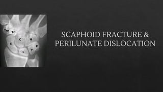
SCAPHOID FRACTURE &
PERILUNATE DISLOCATION
INTRODUCTION
➢ First described by Cousin & Destot in 1889
-Position of the scaphoid on the radial side of the wrist,
as the proximal extension of the thumb ray, makes it vulnerable
to injury.
Incidence:
➢ 10-15% of the hand and wrist fractures.
➢ It’s the most frequently fractured carpal bone around 70%
➢ Of these 70% occurs in waist.
ANATOMY
◈ Derived from greek word “skaphos” meaning boat
◈ Boat or cashew shaped bone
◈ Parts of the scaphoid:
proximal pole
waist
distal pole
tubercle
◈ Small irregular S shaped tubular bone, located at
a 45degree plane to the longitudinal &
horizontal axes of the wrist.
◈ Since its 80% of its surface is articular cartilage,
reduced capacity for periosteal reaction &
increased tendency for delayed & non union.
◈ Distal pole is pronated, flexed & ulnarly
angulated with respect to the proximal pole.
◈ ARTICULATIONS: with 5 bones
Proximal surface- radius
Distal surface- split into two separate articular surfaces by a
bony ridge & laterally with trapezoid & trapezium, medially with
capitate
Ulnar surface- lunate
◈ ATTACHMENTS:
- No musculotendinous attachments
Ligamentous
Extrinsic ligaments are
-radioscaphocapitate
-radioscapholunate
-radial collateral ligament
Intrinsic ligaments are
-scaphotrapezium-trapezoid
-scaphocapitate ligament
-scapholunate ligament
Blood supply
✓ Retrograde blood supply via two vascular pedicles
✓ Originating from the scaphoid branches of the
radial artery
✓ Dorsal branch
-70 to 80% (proximal part)
-enters via small foramina present along the
spiral groove & dorsal ridge
✓ Volar branch
-20 to 30% (distal scaphoid)
-enters via the scaphoid tubercle
Note-minimal or no perforating vasculature at waist
MECHANISM OF INJURY
Hyperextension injury
Fall on outstretched hand
Wrist is dorsiflexed to>95degrees
& radially deviated to >10
degrees
Compression occurs dorsally and
tension on palmar surface of
wrist
Bending forces applied to waist and distal pole of
scaphoid as proximal pole is tightly held between
the capitate, dorsal lip of radius and taut palmar
capsule
Leads to fracture
scaphoid most
commonly waist
➢ Degree of force and position of the wrist at the time of injury –
determines the type & severity
➢ Herbert suggested that wrist deviation may predict the location of
the fracture as the line of midcarpal joint crosses the
proximal pole -in radial deviation
distal pole -in ulnar deviation
➢ Waist # -usually due to shear forces
➢ Tubercle # - either compression or avulsion
➢ Waist -60-70%
➢ Proximal 3rd
-25%
➢ Distal 3rd
-10%
CLASSIFICATION
Herbert & fisher
classification
AO classification- based on anatomic location
Scaphoid(72)
Non comminuted(72.A) Comminuted(72.B)
proximal pole(72.A1) 72.B1
waist (72.A2) 72.B2
distal pole(72.A3) 72.B3
Russe classification-based on inclination of the fracture line
Horizontal
oblique
Vertical(unstable)
Mayo classification (based on location & stability)
based on location-5 types
Unstable fracture:
o >1mm of # displacement
o Lateral intrascaphoid angle>35 degrees
o Bone loss or comminution
o Fracture malalignment
o Proximal pole #
o perilunate # dislocation
CLINICAL ASSESSMENT
➢ Diagnosis made by combination of clinical history,
examination & radiographic assessment.
➢ History of hyperextension to wrist following a fall,
sports ,RTA or punch injury
Clinical Symptoms:
Radial sided wrist pain following a fall onto
the outstretched hand, with almost 90% recalling the
hyperextension injury
Swelling
ecchymosis(rarely)
Clinical Signs:
Anatomical snuff box tenderness
Scaphoid tubercle tenderness
Adding longitudinal thumb compression( scaphoid
compression test) : specificity reaches 75%
100%
sensitive
20% specific
Scaphoid shift test( Watson test)
◈ Pressure applied over the
tubercle & wrist moving
from radial to ulnar
deviation
◈ Positive test- if there is a
“clunk” as the scaphoid
subluxes dorsally out of
the scaphoid fossa
◈ Inference –
scapholunate disruption
◈ Reduced thumb
movement
◈ Anatomical snuff box
swelling
◈ Anatomical snuff box
pain in ulnar deviation
◈ Pain on thumb/index
pinch
Clinical prediction rules-for suspected fracture
INVESTIGATIONS
X-ray
➢ Neutral PA and lateral view:
-to assess the carpal alignment & determining the clear #
-often poor for scaphoid # detection due to tubercle overhang
on PA view and overlap on the lateral view
Ulnar deviated PA view: (scaphoid view)
-to detect the proximal pole fractures
-scaphoid fat pad is best visualized
-ulnar deviation rotates the scaphoid
parallel to the long axis of the foream and to
achieve an en face image
45 degree ulnar oblique view:
-in semipronated position
-to detect tubercle fracture,Oblique sulcus
and waist(displaced)
45 degree radial oblique view:
-in semisupinated position
-to detect proximal pole #, humpback
deformity and avulsion fracture
Note-30 to 40% scaphoid # not visualised on initial
assessment & investigation with four view
radiographs
Ziter view:
-PA view of the wrist in ulnar deviation with 20 degree
tube angulation to the elbow
-To detect waist fracture as beam at right angles to long
axis
Soft tissue signs:
@Scaphoid fat pad sign-distortion or loss of
adjacent fat stripes over the radial aspect of the
scaphoid on the PA view
@Pronator fat pad sign- a prominent pronator
quadratus fat pad over the volar aspect of the wrist
on the lateral view
➢ Barton suggested 3 possible reasons why
standard scaphoid radiographs are often
misinterpreted
- Dark line may be formed by the dorsal lip of
the radius overlapping the scaphoid
- White line formed by the proximal end of
the scaphoid tuberosity
- Dorsal ridge of the scaphoid may appear
bent on the semisupinated view
➢ Contralateral wrist views - because of the wide range of
normal alignment
Bone
scan-scintigraphy-Fast and reliable diagnostic tool
-100% sensitivity
Demerits-
Lacks specificity
Little information regarding location
Ultrasound:
-Inter observer variability
-Useful in patients with cortical irregularity and
hemarthrosis
-Little information on structural integrity of
scaphoid
CT SCAN & MRI
◈ For surgical planning & assessment of healing
◈ To diagnose additional bony & ligament injuries
Fracture displacement
step>1mm at dorsal cortices
gap > 1mm in sagittal or coronal views
◈ To measure the scaphoid fracture displacement-3 angles
-Lateral intrascaphoid angle measurement
-Normal 30+/-5degrees,saggital view
>35* -cut off for displacement.
-angle created by lines drawn perpendicular
to the proximal & distal articular surfaces/poles
◈ Dorsal cortical angle (sagittal view)
-Normal-140 degrees;
-Abnormal >160*
-Angle created by tangential lines drawn along the dorsal
cortices of
proximal & distal scaphoid fragments
Scaphoid height to length ratio (sagittal view)
-normal 0.60, abnormal->0.65
-length is determined by a palmar line drawn from the most
proximal to
the most distal edge
-height is the maximal point with a line perpendicular to the
length line
COMPLICATIONS
Malunion
-Humpback deformity (proximal scaphoid rotates dorsally into
extension & distal part faces downward in flexion)
-Wrist pain, reduced wrist extension &diminished grip strength
-Loss of extension is proportional to the angular deformity, which is
best calculated by lateral intrascaphoid angle & height to length ratio
Humpback
deformity
Non union
• Leads to specific type of post traumatic wrist arthrosis
called Scaphoid nonunion advance collapse(SNAC)
• Non union rate –undisplaced waist #-10%
• Displaced waist-50%
• Risk factors: delayed diagnosis & treatment
• Leads to radiocarpal arthritis & secondary mid carpal
arthritis
• Types fibrous nonunion-seen in stable #
sclerotic nonunion-unstable #
• Two patterns of nonunion displacement
-based on fracture line relative to the dorsal apex of
scaphoid ridge
Dorsal displacement-in proximal waist#
Volar displacement-in distal waist #
AVN
◈ Late complication of scaphoid fracture especially those
involving proximal pole fracture
◈ Preiser disease:
Scaphoid avn without a fracture either SL ligament
injury or idiopathic
-Increasing pain and stiffness of the wrist
-Small, deformed proximal pole fragment with cystic changes
& areas of sclerosis
-MRI is useful
Perilunate dislocation
Perilunate dislocation & fracture
dislocation
◈ Introduction:
◈ Most common form of wrist dislocation
◈ Spectrum of injury which include both ligamentous and
osseous disruption
◈ Prefix trans –refer to associated fracture
◈ Prefix peri – dislocation
◈ Usually traumatic, high energy
◈ Occurs when wrist extended and ulnarly deviated lead to
intercarpal supination
◈ Commonly missed ~25% on initial presentation.
Types
◈ Greater arc injury: (perilunate fracture
dislocation)
Ligamentous injuries associated with a
fracture of one or more of the bones around the
lunate.
◈ Common pattern -transscaphoid perilunate
fracture dislocation
-Fracture neck of the capitate
-Sagittal fracture of the triquetrum
◈ Scaphocapitate syndrome:
both capitate & scaphoid fragments are
◈ Lesser arc injury:
◈ Pure ligamentous injuries around the lunate
◈ Disruption of capsular & ligamentous connections of the lunate to
the adjacent carpal bones & radius without fracture
◈ Classically, the distal row dislocates in a dorsal or dorsoradial
direction
◈ SLD or LTD often persists even after relocation lead to recurrence of
instability and late carpal collapse
◈ Fracture dislocations are twice as frequent as a dislocation alone
Classification
Mayfield classification
◈ Progressive perilunate instability from a radial to
ulnar direction
◈ Stage 1:
◈ As the distal carpal row is violently extended
,supinated & ulnarly deviated, STT and SC
ligaments are tightened causing the scaphoid to
extend
◈ As the scaphoid extends, SL ligament transmits
the force to lunate, which can’t rotate as much as
scaphoid because its constrained by the
palmarly located radiolunate & ulnolunate
ligaments leading to scaphoid # or SLD
◈ STAGE II:
◈ If the extension-supination force persists, once the
proximal row has been dislocated, transmissiom of the
force distally to capitate may lead to displacement
through the space of Poirier
◈ Space of Poirier-ligament free area b/w
radioscapholunate ligament & long radiolunate
ligament at the midcarpal joint level; an area of
potential weakness.
◈ STAGE III:
◈ If the force persists, once the capitate is displace
dorsally lunotriquetral ligament disruption will occur
◈ STAGE IV:
◈ If the force continues,the dorsally displaced capitate is pulled
proximally.
◈ Pressure is applied onto the drsal aspect of lunate,forcing it to
dislocate
◈ As the palmar ligaments are much stronger than the dorsal capsule,
dislocation never involves a pure palmar displacement
◈ Note-lunate dislocation is the end stage of progressive perilunate
dislocation
Other classification system
◈ WITVOET & ALLIEU CLASSIFICATION:
◈ Grade I - lunate appears normally
aligned
◈ Grade II - rotated palmarly <90degrees
◈ Grade III ->90 degrees but still attached
to the radius by its palmar ligaments
◈ Grade IV –totally enucleated
◈ HERZBERG et al – three stage classification
◈ Stage I - dorsal dislocation of capitate but lunate
remains in fossa
◈ Stage II A - dorsal dislocation of capitate + lunate
dislocation ,rotation <90 degrees
◈ Stage II B - dislocation of capitate+lunate ,rotation >90
degrees
Clinical features
◈ Often young male, high energy trauma
◈ h/o hyperextension injury
◈ Wrist pain, swelling & deformity
◈ Signs- tenderness distal to lister tubercle
◈ marked prominence of entire carpus dorsally
◈ compressive test-palpable and audible snap, click or clunk
◈ Midcarpal shift test:
◈ Pressure applied over dorsum of the capitate, wrist moving from radial
to ulnar deviation
◈ Positive-clunk presents as the lunate reduces from the palmar flexed
position
◈ 1/3rd
associated with polytrauma
◈ 16% -median nerve symptoms and signs
◈ Other features- ulnar neuropathy, arterial
injury or tendon disruption
◈ Late presentations:
increasing nerve symptoms or tendon
rupture than wrist deformity because to
which patient has often become accustomed
Investigations
◈ Xray
◈ neutral PA and lateral view
◈ In PA view –normally the anatomic axis of forearm will pass through the
head & base of 3rd
metacarpal, capitate, radial aspect of lunate, center of the
lunate fossa of radius
◈ In lateral view-line will pass through the longitudinal axis of index finger
metacarpal, capitate, lunate and the radius with the scaphoid on an axis at a
45* angle to this line
◈ Normal joint space (b/w carpal bones, carpal & metacarpals, carpal bones
&radius)
◈ < 2mm –normal
◈ >3mm -suspected ligament disruption
◈ >5mm -diagnostic
Spilled tea spot sign
◈ Due to palmar rotation of the lunate & disruption of lunate –capitate
articulation
Piece of pie sign
◈ Triangular appearance of lunate secondary to rotation
Gilula lines
Arc 1- along the proximal articular surface of
proximal carpal row
Arc 2-along the distal articular surface
Arc 3-proximal cortical margins of the capitate &
hamate
Normally - smooth curves
Broken arc-diagnostic of fracture and or
dislocation
Scapholunate angle
◈ Angle created by the longitudinal axes
of the scaphoid and the lunate
◈ Long axis of scaphoid –is a line
tangential to the palmar convex surface
of the proximal & distal poles
◈ Long axis of lunate- line perpendicular
to the line connecting the dorsal &
palmar lips of the lunate.
◈ Normal range- 30 to 60 degrees
>60 degree –DISI pattern
<30 degree –VISI pattern
Carpal-height ratio
◈ Carpal height/length of third metacarpal
◈ Carpal height(L2) is the distance b/w the base
of 3rd
metacarpal to subchondral sclerotic line
of distal radius
◈ Normal ratio 0.5
◈ <0.45 indicates carpal collapse
◈ Alternate method - Height of capitate is used
instead of third metacarpal
◈ Stress radiographs- clenched fist view
To evaluate the widening of scapholunate interval
Often performed bilaterally
◈ CT & MRI to determine the extent of lesion
◈ Arthroscopy and arthrography
Thank you

More Related Content

What's hot

Scaphoid fracture and nonunion
Scaphoid fracture and nonunion Scaphoid fracture and nonunion
Scaphoid fracture and nonunion adityachakri
 
Scaphoid fractures
Scaphoid fracturesScaphoid fractures
Scaphoid fracturesKaushik Ys
 
Three column fixation for complex PROXIMAL TIBIA FRACTURES
Three column fixation for complex PROXIMAL TIBIA FRACTURESThree column fixation for complex PROXIMAL TIBIA FRACTURES
Three column fixation for complex PROXIMAL TIBIA FRACTURESLokesh Sharoff
 
Evolution of Intramedullary Nails
Evolution of Intramedullary NailsEvolution of Intramedullary Nails
Evolution of Intramedullary NailsPrateek Goel
 
Poller or blocking screw
Poller or blocking screwPoller or blocking screw
Poller or blocking screwAvik Sarkar
 
Jose Austine- Orthopaedic evaluation of cerebral palsy
Jose Austine- Orthopaedic evaluation of cerebral palsyJose Austine- Orthopaedic evaluation of cerebral palsy
Jose Austine- Orthopaedic evaluation of cerebral palsyJose Austine
 
Principles of deformity correction
Principles of deformity correctionPrinciples of deformity correction
Principles of deformity correctionAbdulla Kamal
 
Wrist arthroscopy metsovo 2011
Wrist arthroscopy metsovo 2011Wrist arthroscopy metsovo 2011
Wrist arthroscopy metsovo 2011Nikos Darlis
 
Posterolateral corner injuries of knee joint
Posterolateral corner injuries of knee joint Posterolateral corner injuries of knee joint
Posterolateral corner injuries of knee joint Samir Dwidmuthe
 
Pfn biomechanics
Pfn biomechanicsPfn biomechanics
Pfn biomechanicsshivlata
 
Knee stiffness dr anil k jain
Knee stiffness dr anil k jainKnee stiffness dr anil k jain
Knee stiffness dr anil k jainvaruntandra
 
Carpal instability and perilunate dislocation
Carpal instability and perilunate dislocationCarpal instability and perilunate dislocation
Carpal instability and perilunate dislocationdhidhi george
 
Scaphoid fracture gaju
Scaphoid fracture gajuScaphoid fracture gaju
Scaphoid fracture gajuRem Kulung
 

What's hot (20)

Scaphoid fracture and nonunion
Scaphoid fracture and nonunion Scaphoid fracture and nonunion
Scaphoid fracture and nonunion
 
Scaphoid fractures
Scaphoid fracturesScaphoid fractures
Scaphoid fractures
 
Carpal instability
Carpal instabilityCarpal instability
Carpal instability
 
Three column fixation for complex PROXIMAL TIBIA FRACTURES
Three column fixation for complex PROXIMAL TIBIA FRACTURESThree column fixation for complex PROXIMAL TIBIA FRACTURES
Three column fixation for complex PROXIMAL TIBIA FRACTURES
 
Scaphoid fracture
Scaphoid fractureScaphoid fracture
Scaphoid fracture
 
Evolution of Intramedullary Nails
Evolution of Intramedullary NailsEvolution of Intramedullary Nails
Evolution of Intramedullary Nails
 
Poller or blocking screw
Poller or blocking screwPoller or blocking screw
Poller or blocking screw
 
Jose Austine- Orthopaedic evaluation of cerebral palsy
Jose Austine- Orthopaedic evaluation of cerebral palsyJose Austine- Orthopaedic evaluation of cerebral palsy
Jose Austine- Orthopaedic evaluation of cerebral palsy
 
Principles of deformity correction
Principles of deformity correctionPrinciples of deformity correction
Principles of deformity correction
 
Perilunate injuries
Perilunate injuriesPerilunate injuries
Perilunate injuries
 
Wrist arthroscopy metsovo 2011
Wrist arthroscopy metsovo 2011Wrist arthroscopy metsovo 2011
Wrist arthroscopy metsovo 2011
 
Scaphoid fracture
Scaphoid fractureScaphoid fracture
Scaphoid fracture
 
Posterolateral corner injuries of knee joint
Posterolateral corner injuries of knee joint Posterolateral corner injuries of knee joint
Posterolateral corner injuries of knee joint
 
Pfn biomechanics
Pfn biomechanicsPfn biomechanics
Pfn biomechanics
 
Locking plates
Locking platesLocking plates
Locking plates
 
Knee stiffness dr anil k jain
Knee stiffness dr anil k jainKnee stiffness dr anil k jain
Knee stiffness dr anil k jain
 
Scaphoid fracture and hcs
Scaphoid fracture and hcsScaphoid fracture and hcs
Scaphoid fracture and hcs
 
Distal radius
Distal radiusDistal radius
Distal radius
 
Carpal instability and perilunate dislocation
Carpal instability and perilunate dislocationCarpal instability and perilunate dislocation
Carpal instability and perilunate dislocation
 
Scaphoid fracture gaju
Scaphoid fracture gajuScaphoid fracture gaju
Scaphoid fracture gaju
 

Similar to SCAPHOID FRACTURE & PERILUNATE DISLOCATION GUIDE

Wrist disorders
Wrist disordersWrist disorders
Wrist disordersyasim786
 
Radio-anatomy and Radio-pathology of hand and wrist.pdf
Radio-anatomy and Radio-pathology of hand and wrist.pdfRadio-anatomy and Radio-pathology of hand and wrist.pdf
Radio-anatomy and Radio-pathology of hand and wrist.pdfYudHaBaddOn
 
CRANIOVERTEBRAL JUNCTION ANATOMY, CRANIOMETRY, ANAMOLIES AND RADIOLOGY dr sum...
CRANIOVERTEBRAL JUNCTION ANATOMY, CRANIOMETRY, ANAMOLIES AND RADIOLOGY dr sum...CRANIOVERTEBRAL JUNCTION ANATOMY, CRANIOMETRY, ANAMOLIES AND RADIOLOGY dr sum...
CRANIOVERTEBRAL JUNCTION ANATOMY, CRANIOMETRY, ANAMOLIES AND RADIOLOGY dr sum...SUMIT KUMAR
 
Missed fractures in Emergency Department
Missed fractures in Emergency DepartmentMissed fractures in Emergency Department
Missed fractures in Emergency DepartmentLouay Al-Mouazzen
 
Can I Offer More 4
Can I Offer More 4Can I Offer More 4
Can I Offer More 4guest609645b
 
Lunate Fracture & Perilunate dislocation of carpals by Dr.@rpan Chaudhary (Re...
Lunate Fracture & Perilunate dislocation of carpals by Dr.@rpan Chaudhary (Re...Lunate Fracture & Perilunate dislocation of carpals by Dr.@rpan Chaudhary (Re...
Lunate Fracture & Perilunate dislocation of carpals by Dr.@rpan Chaudhary (Re...CHAUDHARY ARPAN
 
Distal radius fracture
Distal radius fractureDistal radius fracture
Distal radius fracturesushilonlines
 
Distal radius fracture
Distal radius fractureDistal radius fracture
Distal radius fractureBarun Patel
 
Carpal Bones Fractures.pptx
Carpal Bones Fractures.pptxCarpal Bones Fractures.pptx
Carpal Bones Fractures.pptxBedrumohammed2
 
Supra condylar fractures
Supra condylar fracturesSupra condylar fractures
Supra condylar fracturesDrzameer
 
Scaphoid Fractures_UTSAV
Scaphoid Fractures_UTSAVScaphoid Fractures_UTSAV
Scaphoid Fractures_UTSAVUtsav Agrawal
 
Lateral condyle of humerus fracture in children
Lateral condyle of humerus fracture in childrenLateral condyle of humerus fracture in children
Lateral condyle of humerus fracture in childrenAnilKC5
 
Fractures of distal end radius
Fractures of distal end radiusFractures of distal end radius
Fractures of distal end radiusMahak Jain
 
Clavicular fracture & acj injury
Clavicular fracture & acj injuryClavicular fracture & acj injury
Clavicular fracture & acj injuryomar ababneh
 
Proximal Femur Fractures with NOF & IT
Proximal Femur Fractures with NOF & ITProximal Femur Fractures with NOF & IT
Proximal Femur Fractures with NOF & ITKunal Mondal
 
Distal Radius Fractures(DER) colless.pdf
Distal Radius Fractures(DER) colless.pdfDistal Radius Fractures(DER) colless.pdf
Distal Radius Fractures(DER) colless.pdfgoyalaman2022
 

Similar to SCAPHOID FRACTURE & PERILUNATE DISLOCATION GUIDE (20)

scaphoid and lunate fractures
scaphoid and lunate fracturesscaphoid and lunate fractures
scaphoid and lunate fractures
 
Wrist disorders
Wrist disordersWrist disorders
Wrist disorders
 
Scaphoid fractures
Scaphoid fracturesScaphoid fractures
Scaphoid fractures
 
Radio-anatomy and Radio-pathology of hand and wrist.pdf
Radio-anatomy and Radio-pathology of hand and wrist.pdfRadio-anatomy and Radio-pathology of hand and wrist.pdf
Radio-anatomy and Radio-pathology of hand and wrist.pdf
 
CRANIOVERTEBRAL JUNCTION ANATOMY, CRANIOMETRY, ANAMOLIES AND RADIOLOGY dr sum...
CRANIOVERTEBRAL JUNCTION ANATOMY, CRANIOMETRY, ANAMOLIES AND RADIOLOGY dr sum...CRANIOVERTEBRAL JUNCTION ANATOMY, CRANIOMETRY, ANAMOLIES AND RADIOLOGY dr sum...
CRANIOVERTEBRAL JUNCTION ANATOMY, CRANIOMETRY, ANAMOLIES AND RADIOLOGY dr sum...
 
Missed fractures in Emergency Department
Missed fractures in Emergency DepartmentMissed fractures in Emergency Department
Missed fractures in Emergency Department
 
Can I Offer More 4
Can I Offer More 4Can I Offer More 4
Can I Offer More 4
 
Lunate Fracture & Perilunate dislocation of carpals by Dr.@rpan Chaudhary (Re...
Lunate Fracture & Perilunate dislocation of carpals by Dr.@rpan Chaudhary (Re...Lunate Fracture & Perilunate dislocation of carpals by Dr.@rpan Chaudhary (Re...
Lunate Fracture & Perilunate dislocation of carpals by Dr.@rpan Chaudhary (Re...
 
Distal radius fracture
Distal radius fractureDistal radius fracture
Distal radius fracture
 
Distal radius fracture
Distal radius fractureDistal radius fracture
Distal radius fracture
 
Carpal Bones Fractures.pptx
Carpal Bones Fractures.pptxCarpal Bones Fractures.pptx
Carpal Bones Fractures.pptx
 
Lecture 3 shah radiology in foot and ankle
Lecture 3 shah radiology in foot and ankleLecture 3 shah radiology in foot and ankle
Lecture 3 shah radiology in foot and ankle
 
Supra condylar fractures
Supra condylar fracturesSupra condylar fractures
Supra condylar fractures
 
Scaphoid Fractures_UTSAV
Scaphoid Fractures_UTSAVScaphoid Fractures_UTSAV
Scaphoid Fractures_UTSAV
 
Lateral condyle of humerus fracture in children
Lateral condyle of humerus fracture in childrenLateral condyle of humerus fracture in children
Lateral condyle of humerus fracture in children
 
Fractures of distal end radius
Fractures of distal end radiusFractures of distal end radius
Fractures of distal end radius
 
Clavicular fracture & acj injury
Clavicular fracture & acj injuryClavicular fracture & acj injury
Clavicular fracture & acj injury
 
Proximal Femur Fractures with NOF & IT
Proximal Femur Fractures with NOF & ITProximal Femur Fractures with NOF & IT
Proximal Femur Fractures with NOF & IT
 
distal radius # ppt
 distal radius # ppt distal radius # ppt
distal radius # ppt
 
Distal Radius Fractures(DER) colless.pdf
Distal Radius Fractures(DER) colless.pdfDistal Radius Fractures(DER) colless.pdf
Distal Radius Fractures(DER) colless.pdf
 

Recently uploaded

POST NATAL EXERCISES AND ITS IMPACT.pptx
POST NATAL EXERCISES AND ITS IMPACT.pptxPOST NATAL EXERCISES AND ITS IMPACT.pptx
POST NATAL EXERCISES AND ITS IMPACT.pptxvirengeeta
 
Hematology and Immunology - Leukocytes Functions
Hematology and Immunology - Leukocytes FunctionsHematology and Immunology - Leukocytes Functions
Hematology and Immunology - Leukocytes FunctionsMedicoseAcademics
 
call girls in Connaught Place DELHI 🔝 >༒9540349809 🔝 genuine Escort Service ...
call girls in Connaught Place  DELHI 🔝 >༒9540349809 🔝 genuine Escort Service ...call girls in Connaught Place  DELHI 🔝 >༒9540349809 🔝 genuine Escort Service ...
call girls in Connaught Place DELHI 🔝 >༒9540349809 🔝 genuine Escort Service ...saminamagar
 
PNEUMOTHORAX AND ITS MANAGEMENTS.pdf
PNEUMOTHORAX   AND  ITS  MANAGEMENTS.pdfPNEUMOTHORAX   AND  ITS  MANAGEMENTS.pdf
PNEUMOTHORAX AND ITS MANAGEMENTS.pdfDolisha Warbi
 
Basic principles involved in the traditional systems of medicine PDF.pdf
Basic principles involved in the traditional systems of medicine PDF.pdfBasic principles involved in the traditional systems of medicine PDF.pdf
Basic principles involved in the traditional systems of medicine PDF.pdfDivya Kanojiya
 
History and Development of Pharmacovigilence.pdf
History and Development of Pharmacovigilence.pdfHistory and Development of Pharmacovigilence.pdf
History and Development of Pharmacovigilence.pdfSasikiranMarri
 
Big Data Analysis Suggests COVID Vaccination Increases Excess Mortality Of ...
Big Data Analysis Suggests COVID  Vaccination Increases Excess Mortality Of  ...Big Data Analysis Suggests COVID  Vaccination Increases Excess Mortality Of  ...
Big Data Analysis Suggests COVID Vaccination Increases Excess Mortality Of ...sdateam0
 
Presentation on Parasympathetic Nervous System
Presentation on Parasympathetic Nervous SystemPresentation on Parasympathetic Nervous System
Presentation on Parasympathetic Nervous SystemPrerana Jadhav
 
call girls in paharganj DELHI 🔝 >༒9540349809 🔝 genuine Escort Service 🔝✔️✔️
call girls in paharganj DELHI 🔝 >༒9540349809 🔝 genuine Escort Service 🔝✔️✔️call girls in paharganj DELHI 🔝 >༒9540349809 🔝 genuine Escort Service 🔝✔️✔️
call girls in paharganj DELHI 🔝 >༒9540349809 🔝 genuine Escort Service 🔝✔️✔️saminamagar
 
Report Back from SGO: What’s New in Uterine Cancer?.pptx
Report Back from SGO: What’s New in Uterine Cancer?.pptxReport Back from SGO: What’s New in Uterine Cancer?.pptx
Report Back from SGO: What’s New in Uterine Cancer?.pptxbkling
 
PULMONARY EMBOLISM AND ITS MANAGEMENTS.pdf
PULMONARY EMBOLISM AND ITS MANAGEMENTS.pdfPULMONARY EMBOLISM AND ITS MANAGEMENTS.pdf
PULMONARY EMBOLISM AND ITS MANAGEMENTS.pdfDolisha Warbi
 
Lippincott Microcards_ Microbiology Flash Cards-LWW (2015).pdf
Lippincott Microcards_ Microbiology Flash Cards-LWW (2015).pdfLippincott Microcards_ Microbiology Flash Cards-LWW (2015).pdf
Lippincott Microcards_ Microbiology Flash Cards-LWW (2015).pdfSreeja Cherukuru
 
Biomechanics- Shoulder Joint!!!!!!!!!!!!
Biomechanics- Shoulder Joint!!!!!!!!!!!!Biomechanics- Shoulder Joint!!!!!!!!!!!!
Biomechanics- Shoulder Joint!!!!!!!!!!!!ibtesaam huma
 
See the 2,456 pharmacies on the National E-Pharmacy Platform
See the 2,456 pharmacies on the National E-Pharmacy PlatformSee the 2,456 pharmacies on the National E-Pharmacy Platform
See the 2,456 pharmacies on the National E-Pharmacy PlatformKweku Zurek
 
METHODS OF ACQUIRING KNOWLEDGE IN NURSING.pptx by navdeep kaur
METHODS OF ACQUIRING KNOWLEDGE IN NURSING.pptx by navdeep kaurMETHODS OF ACQUIRING KNOWLEDGE IN NURSING.pptx by navdeep kaur
METHODS OF ACQUIRING KNOWLEDGE IN NURSING.pptx by navdeep kaurNavdeep Kaur
 
call girls in aerocity DELHI 🔝 >༒9540349809 🔝 genuine Escort Service 🔝✔️✔️
call girls in aerocity DELHI 🔝 >༒9540349809 🔝 genuine Escort Service 🔝✔️✔️call girls in aerocity DELHI 🔝 >༒9540349809 🔝 genuine Escort Service 🔝✔️✔️
call girls in aerocity DELHI 🔝 >༒9540349809 🔝 genuine Escort Service 🔝✔️✔️saminamagar
 
Informed Consent Empowering Healthcare Decision-Making.pptx
Informed Consent Empowering Healthcare Decision-Making.pptxInformed Consent Empowering Healthcare Decision-Making.pptx
Informed Consent Empowering Healthcare Decision-Making.pptxSasikiranMarri
 
COVID-19 (NOVEL CORONA VIRUS DISEASE PANDEMIC ).pptx
COVID-19  (NOVEL CORONA  VIRUS DISEASE PANDEMIC ).pptxCOVID-19  (NOVEL CORONA  VIRUS DISEASE PANDEMIC ).pptx
COVID-19 (NOVEL CORONA VIRUS DISEASE PANDEMIC ).pptxBibekananda shah
 
Radiation Dosimetry Parameters and Isodose Curves.pptx
Radiation Dosimetry Parameters and Isodose Curves.pptxRadiation Dosimetry Parameters and Isodose Curves.pptx
Radiation Dosimetry Parameters and Isodose Curves.pptxDr. Dheeraj Kumar
 
PULMONARY EDEMA AND ITS MANAGEMENT.pdf
PULMONARY EDEMA AND  ITS  MANAGEMENT.pdfPULMONARY EDEMA AND  ITS  MANAGEMENT.pdf
PULMONARY EDEMA AND ITS MANAGEMENT.pdfDolisha Warbi
 

Recently uploaded (20)

POST NATAL EXERCISES AND ITS IMPACT.pptx
POST NATAL EXERCISES AND ITS IMPACT.pptxPOST NATAL EXERCISES AND ITS IMPACT.pptx
POST NATAL EXERCISES AND ITS IMPACT.pptx
 
Hematology and Immunology - Leukocytes Functions
Hematology and Immunology - Leukocytes FunctionsHematology and Immunology - Leukocytes Functions
Hematology and Immunology - Leukocytes Functions
 
call girls in Connaught Place DELHI 🔝 >༒9540349809 🔝 genuine Escort Service ...
call girls in Connaught Place  DELHI 🔝 >༒9540349809 🔝 genuine Escort Service ...call girls in Connaught Place  DELHI 🔝 >༒9540349809 🔝 genuine Escort Service ...
call girls in Connaught Place DELHI 🔝 >༒9540349809 🔝 genuine Escort Service ...
 
PNEUMOTHORAX AND ITS MANAGEMENTS.pdf
PNEUMOTHORAX   AND  ITS  MANAGEMENTS.pdfPNEUMOTHORAX   AND  ITS  MANAGEMENTS.pdf
PNEUMOTHORAX AND ITS MANAGEMENTS.pdf
 
Basic principles involved in the traditional systems of medicine PDF.pdf
Basic principles involved in the traditional systems of medicine PDF.pdfBasic principles involved in the traditional systems of medicine PDF.pdf
Basic principles involved in the traditional systems of medicine PDF.pdf
 
History and Development of Pharmacovigilence.pdf
History and Development of Pharmacovigilence.pdfHistory and Development of Pharmacovigilence.pdf
History and Development of Pharmacovigilence.pdf
 
Big Data Analysis Suggests COVID Vaccination Increases Excess Mortality Of ...
Big Data Analysis Suggests COVID  Vaccination Increases Excess Mortality Of  ...Big Data Analysis Suggests COVID  Vaccination Increases Excess Mortality Of  ...
Big Data Analysis Suggests COVID Vaccination Increases Excess Mortality Of ...
 
Presentation on Parasympathetic Nervous System
Presentation on Parasympathetic Nervous SystemPresentation on Parasympathetic Nervous System
Presentation on Parasympathetic Nervous System
 
call girls in paharganj DELHI 🔝 >༒9540349809 🔝 genuine Escort Service 🔝✔️✔️
call girls in paharganj DELHI 🔝 >༒9540349809 🔝 genuine Escort Service 🔝✔️✔️call girls in paharganj DELHI 🔝 >༒9540349809 🔝 genuine Escort Service 🔝✔️✔️
call girls in paharganj DELHI 🔝 >༒9540349809 🔝 genuine Escort Service 🔝✔️✔️
 
Report Back from SGO: What’s New in Uterine Cancer?.pptx
Report Back from SGO: What’s New in Uterine Cancer?.pptxReport Back from SGO: What’s New in Uterine Cancer?.pptx
Report Back from SGO: What’s New in Uterine Cancer?.pptx
 
PULMONARY EMBOLISM AND ITS MANAGEMENTS.pdf
PULMONARY EMBOLISM AND ITS MANAGEMENTS.pdfPULMONARY EMBOLISM AND ITS MANAGEMENTS.pdf
PULMONARY EMBOLISM AND ITS MANAGEMENTS.pdf
 
Lippincott Microcards_ Microbiology Flash Cards-LWW (2015).pdf
Lippincott Microcards_ Microbiology Flash Cards-LWW (2015).pdfLippincott Microcards_ Microbiology Flash Cards-LWW (2015).pdf
Lippincott Microcards_ Microbiology Flash Cards-LWW (2015).pdf
 
Biomechanics- Shoulder Joint!!!!!!!!!!!!
Biomechanics- Shoulder Joint!!!!!!!!!!!!Biomechanics- Shoulder Joint!!!!!!!!!!!!
Biomechanics- Shoulder Joint!!!!!!!!!!!!
 
See the 2,456 pharmacies on the National E-Pharmacy Platform
See the 2,456 pharmacies on the National E-Pharmacy PlatformSee the 2,456 pharmacies on the National E-Pharmacy Platform
See the 2,456 pharmacies on the National E-Pharmacy Platform
 
METHODS OF ACQUIRING KNOWLEDGE IN NURSING.pptx by navdeep kaur
METHODS OF ACQUIRING KNOWLEDGE IN NURSING.pptx by navdeep kaurMETHODS OF ACQUIRING KNOWLEDGE IN NURSING.pptx by navdeep kaur
METHODS OF ACQUIRING KNOWLEDGE IN NURSING.pptx by navdeep kaur
 
call girls in aerocity DELHI 🔝 >༒9540349809 🔝 genuine Escort Service 🔝✔️✔️
call girls in aerocity DELHI 🔝 >༒9540349809 🔝 genuine Escort Service 🔝✔️✔️call girls in aerocity DELHI 🔝 >༒9540349809 🔝 genuine Escort Service 🔝✔️✔️
call girls in aerocity DELHI 🔝 >༒9540349809 🔝 genuine Escort Service 🔝✔️✔️
 
Informed Consent Empowering Healthcare Decision-Making.pptx
Informed Consent Empowering Healthcare Decision-Making.pptxInformed Consent Empowering Healthcare Decision-Making.pptx
Informed Consent Empowering Healthcare Decision-Making.pptx
 
COVID-19 (NOVEL CORONA VIRUS DISEASE PANDEMIC ).pptx
COVID-19  (NOVEL CORONA  VIRUS DISEASE PANDEMIC ).pptxCOVID-19  (NOVEL CORONA  VIRUS DISEASE PANDEMIC ).pptx
COVID-19 (NOVEL CORONA VIRUS DISEASE PANDEMIC ).pptx
 
Radiation Dosimetry Parameters and Isodose Curves.pptx
Radiation Dosimetry Parameters and Isodose Curves.pptxRadiation Dosimetry Parameters and Isodose Curves.pptx
Radiation Dosimetry Parameters and Isodose Curves.pptx
 
PULMONARY EDEMA AND ITS MANAGEMENT.pdf
PULMONARY EDEMA AND  ITS  MANAGEMENT.pdfPULMONARY EDEMA AND  ITS  MANAGEMENT.pdf
PULMONARY EDEMA AND ITS MANAGEMENT.pdf
 

SCAPHOID FRACTURE & PERILUNATE DISLOCATION GUIDE

  • 2. INTRODUCTION ➢ First described by Cousin & Destot in 1889 -Position of the scaphoid on the radial side of the wrist, as the proximal extension of the thumb ray, makes it vulnerable to injury. Incidence: ➢ 10-15% of the hand and wrist fractures. ➢ It’s the most frequently fractured carpal bone around 70% ➢ Of these 70% occurs in waist.
  • 4. ◈ Derived from greek word “skaphos” meaning boat ◈ Boat or cashew shaped bone ◈ Parts of the scaphoid: proximal pole waist distal pole tubercle
  • 5. ◈ Small irregular S shaped tubular bone, located at a 45degree plane to the longitudinal & horizontal axes of the wrist. ◈ Since its 80% of its surface is articular cartilage, reduced capacity for periosteal reaction & increased tendency for delayed & non union. ◈ Distal pole is pronated, flexed & ulnarly angulated with respect to the proximal pole.
  • 6. ◈ ARTICULATIONS: with 5 bones Proximal surface- radius Distal surface- split into two separate articular surfaces by a bony ridge & laterally with trapezoid & trapezium, medially with capitate Ulnar surface- lunate
  • 7. ◈ ATTACHMENTS: - No musculotendinous attachments Ligamentous Extrinsic ligaments are -radioscaphocapitate -radioscapholunate -radial collateral ligament Intrinsic ligaments are -scaphotrapezium-trapezoid -scaphocapitate ligament -scapholunate ligament
  • 8. Blood supply ✓ Retrograde blood supply via two vascular pedicles ✓ Originating from the scaphoid branches of the radial artery ✓ Dorsal branch -70 to 80% (proximal part) -enters via small foramina present along the spiral groove & dorsal ridge ✓ Volar branch -20 to 30% (distal scaphoid) -enters via the scaphoid tubercle Note-minimal or no perforating vasculature at waist
  • 10. Hyperextension injury Fall on outstretched hand Wrist is dorsiflexed to>95degrees & radially deviated to >10 degrees Compression occurs dorsally and tension on palmar surface of wrist Bending forces applied to waist and distal pole of scaphoid as proximal pole is tightly held between the capitate, dorsal lip of radius and taut palmar capsule Leads to fracture scaphoid most commonly waist
  • 11. ➢ Degree of force and position of the wrist at the time of injury – determines the type & severity ➢ Herbert suggested that wrist deviation may predict the location of the fracture as the line of midcarpal joint crosses the proximal pole -in radial deviation distal pole -in ulnar deviation ➢ Waist # -usually due to shear forces ➢ Tubercle # - either compression or avulsion ➢ Waist -60-70% ➢ Proximal 3rd -25% ➢ Distal 3rd -10%
  • 14. AO classification- based on anatomic location Scaphoid(72) Non comminuted(72.A) Comminuted(72.B) proximal pole(72.A1) 72.B1 waist (72.A2) 72.B2 distal pole(72.A3) 72.B3 Russe classification-based on inclination of the fracture line Horizontal oblique Vertical(unstable)
  • 15. Mayo classification (based on location & stability) based on location-5 types Unstable fracture: o >1mm of # displacement o Lateral intrascaphoid angle>35 degrees o Bone loss or comminution o Fracture malalignment o Proximal pole # o perilunate # dislocation
  • 17. ➢ Diagnosis made by combination of clinical history, examination & radiographic assessment. ➢ History of hyperextension to wrist following a fall, sports ,RTA or punch injury Clinical Symptoms: Radial sided wrist pain following a fall onto the outstretched hand, with almost 90% recalling the hyperextension injury Swelling ecchymosis(rarely)
  • 18. Clinical Signs: Anatomical snuff box tenderness Scaphoid tubercle tenderness Adding longitudinal thumb compression( scaphoid compression test) : specificity reaches 75% 100% sensitive 20% specific
  • 19. Scaphoid shift test( Watson test) ◈ Pressure applied over the tubercle & wrist moving from radial to ulnar deviation ◈ Positive test- if there is a “clunk” as the scaphoid subluxes dorsally out of the scaphoid fossa ◈ Inference – scapholunate disruption
  • 20. ◈ Reduced thumb movement ◈ Anatomical snuff box swelling ◈ Anatomical snuff box pain in ulnar deviation ◈ Pain on thumb/index pinch
  • 21. Clinical prediction rules-for suspected fracture
  • 23. X-ray ➢ Neutral PA and lateral view: -to assess the carpal alignment & determining the clear # -often poor for scaphoid # detection due to tubercle overhang on PA view and overlap on the lateral view Ulnar deviated PA view: (scaphoid view) -to detect the proximal pole fractures -scaphoid fat pad is best visualized -ulnar deviation rotates the scaphoid parallel to the long axis of the foream and to achieve an en face image
  • 24. 45 degree ulnar oblique view: -in semipronated position -to detect tubercle fracture,Oblique sulcus and waist(displaced) 45 degree radial oblique view: -in semisupinated position -to detect proximal pole #, humpback deformity and avulsion fracture Note-30 to 40% scaphoid # not visualised on initial assessment & investigation with four view radiographs
  • 25. Ziter view: -PA view of the wrist in ulnar deviation with 20 degree tube angulation to the elbow -To detect waist fracture as beam at right angles to long axis Soft tissue signs: @Scaphoid fat pad sign-distortion or loss of adjacent fat stripes over the radial aspect of the scaphoid on the PA view @Pronator fat pad sign- a prominent pronator quadratus fat pad over the volar aspect of the wrist on the lateral view
  • 26. ➢ Barton suggested 3 possible reasons why standard scaphoid radiographs are often misinterpreted - Dark line may be formed by the dorsal lip of the radius overlapping the scaphoid - White line formed by the proximal end of the scaphoid tuberosity - Dorsal ridge of the scaphoid may appear bent on the semisupinated view ➢ Contralateral wrist views - because of the wide range of normal alignment
  • 27. Bone scan-scintigraphy-Fast and reliable diagnostic tool -100% sensitivity Demerits- Lacks specificity Little information regarding location Ultrasound: -Inter observer variability -Useful in patients with cortical irregularity and hemarthrosis -Little information on structural integrity of scaphoid
  • 28. CT SCAN & MRI ◈ For surgical planning & assessment of healing ◈ To diagnose additional bony & ligament injuries Fracture displacement step>1mm at dorsal cortices gap > 1mm in sagittal or coronal views ◈ To measure the scaphoid fracture displacement-3 angles -Lateral intrascaphoid angle measurement -Normal 30+/-5degrees,saggital view >35* -cut off for displacement. -angle created by lines drawn perpendicular to the proximal & distal articular surfaces/poles
  • 29. ◈ Dorsal cortical angle (sagittal view) -Normal-140 degrees; -Abnormal >160* -Angle created by tangential lines drawn along the dorsal cortices of proximal & distal scaphoid fragments Scaphoid height to length ratio (sagittal view) -normal 0.60, abnormal->0.65 -length is determined by a palmar line drawn from the most proximal to the most distal edge -height is the maximal point with a line perpendicular to the length line
  • 31. Malunion -Humpback deformity (proximal scaphoid rotates dorsally into extension & distal part faces downward in flexion) -Wrist pain, reduced wrist extension &diminished grip strength -Loss of extension is proportional to the angular deformity, which is best calculated by lateral intrascaphoid angle & height to length ratio Humpback deformity
  • 32. Non union • Leads to specific type of post traumatic wrist arthrosis called Scaphoid nonunion advance collapse(SNAC) • Non union rate –undisplaced waist #-10% • Displaced waist-50% • Risk factors: delayed diagnosis & treatment • Leads to radiocarpal arthritis & secondary mid carpal arthritis • Types fibrous nonunion-seen in stable # sclerotic nonunion-unstable # • Two patterns of nonunion displacement -based on fracture line relative to the dorsal apex of scaphoid ridge Dorsal displacement-in proximal waist# Volar displacement-in distal waist #
  • 33. AVN ◈ Late complication of scaphoid fracture especially those involving proximal pole fracture ◈ Preiser disease: Scaphoid avn without a fracture either SL ligament injury or idiopathic -Increasing pain and stiffness of the wrist -Small, deformed proximal pole fragment with cystic changes & areas of sclerosis -MRI is useful
  • 35. Perilunate dislocation & fracture dislocation ◈ Introduction: ◈ Most common form of wrist dislocation ◈ Spectrum of injury which include both ligamentous and osseous disruption ◈ Prefix trans –refer to associated fracture ◈ Prefix peri – dislocation ◈ Usually traumatic, high energy ◈ Occurs when wrist extended and ulnarly deviated lead to intercarpal supination ◈ Commonly missed ~25% on initial presentation.
  • 36. Types ◈ Greater arc injury: (perilunate fracture dislocation) Ligamentous injuries associated with a fracture of one or more of the bones around the lunate. ◈ Common pattern -transscaphoid perilunate fracture dislocation -Fracture neck of the capitate -Sagittal fracture of the triquetrum ◈ Scaphocapitate syndrome: both capitate & scaphoid fragments are
  • 37. ◈ Lesser arc injury: ◈ Pure ligamentous injuries around the lunate ◈ Disruption of capsular & ligamentous connections of the lunate to the adjacent carpal bones & radius without fracture ◈ Classically, the distal row dislocates in a dorsal or dorsoradial direction ◈ SLD or LTD often persists even after relocation lead to recurrence of instability and late carpal collapse ◈ Fracture dislocations are twice as frequent as a dislocation alone
  • 39. Mayfield classification ◈ Progressive perilunate instability from a radial to ulnar direction ◈ Stage 1: ◈ As the distal carpal row is violently extended ,supinated & ulnarly deviated, STT and SC ligaments are tightened causing the scaphoid to extend ◈ As the scaphoid extends, SL ligament transmits the force to lunate, which can’t rotate as much as scaphoid because its constrained by the palmarly located radiolunate & ulnolunate ligaments leading to scaphoid # or SLD
  • 40. ◈ STAGE II: ◈ If the extension-supination force persists, once the proximal row has been dislocated, transmissiom of the force distally to capitate may lead to displacement through the space of Poirier ◈ Space of Poirier-ligament free area b/w radioscapholunate ligament & long radiolunate ligament at the midcarpal joint level; an area of potential weakness. ◈ STAGE III: ◈ If the force persists, once the capitate is displace dorsally lunotriquetral ligament disruption will occur
  • 41. ◈ STAGE IV: ◈ If the force continues,the dorsally displaced capitate is pulled proximally. ◈ Pressure is applied onto the drsal aspect of lunate,forcing it to dislocate ◈ As the palmar ligaments are much stronger than the dorsal capsule, dislocation never involves a pure palmar displacement ◈ Note-lunate dislocation is the end stage of progressive perilunate dislocation
  • 42. Other classification system ◈ WITVOET & ALLIEU CLASSIFICATION: ◈ Grade I - lunate appears normally aligned ◈ Grade II - rotated palmarly <90degrees ◈ Grade III ->90 degrees but still attached to the radius by its palmar ligaments ◈ Grade IV –totally enucleated
  • 43. ◈ HERZBERG et al – three stage classification ◈ Stage I - dorsal dislocation of capitate but lunate remains in fossa ◈ Stage II A - dorsal dislocation of capitate + lunate dislocation ,rotation <90 degrees ◈ Stage II B - dislocation of capitate+lunate ,rotation >90 degrees
  • 44. Clinical features ◈ Often young male, high energy trauma ◈ h/o hyperextension injury ◈ Wrist pain, swelling & deformity ◈ Signs- tenderness distal to lister tubercle ◈ marked prominence of entire carpus dorsally ◈ compressive test-palpable and audible snap, click or clunk ◈ Midcarpal shift test: ◈ Pressure applied over dorsum of the capitate, wrist moving from radial to ulnar deviation ◈ Positive-clunk presents as the lunate reduces from the palmar flexed position
  • 45. ◈ 1/3rd associated with polytrauma ◈ 16% -median nerve symptoms and signs ◈ Other features- ulnar neuropathy, arterial injury or tendon disruption ◈ Late presentations: increasing nerve symptoms or tendon rupture than wrist deformity because to which patient has often become accustomed
  • 46. Investigations ◈ Xray ◈ neutral PA and lateral view ◈ In PA view –normally the anatomic axis of forearm will pass through the head & base of 3rd metacarpal, capitate, radial aspect of lunate, center of the lunate fossa of radius ◈ In lateral view-line will pass through the longitudinal axis of index finger metacarpal, capitate, lunate and the radius with the scaphoid on an axis at a 45* angle to this line ◈ Normal joint space (b/w carpal bones, carpal & metacarpals, carpal bones &radius) ◈ < 2mm –normal ◈ >3mm -suspected ligament disruption ◈ >5mm -diagnostic
  • 47. Spilled tea spot sign ◈ Due to palmar rotation of the lunate & disruption of lunate –capitate articulation
  • 48. Piece of pie sign ◈ Triangular appearance of lunate secondary to rotation
  • 49. Gilula lines Arc 1- along the proximal articular surface of proximal carpal row Arc 2-along the distal articular surface Arc 3-proximal cortical margins of the capitate & hamate Normally - smooth curves Broken arc-diagnostic of fracture and or dislocation
  • 50. Scapholunate angle ◈ Angle created by the longitudinal axes of the scaphoid and the lunate ◈ Long axis of scaphoid –is a line tangential to the palmar convex surface of the proximal & distal poles ◈ Long axis of lunate- line perpendicular to the line connecting the dorsal & palmar lips of the lunate. ◈ Normal range- 30 to 60 degrees
  • 51. >60 degree –DISI pattern <30 degree –VISI pattern
  • 52. Carpal-height ratio ◈ Carpal height/length of third metacarpal ◈ Carpal height(L2) is the distance b/w the base of 3rd metacarpal to subchondral sclerotic line of distal radius ◈ Normal ratio 0.5 ◈ <0.45 indicates carpal collapse ◈ Alternate method - Height of capitate is used instead of third metacarpal
  • 53. ◈ Stress radiographs- clenched fist view To evaluate the widening of scapholunate interval Often performed bilaterally ◈ CT & MRI to determine the extent of lesion ◈ Arthroscopy and arthrography