Classification and Management of Odontogenic Tumors
•
145 likes•21,602 views

Oral & Maxillofacial Surgery Fifth Year

Recommended
More Related Content
What's hot
What's hot (20)
Similar to Classification and Management of Odontogenic Tumors
Similar to Classification and Management of Odontogenic Tumors (20)
More from IAU Dent
More from IAU Dent (20)
Recently uploaded
Recently uploaded (20)
Classification and Management of Odontogenic Tumors
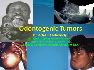
- 1. Dr. Adel I. Abdelhady BDS, MSC, (Egypt) PhD. (Egypt, USA) Oral and Maxillofacial Surgery Dept. College of Dentistry, Dammam University, KSA
- 2. WELLCOME BACK قال رسول اللة صلى اللة علية وسلم من سلك طريقا يلتمس فية علما سهل اللة لة بة طريقا الى الجنة . ان المالئكة لتضع اجنحتها رحمة بطالب العلم انتم وصية رسول اللة صلى اللة علية وسلم
- 3. Duration 3 credit hours 1 hour lecture and 2 hours clinical Assessment 1. Continuous assessment: ( Quizzes + Med term exam + Attendance & activities) 45% 2. Final Written exam: 35 % 3. Final clinical exam: 20% 10% 4. OSCE
- 4. Text Book Resources Peterson L, Ellis E, Hupp, J and Tucker, M. Contemporary Oral And Maxillofacial Surgery Mosby, C.V.Co.Ltd, 4th Edition 2003. Pedlar & Frame. Oral & Maxillofacial Surgery – An objective based textbook, Churchill Livingstone 1st edition, 2001. Kwon and Laskin. Clinician's manual of Oral and Maxillofacial Surgery. Quintessence Publishing Co, Inc. 3rd Edition, 2001. Killey & Kay’s. Outline of Oral Surgery. Part 1. 5th Edition, 1999.
- 6. 1-Odontogenic Tumors I.Tumors of odontogenic epithelium: Ameloblastoma 1. Malignant ameloblastoma 2. Ameloblastic carcinoma B. Clear cell odontogenic carcinoma C. Adenomatoid odontogenic tumor D.Calcifying epithelial odontogenic tumor E.Squamous odontogenic tumor II. Mixed odontogenic tumors: A. Ameloblastic fibroma B.Ameloblastic fibro-odontoma C.Ameloblastic fibrosarcoma D.Odontoameloblastoma E.Compound odontoma F. Complex odontoma III. Tumors of odontogenic ectomesenchyme: A.Odontogenic fibroma B.Granular cell odontogenic tumor C.Odontogenic myxoma D.Cementoblastoma
- 7. 2. Management of Oral Cancer (2h) Students should display a knowledge and understanding of the following: Epidemiology. Risk factors. Clinical presentation. When to refer. Special investigations including the use of plain radiographs, the value and use of CT scanning, magnetic resonance imaging, the types and use of biopsy. Soft tissue surgery for benign, locally invasive and malignant lesions. Methods of treatment including surgery, radiotherapy, chemotherapy. Types of reconstructive procedures and complications of treatment.
- 8. 3-Cysts of jaws Odontogenic Developmental Dentigerous Primordial Eruption Gingival Odontogenic keratocyst Calcifying epithelial odontogenic cyst Gorlin’s cyst Inflammatory Radicular cyst Residual cyst Inflammatory lateral periodontal cyst Non-Odontogenic Fissural cysts Nasopalatine Nasolabial Median Palatine Globulo-maxillary Median mandibular Bone cysts or Pseudocysts Solitary bone cyst Aneurysmal bone cyst Traumatic bone cyst Soft tissue cysts Salivary cysts (mucocele) Gingival cysts (odontogenic) Dermoid, Epidermoid Branchial cleft cyst Thyroglossal duct cyst
- 9. 2. Salivary Gland Disease - Surgical Considerations (2 H) Students should display a knowledge and understanding of: The role of saliva and its relation to the problems with disease. Minor salivary glands including anatomy and presentation of disease. Major salivary glands including the role of investigations including salivary flow - cannulation - sialogram - scintiscanning - CT and MRI Conditions that may affect salivary glands including: - calculi - sialoadenitis - tumours - cystic lesions
- 10. 5. Temporomandibular Joint Disorders (3 H) Students should display a knowledge and understanding of the: The importance of a full and systematic history of present complaint including previous treatment. Method of examination to differentiate between temporomandibular joint disorders, muscular discomfort and atypical facial pain. Special investigations required. Value of conservative/surgery/drug therapy for TM joint disorders. The use of a conservative regime of soft diet, simple jaw exercises, jaw rest, physiotherapy: - the use of splints - the value of surgery - the use of drug therapy - THE ART OF REASSURANCE Conditions that may affect the temporomandibular joint including: TMJ dysfunction syndrome TMJ arthritis and other degenerative disorders TMJ ankylosis TMJ dislocation
- 11. 6. Facial deformities and osteotomies (3 H) Students should display a knowledge and understanding of the Anatomy of both maxilla and mandible The causes of dentofacial deformities. Orthodontic oral and Maxillofacial surgery interface in the treatment of facial deformity Pre operative orthodontic and surgical planning Management of basic mandibular and maxillary deficiencies: split osteotomy, vertical sub sigmoid osteotomy (mandible), maxilla Le Fort 1 osteotomy The concept of distraction osteogenesis and its application in management of the facial deformities
- 12. 7. Cleft Lip and Palate (2 H) Students should: Understand the embryology of the maxilla, mandible, nose, lip and palate. Know the aetiology and incidence of cleft lip and palate Understand the dental problems associated with cleft lip and palate. Feeding Ear Problems Speech Difficulties Dental Problems Malocclusion Nasal Deformity Understand the treatment and appropriate timing of cleft lip and palate patients. Appreciate the importance of maxilla augmentation and using the distraction osteogenesis concept in management of maxillary deficiency in cleft lip and palate patients.
- 14. Dr. Adel I. Abdelhady BDS, MSC, (Egypt) PhD. (Egypt, USA) Oral and Maxillofacial Surgery Dept. College of Dentistry, Dammam University, KSA
- 15. By the end of the lecture(s) Students should display a knowledge and understanding of : 1.Classification the odontogenic tumors 2.familiar with the different types of odontogenic tumor according to its origin 3.Examine and diagnose patient complained from facial swelling 4. Establish a D.D. of the mandibular as will as maxillary swelling 5. Differentiate between different types of tumor’s biological behaviors and accordingly select the suitable management technique (s)
- 16. Classification of Odontogenic Tumors From Neville, et al. I.Tumors of odontogenic epithelium: A. Ameloblastoma 1. Malignant ameloblastoma 2. Ameloblastic carcinoma B. Clear cell odontogenic carcinoma C. Adenomatoid odontogenic tumor D.Calcifying epithelial odontogenic tumor E.Squamous odontogenic tumor II. Mixed odontogenic tumors: A. Ameloblastic fibroma B.Ameloblastic fibro-odontoma C.Ameloblastic fibrosarcoma D.Odontoameloblastoma E.Compound odontoma F. Complex odontoma III. Tumors of odontogenic ectomesenchyme: A.Odontogenic fibroma B.Granular cell odontogenic tumor C.Odontogenic myxoma D.Cementoblastoma
- 17. Biological Classification of Odontogenic Tumors* BENIGN, NO RECURRENCE POTENTIAL Adenomatoid odontogenic tumor Squamous odontogenic tumor Cementoblastoma Periapical cementoosseous dysplasia Odontoma BENIGN, SOME RECURRENCE POTENTIAL Cystic ameloblastoma (unicystic) Calcifying epithelial odontogenic tumor Central odontogenic fibroma Florid cementoosseous dysplasia Ameloblastic fibroma and fibroodontoma *From Regezi, et al. • BENIGN AGGRESSIVE • Ameloblastoma • Clear cell odontogenic tumor (some consider this a carcinoma) • Odontogenic ghost cell tumor (COC, solid type) • Odontogenic myxoma • Odontoameloblastoma • MALIGNANT • Malignant ameloblastoma • Ameloblastic carcinoma • Primary intraosseous carcinoma • Odontogenic ghost cell carcinoma • Ameloblastic fibrosarcoma
- 19. Ameloblastoma The ameloblastoma is the most common odontogenic tumor. It is a benign but locally invasive neoplasm derived from odontogenic epithelium tumor arising from the dental lamina, Hertwig sheath, the enamel organ, or the lining of dental follicles/dentigerous cysts . It has three different clinico-pathologic subtypes: Multicystic , solid (86%), Unicystic (13%) and Peripheral (extraosseus – 1%). It usually occurs in the 4th and 5th decades without a gender predilection.
- 20. Ameloblastoma It is typically slow-growing, locally invasive and runs a benign course. WHO described it as being a benign tumor that is “usually unicentric,(unilocular) non-functional, intermittent in growth, anatomically benign and clinically persistent.” It may develop from cell rests of the enamel organ; from the developing enamel organ; from the lining of odontogenic cysts or from the basal cells of the oral mucosa.
- 21. It may arise from the lining of a dentigerous cyst but more often arises independently of impacted teeth. It is characterized by a progressive growth rate and , when untreated, may reach enormous proportions. Early symptoms are often absent, but late symptoms may include a painless swelling, loose teeth, malocclusion, or nasal obstruction
- 22. Ameloblastoma Ameloblastomas occur in 3 different clinico- radiographic situations requiring different therapeutic considerations and having different prognoses. Conventional Solid/Multicystic or multilocular (86 % of all cases) Unicystic (13 % of all cases) Peripheral or Extraosseous (1 % of all cases)
- 23. Maxillary tumors frequently perforate into the antrum and may grow freely, with extension into the nasal cavity, ethmoid sinuses, and skull base.
- 25. Solid or Multicystic Ameloblastoma: Clinical Features Patient Age: Approximately equal frequency from the third through the seventh decades. Sex Predilection: Approximately equal. Location: 80 % in mandible; 70 % in posterior regions. Radiographic Appearance: Radiolucent lesion which is usually well-circumscribed; it may be unilocular or multilocular (soap-bubble, honeycomb); occasionally an ameloblastoma will be ill-defined with a ragged border. Well- or ill- Defined. Small or Large, may contain Tooth. Root Resorption & tooth Displacement. Distortion of Anatomic features
- 26. Solid or Multicystic Ameloblastoma: Histologic Features There are several microscopic subtypes but these generally have little bearing on the behavior of the tumor. The follicular and plexiform types are the most common. The follicular type is composed of islands of epithelium which resemble the enamel organ in a mature fibrous connective tissue stoma.
- 27. Solid or Multicystic Ameloblastoma: Histologic Features The plexiform type is composed of long, anastomosing cords or larger sheets of odontogenic epithelium. Its stroma tends to be loose and more vascular. The acanthomatous type shows evidence of extensive squamous metaplasia with keratin formation in the island of odontogenic epithelium.
- 28. Solid or Multicystic Ameloblastoma: Histologic Features In the granular cell type there is transformation of groups of epithelial cells to granular cells; the nature of the granular change is unknown. This type is more common in young patients and has been shown to be clinically aggressive.
- 29. Solid or Multicystic Ameloblastoma: Histologic Features The desmoplastic form is composed of islands/cords of odontogenic epithelium in a very dense collagenous stroma. It has a predilection for the anterior maxilla and because of the dense connective tissue may appear as a radiolucent-radiopaque lesion. The basaloid type is the least common and is composed of uniform basaloid cells with no stellate reticulum.
- 30. Solid or Multicystic Ameloblastoma: Additional Features In some studies solid/multicystic ameloblastomas are reported to be more common in Blacks. While lesions are generally asymptomatic, ameloblastomas may cause paresthesia, pain particularly if infected and they can erode the cortical plates of bone.
- 31. Radiographic findings Radiographs classically show a well-circumscribed, expansile soap-bubble radiolucency with clearly demarcated borders. However, the unilocular lesion is indistinguishable from an odontogenic cyst. The extent of root resorption may indicate a neoplastic process. Well-circumscribed, soap-bubble Unilocular – often confused with odontogenic cysts Root resorption – associated with malignancy
- 32. Ameloblastoma VIP What are the main points about this tumor? It is Unencapsulated and infiltrates surrounding bone marrow. Even though they are locally infiltrative, they do Not metastasize. They may occur in any part of both jaws but most are in the middle and posterior regions of the mandible. Ameloblastomas are always purely radiolucent and may be unilocular but frequently become multilocular as they increase in size.
- 33. Solid or Multicystic Ameloblastoma: Treatment •General Principles: Treatments have ranged from simple enucleation and curettage to en bloc resection. behavior & Histologic assessment. Marginal resection is the most •" The Goal is Total Removal (? Pt Med Status). widely used method of •" Curettage & Enucleation should be treatment with the least recurrences reported (up to 15 Abandoned ?! Curettage can disperse tumor %). cells into uninvolved areas. •Microscopic Infiltration of Bone beyond the Most surgeons advocate a margin of at least 1.0 cm Image Margin. beyond the radiographic limits •" Safety Margin must be employed as follows: of the tumor as the tumor often extends beyond the apparent •2 cm for Solid & Multicystic 1-1.5 cm for radiologic/clinical margins. Unicystic & Peripheral. •" Treatment must be Guided by Biologic •" Post Treatment F Up for 15-20 Yrs.
- 34. Solid or Multicystic Ameloblastoma: Prognosis Treatment with curettage has resulted in recurrence rates ranging from 55-90 %. Treatment with marginal resection has resulted in approximately a 15 % recurrence rate. Ameloblastomas of this type arising in the maxilla are particularly dangerous as it is often difficult in getting adequate margins. Rarely is an ameloblastoma life threatening.
- 35. Treatment According to growth characteristics and type Unicystic Complete removal Peripheral ostectomies if extension through cyst wall Classic infiltrative (aggressive) Mandibular – adequate normal bone around margins of resection Maxillary – more aggressive surgery, 1.5 cm margins Ameloblastic carcinoma Radical surgical resection (like SCCa) Neck dissection for LAN
- 36. The treatment of the classic infiltrative, more aggressive ameloblastoma should not be taken lightly. Mandibular resection must include an adequate zone of normal-appearing bone around the main tumor mass. Extension of tumor into surrounding soft tissues is an ominous sign and demands surgery in these areas as vigorous as within the confines of the bone. Maxillary ameloblastomas require more aggressive initial management with at least a 1.5 cm margin of radiographically normal bone. Postoperative follow-up is critical for a minimum of 10-15 years. Ameloblastic carcinoma should be treated with radical surgical resection as for squamous cell carcinoma, with neck dissection reserved for apparent lymphadenopathy.
- 37. Types of Surgical Operations Used for the Removal of Jaw Tumors
- 38. Common types of mandibular resection A.Marginal or segmental
- 39. Types of Jaw Tumors and Primary Treatment Modalities
- 40. Types of surgical operations used for the removal of jaw tumours A. Enucleation and/or curettage: Local removal of tumour by instrumentation in direct contact with the lesion. Used for very benign types of lesions.
- 41. Tumours treated by Enucleation and/or curettage • Odontogenic tumours: Odontoma Ameloblastic fibroma Ameloblastic fibroodontoma Adenomatoid odontogenic tumour Calcifying odontogenic cyst Cementoblastoma Central cementifying fibroma • Other lesions • Fibroosseous lesions Central ossifying fibroma Fibrous dysplasia (if indicated) Cherubism Central giant cell granuloma Aneurismal bone cyst Osteoma Osteoid osteoma osteoblastoma Hemangioma Eosinophilic granuloma Neurilemmoma Neurofibroma Pigmented neurectodermal tumour
- 42. B. Resection Removal of a tumour by incising through uninvolved tissue around the tumour (en block resection): • Marginal resection: Where there is no disruption of the continuity of the bone. • • • Partial segmental resection: Jaw continuity is disrupted due to full-thickness resection. Total resection: total removal of the involved bone e.g. Maxillectomy and mandibulectomy. Composite resection: this is an ablative procedure used most commonly for malignant tumours where tumour is removed with surrounding bone, soft tissue, lymph nodes and channels.
- 43. Tumour treated by Marginal or segmental resection • Odontogenic tumours: Ameloblastoma Calcifying epithelial odontogenic tumour Myxoma Ameloblastic Odontoma • Fibroosseous lesions Benign chondroblastoma • Other lesions Haemangioma
- 44. Tumour treated by Composite resection • Odontogenic tumours: • Fibroosseous lesions Malignant Ameloblastoma Fibrosarcoma Ameloblastic fibrosarcoma Osteosarcoma Ameloblastic odontosarcoma Chondrosarcoma Primary inraosseous carcinoma Ewing's sarcoma
- 51. Unicystic (unilocular) Ameloblastoma: Clinical Features Patient Age: The patients are younger than those with the solid/multicystic form. 50% are diagnosed during the second decade of life. Sex Predilection: ? Same as for the solid?? Location: 90 % occur in the mandible usually in the posterior region. Radiographic Appearance: Typically appears as a RL around the crown of an unerupted tooth (most commonly a mandibular third molar).
- 52. Unicystic Ameloblastoma: Histologic Features Three histopathologic variants are recognized: Luminal: the tumor is confined to the luminal surface of the cyst. Intraluminal/plexiform: the tumor projects from the cystic lining; sometimes resembles the plexiform type of solid/multicystic ameloblastoma. Mural: the tumor infiltrates the fibrous cystic wall.
- 53. Unicystic (unilocular) Ameloblastoma: Treatment and Prognosis Enucleation of the cyst is probably adequate for the luminal and intraluminal/plexiform types. Treatment of the mural type is controversial with some surgeons believing that local resection is best. 10-20 % recurrence after enucleation and curettage with all unicystic ameloblastomas.
- 55. Peripheral Ameloblastoma These tumors are extraosseous and therefore occupy the lamina propria underneath the surface epithelium but outside of the bone. Histologically, these lesions have the same features as the intraosseous forms of the tumor.
- 56. Peripheral Ameloblastoma: Clinical Features Patient Age: Wide age range but most occur during middle-age. Gender Predilection: This is not known. Location: Posterior gingival/alveolar mucosa is involved most frequently. There is a slight predilection for the mandible. The buccal mucosa has been the site in a few reported cases.
- 61. Peripheral Ameloblastoma: Radiographic & Histologic Features Radiographic Appearance: Although not in bone, a few cases have shown superficial erosion of the alvelolar bone. Histologic Appearance: Islands of ameloblastic epithelium are observed in the lamina propria; plexiform and follicular patterns are the most common; in 50 % of the cases the tumor connects with the basal cell layer of the surface epithelium.
- 62. Peripheral Ameloblastoma: Treatment and Prognosis Unlike its intraosseous counterpart, this tumor has an innocuous clinical behavior. Patients respond well to local surgical excision. Some reports indicate a 25 % recurrence rate but in these cases as second surgical procedure results in cure. There has been a rare malignant change reported.
- 63. Malignant Ameloblastoma and Ameloblastic Carcinoma Less than 1 % of the ameloblastomas show malignant behavior with the development of metastases. Malignant ameloblastoma is a tumor that shows histologic features of the typical (benign) ameloblastoma in both the primary and secondary deposits. Ameloblastic carcinoma is a tumor that shows cytologic features of malignancy in the primary tumor, in recurrence and any metastases.
- 64. Malignant Ameloblastoma & Ameloblastic Carcinoma: Clinical Patients range in age from 4-75 with a mean of 30 years. Metastasis has occurred from 1-30 years after the initial treatment. Metastases most often occur in the lungs and in the case of malignant ameloblastoma raises the question of aspiration during surgery. Spread has also occurred to the cervical lymph nodes and to vertebrae and viscera.
- 65. Malignant Ameloblastoma & Ameloblastic Carcinoma: X-Ray With the malignant ameloblastoma, the appearance is similar to the typical solid/multicystic ameloblastoma. The ameloblastic carcinoma is often more aggressive with the lesion appearing as an illdefined radiolucency with cortical destruction.
- 66. Malignant Ameloblastoma & Ameloblastic Carcinoma: Histology With the malignant ameloblastoma, both the primary and metastases show no microscopic features that differ from those of the typical solid/multicystic ameloblastoma. The ameloblastic carcinoma shows cytological features of malignancy in addition to a pattern of an ameloblastoma.
- 67. Malignant Ameloblastoma & Ameloblastic Carcinoma: Treatment & Prognosis Long-term follow-up does not permit accurate assumptions to be made but prognosis appears to be poor. Approximately 50 % of the patients with documented metastases and long-term follow-up have died as the result of their disease.
- 68. Adenomatoid Odontogenic Tumor (AOT) Formerly called an adenoameloblastoma, a somewhat deceptive term that should be discarded, the AOT represents about 3-7 % of all odontogenic tumors. This epithelial tumor has an inductive effect on the odontogenic ectomesenchyme with dentinoid frequently being produced.
- 70. Adenomatoid Odontogenic Tumor (AOT) This is a tumor mostly of teenagers. It occurs in the middle and anterior portions of the jaws in contrast to ameloblastoma which is found mostly in the posterior segment. Two-thirds occur in the maxilla and it is more common in females. This tumor is encapsulated and is treated by curettage with a recurrence rate approaching zero. The radiographic appearance is a unilocular radiolucency, often around the crown of an unerupted tooth in which case they resemble a dentigeous cyst
- 71. AOT: Clinical Features Patient Age: The peak age is in the second decade with a mean around 17 years. Gender Predilection: Females, 2:1. Location: Sixty-five percent of the AOTs occur in the maxilla with 65 % occurring in the canine region. Seventy-five percent of the cases are associated with the crown of an unerupted tooth. On rare occasion the lesion is extraosseous.
- 72. AOT: Radiographic and Additional Features AOTs typically appear as pericoronal radiolucencies, which may have radiopaque material (“snowflake” calcifications) within the lucency. These lesions are frequently asymptomatic and therefore are discovered upon routine radiographic examination. AOTs may also block the eruption of a permanent tooth and be discovered when radiographs are taken to “search for” the unerupted tooth.
- 73. AOT: Histologic Features The lesion is usually surrounded by a thick, fibrous capsule. The tumor is composed of spindle-shaped epithelial cells that form sheets, strands or whorled masses with little connective tissue. The epithelial cells may form rosette-like structures, tubular or duct-like structures may be prominent or absent. Calcifications may be observed in the tumor mass.
- 74. AOT: Treatment and Prognosis Enucleation is the treatment of choice as the tumor is easily removed from the bone. AOTs seldom recur.
- 76. Calcifying Epithelial Odontogenic Tumor (CEOT; Pindborg Tumor) Pindborg tumor accounts for < 1 % of all odontogenic tumors. It is clearly of odontogenic origin but its histogenesis is uncertain. The tumor cells are said to resemble cells of the stratum intermedium.
- 78. CEOT This tumor forms an amorphous material that is said to be amyloid or amyloid-like Whatever it is, it calcifies in a concentrically lamellated “tree-ring” pattern calcifications. This explains the name of calcifying epithelial odontogenic tumor. Calcifying epithelial odontogenic tumor in the body of the mandible. It appears as a radiolucent lesion with smokey dense areas.
- 79. CEOT: Clinical Features Patient Age: Patients ages range from the second to the tenth decades with a mean around 40 years. Gender Predilection: There is no reported sex predilection. Location: 75 % of the CEOTs occur in the mandible with most occurring in the posterior region. A rare peripheral CEOT does occur.
- 80. CEOT: Radiographic Features CEOTs occur as radiolucent lesions with/without opaque foci. They are usually well-circumscribed and may be unilocular or multilocular. Slightly over 50 % of the CEOTs are associated with an unerupted tooth.
- 81. CEOT: Histologic Features This lesion is typically composed of islands, sheets or strands of polyhedral epithelial cells in a fibrous stroma. Areas of amorphous, eosinophilic, hyalinized extracellular material may be scattered throughout. Cells outlines are distinct and intercellular bridges may be seen. Nuclei show considerable variation with giant nuclei and pleomorphism observed. Calcifications may be noted as well as amyloid-like material. Liesegang rings also may be present.
- 82. CEOT: Additional Features, Treatment and Prognosis Bony lesions most commonly present as painless, slow-growing swellings. Peripheral lesions typically appear as non-specific sessile gingival masses. Conservative local resection is the treatment of choice as these lesions are typically less aggressive than the ameloblastoma. With this treatment the recurrence rate is approximately 15 % and the overall prognosis is good.
- 83. Calcifying Epithelial Odontogenic Tumor (CEOT; Pindborg Tumor)
- 84. This group of tumors is composed of proliferating odontogenic epithelium in a cellular ectomesenchyme resembling the dental papilla.
- 85. Ameloblastic Fibroma Clinical Features This true mixed odontogenic tumor is more common in patients in the first and second decades of life with a mean of 14 years seldom is it seen beyond age 20. It is slightly more common in males than females. Approximately 70 % of the ameloblastic fibromas occur in the posterior mandible. Local swelling or failure of teeth to erupt on time or in proper alignment may call attention to the tumor.
- 86. Ameloblastic Fibroma: Radiographic Features Generally, these lesions appear as either a unilocular or multilocular radiolucency. They tend to be well-defined and may have a sclerotic border. Approximately, 50 % are associated with an unerupted tooth.
- 87. Ameloblastic Fibroma: Additional Features, Treatment and Prognosis The tumor is often encapsulated with small tumors usually being asymptomatic. Larger tumors produce swelling, which can expand the cortex and be quite pronounced. Most ameloblastic fibroma are treated by conservative surgical excision; however, a 20 % recurrence rate has led some surgeons to recommend a more aggressive approach.
- 90. Ameloblastic Fibro-odontoma This lesion is defined as a tumor with general features of an ameloblastic fibroma but containing enamel and dentin. Some investigators believe that this entity is but a stage in the development of an odontoma ; however, most agree that progressive destructive tumors are true neoplasms.
- 91. Ameloblastic Fibro-odontoma: Clinical and Radiographic Features Patient Age: Most common in the 5-12 year age range with a mean of 10 years. Gender Predilection: None. Location: It is more common in the premolar/molar regions of both jaws. Radiographic Features: Usually appears as a welldefined unilocular or rarely multilocular radiolucency with variable amounts of calcified material which is radiopaque. Therefore, it may appear as a mixed, radiolucent-radiopaque lesion.
- 92. Ameloblastic Fibro-odontoma: Histologic Features The soft tissue component is identical to the ameloblastic fibroma. The calcified portion consists of foci of enamel and dentin matrix formation in close relationship to the epithelial structures.
- 93. Ameloblastic Fibro-odontoma: Treatment and Prognosis The ameloblastic fibro-odontoma is usually treated by conservative curettage with the lesion separating easily from the surrounding bone. Prognosis is excellent and recurrence is unusual.
- 95. Odontoma The tumors in which odontogenic differentiation is fully expressed are the odontomas. In these tumors, the epithelium an ectomesenchyme realize their potential and make enamel and dentin respectively. As a result, these tumors are mostly radiodense. In the compound odontoma, multiple small and malformed tooth-like structures are formed creating a “bag of marbles” radiographic appearance E D P Compound odontoma, photomicrograph of decalcifed specimen. Note the structure that resembles a tooth with a pulp (P), a surrounding mantle of dentin (D) capped by enamel (E).
- 96. Odontoma The odontoma is the most common odontogenic tumor. It is not a true neoplasm but rather is considered to be a developmental anomaly (hamartoma). Two types of odontomas are recognized: Compound: this type of odontoma is composed of multiple small tooth-like structures. Complex: this lesion is composed of a conglomerate mass of enamel and dentin, which bears no anatomic resemblance to a tooth.
- 97. Odontoma: Clinical Features Patient Age: Most cases are recognized during the second decade of life with a mean of 14 years. Gender Predilection: Approximately equal. Location: Somewhat more common in the maxilla. The compound type is more often in the anterior maxilla while the complex type occurs more often in the posterior regions of either jaw.
- 98. Odontoma: Clinical Features Both types of odontoma are found in the early years, usually in the teens or early twenties. Many are associated with an unerupted tooth. Although they may reach a large size, they do eventually cease growing in contrast to true neoplasms which show continuous growth.
- 99. Odontoma: Radiographic Features Early lesions are radiolucent with smooth, well- defined contours. Later a well-defined radiopaque appearance develops. The compound type shows apparent tooth shapes while the complex type appears as a uniform opaque mass with no apparent tooth shapes present.
- 100. Odontoma: Additional Features Most odontomas are small and do not exceed the size of a normal tooth in the region. However, large ones do occur and these may cause expansion of the jaw. Most odontomas are asymptomatic and as a result are discovered upon routine radiographic examination. Odontomas may block the eruption of a permanent tooth and in these cases are often discovered when “searching for” the “missing” tooth radiographically.
- 101. Odontoma: Histologic Features The compound odontoma is composed of enamel, dentin and cementum arrange in recognizable tooth forms; some enamel matrix may be retained in immature and hypomineralized specimens. The complex odontoma is composed of enamel, dentin and cementum but these tissues are arranged in a random manner that bears no morphological resemblance to a tooth.
- 102. Odontoma: Treatment and Prognosis Odontomas are treated by simple local excision and the prognosis is excellent.
- 103. ? ?
- 104. Odontogenic Myxoma Myxomas arise from altered fibroblasts or myofibroblasts that produce an excess of mucopolysaccharides and are usually incapable of producing mature collagen, tissue that is cell-poor and rich in mucopolysaccharide Clinical Features: Myxomas are benign, sometimes locally aggressive tumors that may arise in bone or soft tissue. Odontogenic myxomas occur primarily in individuals between 20-40 years of age and exhibit a male:female ratio of 1:1 to 1:2. They are rarely seen in patients less than 10 or over the age of 50 years. The mandible is more frequently involved (55-65% of cases) than the maxilla, especially the posterior portion.
- 105. Odontogenic Myxoma Myxoma is a tumor that occurs over a wide age range but most occur in the second and third decades. It is an unencapsulated, locally infiltrating tumor Like many odontogenic tumors, when small it is ordinarily unilocular but becomes multilocular as it enlarges. Since it does not produce a calcified matrix material, it is purely radiolucent.
- 107. Odontogenic Myxoma: Clinical and Radiographic Features Patient Age: 10-50 years with a mean around 30 years. Gender Predilection: Reported to be about equal. Location: May occur in any area of the jaws but more common in the mandible. Radiographic Appearance: In bone it typically presents as a well circumscribed, unilocular radiolucency or as a multilocular, expanding and destructive lesion replacing a large area of the jaw. Maxillary lesions may extend into the maxillary sinus, nasal cavity, orbit or cranial cavity. Teeth may be displaced or their roots may be resorbed resulting in loosening of the teeth.
- 108. Odontogenic Myxoma
- 109. Odontogenic Myxoma: Histologic Features The tumor is composed of loosely arranged stellate, spindle-shaped and round cells in an abundant, loose myxoid stroma with few collagen bundles. Epithelial cells are not required for diagnosis. The odontogenic myxoma may be confused with a chrondromyxoid fibroma or with myxoid change in an enlarged dental follicle or papilla.
- 110. Odontogenic Myxoma: Treatment and Prognosis Small odontogenic myxomas are treated by curettage, while larger lesions may require surgical resection. Odontogenic myxomas are not encapsulated and tend to infiltrate adjacent tissues. Recurrence rates of up to 25 % are reported. Overall, the prognosis is good for most odontogenic myxomas.
- 111. Odontogenic Myxoma