
Recommended
More Related Content
What's hot
What's hot (20)
Viewers also liked
Similar to Fluorescence Microscopy
Similar to Fluorescence Microscopy (20)
More from VIVEK KUMAR SINGH
Recently uploaded
Recently uploaded (20)
Fluorescence Microscopy
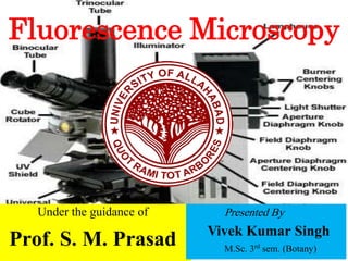
- 1. Fluorescence Microscopy Under the guidance of Prof. S. M. Prasad Presented By Vivek Kumar Singh M.Sc. 3rd sem. (Botany)
- 2. What is Fluorescence Microscopy..? A fluorescence microscope is an optical microscope that uses fluorescence and phosphorescence instead of, or in addition to, reflection and absorption to study properties of organic or inorganic substances.
- 3. Fluorescence- Fluorescence is the emission of light by a substance that has absorbed light or other electromagnetic radiation. Phosphorescence Phosphorescence is a specific type of photoluminescence related to fluorescence. Unlike fluorescence, a phosphorescent material does not immediately re-emit the radiation it absorbs.
- 4. Discovery British scientist Sir George G. Stokes first described fluorescence in 1852. He observed that the mineral fluorspar emitted red light when it was illuminated by ultraviolet excitation. Stokes noted that fluorescence emission always occurred at a longer wavelength than of the excitation light. This shift towards longer wavelength is known as Stokes Shift.
- 5. The "fluorescence microscope" refers to any microscope that uses fluorescence to generate an image, whether it is a more simple set up like an epifluorescence microscope, or A more complicated design such as a confocal microscope, which uses optical sectioning to get better resolution of the fluorescent image.
- 6. On 8 October 2014, the Nobel Prize in Chemistry was awarded to Eric Betzig, William Moerner and Stefan Hell for "the development of super-resolved fluorescence microscopy," which brings "optical microscopy into the nanodimension".
- 7. Principle The specimen is illuminated with light of a specific wavelength. Which is absorbed by the fluorophores, causing them to emit light of longer wavelengths (i.e., of a Principle different color than the absorbed light). The illumination light is separated from the much weaker emitted fluorescence through the use of a spectral emission filter.
- 8. Dichroic filter A dichroic filter or thin- film filter, is a very accurate color filter used to selectively pass light of a small range of colors while reflecting other colors.
- 9. A fluorophore is a fluorescent chemical compound that can re-emit light upon light excitation. Fluorophores typically contain several combined aromatic groups, or plane or cyclic molecules with several π bonds. Fluorophore
- 10. Typical components of a fluorescence microscope are a light source, (xenon arc lamp or mercury-vapor lamp or high-power LEDs and lasers), the excitation filter, the dichroic mirror and the emission filter.
- 11. Most fluorescence microscopes in use are epifluorescence microscopes, where excitation of the fluorophore and detection of the fluorescence are done through the same light path (i.e. through the objective). These microscopes are widely used in biology and are the basis for more advanced microscope designs.
- 12. Epifluorescence microscopy The majority of fluorescence microscopes, especially those used in the life sciences, are of the epifluorescence design shown in the diagram. Light of the excitation wavelength is focused on the specimen through the objective lens. The fluorescence emitted by the specimen is focused to the detector by the objective. Since most of the excitation light is transmitted through the specimen, only reflected excitatory light reaches the objective together with the emitted light.
- 16. Light Source Four main types of light source are used, including xenon arc lamps or mercury-vapor lamps with an excitation filter, lasers, and high- power LEDs. Lasers are mostly used for complex fluorescence microscopy techniques, while xenon lamps, and mercury lamps, and LEDs with a dichroic excitation filter are commonly used for wide field epifluorescence microscopes.
- 17. Xenon arc Lamp A xenon arc lamp is a specialized type of gas discharge lamp, an electric light that produces light by passing electricity through ionized xenon gas at high pressure. It produces a bright white light that closely mimics natural sunlight.
- 18. Biological fluorescent stains Many fluorescent stains have been designed for a range of biological molecules. Some of these are small molecules which are intrinsically fluorescent and bind a biological molecule of interest. Major examples of these are nucleic acid stains like DAPI and Hoechst.
- 19. DAPI (4',6-diamidino-2-phenylindole) is a fluorescent stain that binds strongly to A- T rich regions in DNA and Hoechst. Hoechst stains are part of a family of blue fluorescent dyes used to stain DNA
- 20. A major example of fluorescent stain is phalloidin which is used to stain actin fibres in mammalian cells. There are many fluorescent molecules called fluorophores or fluorochromes such as fluorescein, Alexa Fluors or DyLight 488, which can be chemically linked to a different molecule which binds the target of interest within the sample.
- 22. References • Wikipedia, the free encyclopedia • https://www.microscopyu.com/.../fluorescence/intr oduction-to-fluorescence-microsco..
- 23. Thank You