Automation in hematology part 1
•Download as PPTX, PDF•
125 likes•31,775 views

Automation in hematology part 1

Recommended
More Related Content
What's hot
What's hot (20)
Viewers also liked
Viewers also liked (20)
Similar to Automation in hematology part 1
Similar to Automation in hematology part 1 (20)
More from Dr. Varughese George
More from Dr. Varughese George (20)
Recently uploaded
Recently uploaded (20)
Automation in hematology part 1
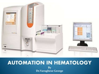
- 1. AUTOMATION IN HEMATOLOGY By Dr.Varughese George
- 2. PART 1
- 3. I N D E X PART 1 I. Necessity for Automation. II. Advantages & Disadvantages of Automation. III. Types of Automated Hematology Analyzers. IV. Principles involved in Automation. V. Pentra ES 60 Haematology Analyzer. VI. Pentra DF Nexus Haematology Analyzer. PART 2 I. Histograms. II. Flags III. Quality Control
- 4. Necessity for Automation Cell counts Dx of Hemoglobinopathies Immunophenotyping Dx of Leukemias & Lymphomas Coagulation Abnormalities.
- 5. Inventor of the first automated analyzer for counting and sizing cells based on his famous ‘Coulter Principle’ The man who started it all- Wallace H. Coulter (1913 –1998)
- 6. I N D E X PART 1 I. Necessity for Automation. II. Advantages & Disadvantages of Automation. III. Types of Automated Hematology Analyzers. IV. Principles involved in Automation. V. Pentra ES 60 Haematology Analyzer. VI. Pentra DF Nexus Haematology Analyzer. PART 2 I. Histograms. II. Flags III. Quality Control
- 7. Automation Advantages Disadvantages Speed & Efficient Handling Accuracy & Precision Multiple Tests on Single Platform Significant Reduction of labor. Flagging RBC Morphology Erroneous results Expensive
- 8. I N D E X PART 1 I. Necessity for Automation. II. Advantages & Disadvantages of Automation. III. Types of Automated Hematology Analyzers. IV. Principles involved in Automation. V. Pentra ES 60 Haematology Analyzer. VI. Pentra DF Nexus Haematology Analyzer. PART 2 I. Histograms. II. Flags III. Quality Control
- 9. Types of Automated Hematology Analyzers Semi-automated analyzers Fully automated analyzers Measures only few parameters Some steps like dilution of blood is carried out manually Measures multiple parameters. Requires only anticoagulated blood samples.
- 10. Components of a cell counter HYDRAULICS Aspirating unit. Dispensers. Diluters. Mixing chambers. Aperture bath. Hemoglobinometer. PNEUMATICS Vacuums & Pressures for operating valves. ELECTRICALS Analyzers & Computing circuitary.
- 11. I N D E X PART 1 I. Necessity for Automation. II. Advantages & Disadvantages of Automation. III. Types of Automated Hematology Analyzers. IV. Principles involved in Automation. V. Pentra ES 60 Haematology Analyzer. VI. Pentra DF Nexus Haematology Analyzer. PART 2 I. Histograms. II. Flags III. Quality Control
- 12. Principles of working of an automated blood analyzer Electrical Impedance. Light Scatter. Fluorescence. Light Absorption. Electrical Conductivity.
- 13. Electrical impedance Cell counting & sizing is based on the Coulter principle - detection & measurement of changes in electrical impedance (resistance) produced by a blood cell as it passes through an electrical field. Blood cells are poor conductors of electricity but are suspended in an electrically conductive diluent. 2 chambers filled with a conductive buffered electrolyte solution separated by a glass tube having a small aperture. A DC current is generated between two electrolytes.
- 14. Electrical impedance As a cell passes through the aperture, flow of current is impeded and a voltage pulse is generated. The no: of pulses indicate the no: of the blood cells. The amplitude (height) of each pulse is proportional to the cell volume. The requisite condition for cell counting by this method is high dilution of sample
- 15. Variables measured by using electrical impedance RBC •RBC Count •MCV •Size distribution histogram •RDW •Hematocrit •MCH •MCHC WBC •Total Count •3 part differential Lymphocyte Mononuclear cells Granulocyte Platelets •Platelet count •Platelet histograms giving MPV PDW
- 16. Optical light scatter Each cell flows in a single line through a flow cell. A LASER device is focused on the flow cell. As LASER light beam strikes a cell, it is scattered in various directions. Photodetectors capture the light. Forward Scatter Light (FALS) ∝ to cell size. Side Scatter Light (SS) (90°) corresponds to nuclear complexity & granularity of cytoplasm. Used to distinguish between granulocytes, lymphocytes & monocytes.
- 17. Variables measured by using OPTICAL LIGHT SCATTER • RBC Count • The 5 part differential Neutrophils Eosinophils Basophils Lymphocytes Monocytes • Mean Cell Volume
- 18. Flow Cytometry Measures multiple cellular & fluorescent properties of cells when they flow as a single cell suspension through a laser beam. Provides the following information about a cell: • Cell size (forward scatter) • Internal complexity or granularity (side scatter) • Relative fluorescence intensity.
- 19. Components of Flow Cytometry Fluidics (The Flow System) The sample is injected into a stream of sheath fluid within the flow chamber. They are forced into the center of the stream forming a single file by the principle of HYDRODYNAMIC FOCUSING. ‘Only 1 cell or particle can pass through the LASER Beam@ a given moment.’ The sample pressure is always > than the sheath pressure ensuring a high flow rate, thus allowing more cells to enter the stream@a given moment. High Flow rate used for immunophenotyping analysis of cells. Low Flow rate used for DNA Analysis.
- 20. Components of Flow Cytometry Optics Following cell delivery, a light source like the Argon- ion LASER is required to excite the cells. When light from a Laser Beam intersects a cell at the ‘interrogation point’, 2 events occur - Light Scattering Fluorescence (Emission of Light ) Light Scattered in the forward direction is detected in Forward Scatter Channel ∝ to cell size and that scattered@90° to axis of Laser path is detected in Side Scatter Channel ∝ to granularity of cell. The cells tagged with fluorescence emit a momentary pulse of fluorescence. A system of optical mirrors and filters then direct the specified wavelengths of light to the designated photodetectors.
- 21. Components of Flow Cytometry Electronics The photodetectors - photodiodes and photomultiplier tubes convert the optical signals (photons) to corresponding electronic signals(electrons). The electronic signal produced is proportional to the amount of light striking a cell. The electric current travels to the amplifier and is converted to a voltage pulse The voltage pulse is assigned a digital value representing a channel by the Analog-to Digital Converter (ADC) . The channel no: is transferred to the computer which displays it to the appropriate position on the data plot.
- 22. Data Analysis Data is collected and stored in the computer – can be displayed in various formats. Parameters – Forward Scatter, Side scatter, emitted fluorescence. Data plots – Single Parameter – Histogram Two Parameters – Dot Plot
- 23. Gating A boundary that can be set to restrict the analysis to a specific population within the sample. Could be Inclusive – Selection of events that fall within the boundary. Exclusive - Selection of events that fall outside the boundary. Data selected by the gate is then displayed in subsequent plots.
- 24. Sorting Consists of collecting cells of interest (defined through criteria of size and fluorescence) for further analysis (microscopy /functional/ chemical analysis)
- 25. Common Applications of Flow Cytometry 1. Leukemias and lympomas Immunophenotyping (evaluation of cell surface markers),diagnosis, detection of minimal residual disease, and to identify prognostically important subgroups. 2. Paroxysmal nocturnal hemoglobinuria Deficiency of CD 55 and CD 59. 3. Hematopoietic stem cell transplantation Enumeration of CD34+ stem cells. 4. Feto-maternal hemorrhage Detection and quantitation of foetal hemoglobin in maternal blood sample. 5. Anemias Reticulocyte count. 6. Human immunodeficiency virus infection For enumeration of CD4+ lymphocytes 7. . Histocompatibility cross matching
- 26. Estimation of Reticulocyte Count Estimation based upon uptake of various dyes and fluorochromes by the RNA of reticulocytes. The flourescent cells are enumerated using a flow cytometer. Various dyes used are – Auramine O Thiazole Orange CD4K 530 Oxazine 750 New Methylene Blue. Flow cytometry allows classification of reticulocytes into 3 maturation stages – Low Fluorescence Reticulocytes (LFR). Middle Fluorescence Reticulocytes (MFR). High Fluorescence Reticulocytes (HFR).
- 27. Estimation of reticulocyte count – Reticulocyte parameters Immature Reticulocyte Fraction ( IRF) / Reticulocyte Maturation Index. The immature reticulocyte fraction (IRF) is calculated as Sum of immature reticulocytes (MFR & HFR) Early sensitive marker of erythropoiesis. Early identification of marrow regeneration in patients undergoing BMT/chemotherapy. Reticulocyte Hemoglobin Equivalent (RET-He) / Reticulocyte Hemoglobin Concentration (CHr) Gives the Hb content of freshly produced RBCs. Early detection of Fe deficiency anemia. Monitoring of erythropoietin & Fe therapy. Mean reticulocyte Volume (MCVr) ↑ rapidly following Fe therapy in subjects with depleted Fe stores. ↓ rapidly with development of iron deficient erythropoiesis.
- 28. Other Methods PEROXIDASE based cell counts. Myeloperoxidase is used to count neutrophils. Lymphocytes are not stained. FLUORESCENCE based cell counts. For reticulocyte and platelet count. Best for detecting immature platelets. IMMUNOLOGICAL based cell counts. Accurate platelet count using CD41/CD61 Abs
- 29. I N D E X PART 1 I. Necessity for Automation. II. Advantages & Disadvantages of Automation. III. Types of Automated Hematology Analyzers. IV. Principles involved in Automation. V. Pentra ES 60 Haematology Analyzer. VI. Pentra DF Nexus Haematology Analyzer. PART 2 I. Histograms. II. Flags III. Quality Control
- 30. Pentra ES 60 Haematology Analyzer
- 31. Features of Pentra ES 60 Throughput: Upto 60 samples/hour Reagents: Only 4 onboard reagents and 1 diluent Perfect differentiation of the 5 WBC subpopulations with DHSS* Technology 3 histograms for RBC, BAS/WBC and PLT together with the 5 DIFF Matrix. Basophils counted through specific channel High resolution matrix includes the determination of 2 additional subpopulations (% and #): Atypical Lymphocytes (ALY***) and Large Immature Cells (LIC***)
- 33. Reagents ABX CLEANER (1L) ABX EOSINOFIX (1L) ABX BASOLYSE II (1L) ABX LYSEBIO (0.4 L) ABX DILUENT (20L)
- 34. Parameters 26 Parameters in CBC mode + 5 DIFF mode 12 parameters in CBC mode 26 parameters in 5 DIFF mode = 12 parameters (CBC mode) + 14 parameters (5 WBC sub-populations & ALY, LIC) WBC (White Blood Cell) RBC (Red Blood Cell) HGB (Hemoglobin Concentration) HCT (Hematocrit) MCV (Mean Corpuscular Volume) MCH (Mean Corpuscular Hemoglobin) MCHC (Mean Corpuscular Hemoglobin Concentration) RDW (Red Distribution Width) PLT (Platelets) PCT (Plateletcrit) MPV (Mean Platelet Volume) PDW (Platelets Distribution Width) WBC (White Blood Cell) RBC (Red Blood Cell) HGB (Hemoglobin Concentration) HCT (Hematocrit) MCV (Mean Corpuscular Volume) MCH (Mean Corpuscular Hemoglobin) MCHC (Mean Corpuscular Hemoglobin Concentration) RDW (Red Distribution Width) PLT (Platelets) PCT (Plateletcrit) MPV (Mean Platelet Volume) PDW (Platelets Distribution Width) LYM (Lymphocytes) in % and MON (Monocytes) in % and NEU (Neutrophils) in % and EOS (Eosinophils) in % and BAS (Basophils) in % and ALY (Atypical Lymphocytes) in % and LIC (Large Immature Cells) in % and 5 WBC sub-populations CBC
- 35. Micro-sampling MDSS (Multi-Distribution Sampling System) Micro-sampling: 30 μL in CBC mode and 53 μL in CBC+DIFF mode are aspired Ideal for pediatric, oncology or geriatric sample types or whenever a small sample volume is required The remaining volume may be used for additional analysis such as sedimentation rate, smear…It will avoid to puncture again the patient Blood split into precise aliquots Aliquots distributed directly into pre-heated analysis chambers with a synchronized tangential flow of diluent for appropriate dilutions without viscosity problem Perfect mixing and homogenization of blood with reagents. No sampling shear-valve to distribute blood sample in all appropriate chambers: No maintenance, no clogging Immediate CBC+DIFF test selection without a cleaning cycle in between.
- 36. Estimation of Hemoglobin Sample is diluted with Drabkin’s reagent. Potassium ferricyanide in the reagent converts Hb Fe from ferrous (Fe2+) to Ferric (Fe3+) to form methemoglobin. This methemoglobin combines with potassium cyanide to form the stable cyanmethemoglobin. Hb concentration is measured by a photodetector which reads absorbance of cyanmethemoglobin at 540 nm. Cyanide free reagents like sodium lauryl sulphate are also used
- 37. HGB measurement Spectrophotometry The newest developed reagent for RBC lysis and determination of HGB. ABX Lysebio : cyanide-free ! How does it work ? By action of lysis agent, contained in the reagent, hemoglobin is released. All the heme iron is oxidized and stabilized. Oxidation resulting complexes are measured through the optical part of the first dilution chamber by spectrophotometry at a wavelength of 550nm. Result = Absorbency value x coef. of calibration. Advantage It does not contain cyanide. It could be thrown with the regular wastes (depending of the national regulations).
- 38. WBC and DIFFERENTIAL count WBC/BAS count Electronic Impedance Variation Principle Differentiation between the BAS and the other WBC is obtained by the use of the ABX BASOLYSE II reagent with it specific lysing action. Nucleus of WBC populations are counted between the electronic thresholds from 0 to <BA2>. BAS are counted between the electronic thresholds <BA2> and <BA3>. Results WBC = Number of cells counted within a specified amount of time per volume x WBC calibration coefficient. BAS = Number of cells counted within a specified amount of time per volume x WBC calibration coefficient in a percentage as the total number of leukocytes (BAS and WBC nuclei).
- 39. WBC and DIFFERENTIAL count LMNE count - It stabilizes WBC in their original state : 48 hour post-draw stability Step 1: Cytochemistry 25 μL of whole blood is delivered into the LMNE chamber in a tangential flow of the reagent ABX Eosinofix. The blood sample is incubated at a regulated temperature with ABX Eosinofix during 12 seconds. - It lyses RBC - It stains EOS cytoplasm, granules and nuclei with a specific dye agent : Chlorazol Black Then, the sample is diluted in a current conductor diluent.
- 40. WBC and DIFFERENTIAL count LMNE count Step 2: Flow cytometry The prepared sample is injected through the flow cytometer: DHSS : Double Hydrodynamic Sequential System 1 - Focused flow for impedance measurement Cell volume measurement: The dilution is aspirated through a calibrated aperture. Two electrodes are placed on each side of the aperture. Electric current passes through the electrodes continuously. When a cell passes through the aperture, electric resistance (or impedance) between the 2 electrodes increases proportionately with cell volume. 2- Focused flow for optical detection Analysis of the internal cellular structure by measuring light absorbency of cells.
- 41. WBC and DIFFERENTIAL count LMNE count Step 3: Results are obtained and displayed in LMNE matrix LMNE matrix is obtained from: • Monocytes • Lymphocytes • Neutrophils • Eosinophils - IMPEDANCE measurement - OPTICAL detection 4 sub-populations are perfectly separated because of the high definition system: The quality of the resolution allows the counting of 2 additional sub-populations: • Large Immature Cells (LIC) : myelocytes, promyelocytes, large blasts. • Atypical Lymphocytes (ALY) : large lymphocytes, activated lymphocytes, blasts. BAS are removed in proportion to LMN populations.
- 43. I N D E X PART 1 I. Necessity for Automation. II. Advantages & Disadvantages of Automation. III. Types of Automated Hematology Analyzers. IV. Principles involved in Automation. V. Pentra ES 60 Haematology Analyzer. VI. Pentra DF Nexus Haematology Analyzer. PART 2 I. Histograms. II. Flags III. Quality Control
- 44. Pentra DF Nexus Haematology Analyzer
- 45. Features of Pentra DF Nexus Methods of measurement: cytochemistry , impedance and flow cytometry. Balance concept : automatic control of the leucocyte count based on 3 independent principles. DHSS: focused flow cytometry and sequential measurement (impedance and absorbance). Automatic reflex testing: selective and programmable (hematology parameters, alarms and flags, demography). Rack rotation mixing of samples: smooth and efficient. Quality: complete traceability for each run in agreement with the accreditation requirements
- 46. Components of Pentra DF Nexus
- 47. Reagents Pentra DF Nexus must be used exclusively with the following reagents: ■ ABX Diluent (10 Liters or 20 Liters): for RBC/PLT dilution, sleeving and cleaning. ■ ABX Cleaner (1 Liter, integrated): for cleaning. ■ ABX Basolyse (5 liters): for BAS count. ■ ABX Leucodiff (1 Liter, integrated): for LMNE and immatures differentiation. ■ ABX Lysebio (1 Liter, integrated): for hemoglobin measurement. ■ ABX Minoclair (0.5 Liter, non-integrated): for concentrated cleaning procedure.
- 52. Parameters
- 53. Sampling principles for RBC & platelets
- 56. Sampling principles for WBC Count & Hb measurement
- 57. Sampling principles for Basophil count
- 58. Sampling principles for Differential Count
- 61. Measurement of Parameters Directly Measured Derived from Histogram Calculated Hemoglobin Mean Cell Volume (MCV) Hematocrit RBC Count Red Cell Distribution Width (RDW) Mean Cell Hemoglobin (MCH) WBC Count Differential Leukocyte Count (DLC) Mean Cell Hemoglobin Concentration (MCHC) Platelet Count Platelet Distribution Width Reticulocyte Count
- 63. END OF PART 1