3. Introduction
• plays an integral role in evaluation of the patient with
neuromuscular diseases.
• provides definitive diagnosis of a wide range of
myopathies and denervating disorders.
• involves obtaining a tissue sample that will be processed
and examined under a light microscope for pathological
alterations in muscle, connective tissue, and blood
vessels.
• requires to be handled, processed, and stained by a skilled
histotechnologist and interpreted by a specialist
pathologist.
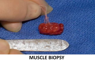
Muscle Biopsy
4. History of Muscle Biopsy
1860 - Duchenne introduced a needle
with a trocar to obtain skeletal muscle
from living subjects through
percutaneous biopsy
1962 - Bergström introduced a
percutaneous biopsy needle similar to
that described by Duchenne in which
gained popularity through widespread
use in diagnosis and research
5. History of Muscle Biopsy
• 1970 - Victor Dubowitz
introduced enzyme histochemical
methods which revolutionised the
role of muscle biopsy for the
diagnosis of various primary and
secondary muscle diseases.
• 1979 - Henriksson designed the
Weil-Blakesley conchotome similar
to the Bergström needle which
doesn’t require a sharp trocar for
aid muscle penetration
6. Indications for muscle biopsy
1. Inflammatory muscle disease before beginning
treatment.
2. Proximal weakness of uncertain cause - myopathic or
neurogenic in adults/children, including 'floppy baby'
syndromes.
3. Hereditary myopathies and muscular dystrophies.
4. To exclude treatable disorder, e.g. polymyositis, in
patients in whom motor neuron disease is suspected.
5. Suspected metabolic myopathies, particularly in
patients with muscle cramps, stiffness or tenderness.
7. Indications for muscle biopsy
6. Autoimmune vasculitis, especially polyarteritis nodosa,
even in the absence of muscular symptoms.
7. Other systemic disorders, e.g. sarcoidosis, infestations.
8. To assess the effects of steroid treatment in the
management of polymyositis, particularly in relation to
the development of steroid myopathy.
9. Occasionally in carrier detection in female siblings or
other close female relatives of boys with Duchenne
dystrophy.
10. Diagnosis of malignant hyperpyrexia syndrome by in vitro
test.
11. Research; e.g. exercise physiology, pathological and
immunological studies,etc.
9. Ideal sites for muscle biopsy
• Muscle which is moderately involved by disease process.
• Muscle belly
• Avoid tendon insertion sites
• Vastus lateralis, biceps,gatrocnemius for proximal
myopathies/generalised systemic disease.
• Avoid deltoid/muscles that are site of
electromyogram needle/recent injections/previous
surgeries.
• Imaging used to select pathological muscle site in
difficult cases e.g tibialis anterior
10. Techniques of Muscle Biopsy
Open Biopsy
•Indicated for disorders with patchy pathology.
•Invasive procedure
• A bigger piece of muscle is available.
•Scarring
•Risk of conscious sedation
Needle Biopsy
•Safe and less invasive procedure.
•Free of any complications.
•Scar is often almost invisible
•More amenable to repeated sampling.
11. Bergström needle biopsy
Components of the biopsy needle:
(a) outer cannula
(b) inner trocar
(c) plunger
Assembled for biopsy, with cutting cannula inserted within
the trocar. A syringe can be connected to the cutting
cannula to increase the yield of tissue by suction.
The inner trocar of the Bergström needle (5 mm)
withdrawn approximately 1 cm opens the window of the
outer cannula.
13. Spring loaded micro-biopsy system
Spring loaded micro-biopsy system consisting of trigger housing,
biopsy needle and the insertion cannula (not shown)
15. Tissue Adequacy
• Open biopsy
– for an adult minimal aggregate of
muscle tissue measuring 1.0 cm cube
(sugar cube size) is required.
– for a child, a minimal aggregate of
0.5 cm cubed (pencil eraser head size)
required.
• Needle biopsy
- one core of muscle can yield a 0.5 cm
cubed aggregate of tissue that is
sufficient for diagnosis.
16. Tissue Transportation
– Muscle may be saved in saline
moistened gauze for several
hours.
– Always keep the specimen cool.
– Muscle should NOT be immersed
in saline, fixative or other liquids.
– Frozen muscle may be safely
shipped "overnight”
with adequate dry ice.
18. Tissue Processing
• Orientation of the fibres is of utmost importance since most of the
information is provided by transverse sections.
• For electron microscopy, 2-3 mm fragments are kept in cacodylate
buffered glutaraldehyde and preserved at 4°C.
• For cryosections,
– fresh frozen in isopentane cooled in liquid N2 (-170°C to -180°C)
– sections are cut in cryostat at -18°C to -20°C.
– sections are stained with Hematoxylin and Eosin (H&E), Masson
trichrome, Modified Gomori’s trichrome (MGT).
– various enzyme histochemial stains done include
• Myosine adenosine triphosphatase (ATPase) preincubated at pH 9.4, 4.6
and 4.3.
• Succinate dehydrogenase (SDH)
• Nicotinamide adenine dinucleotide-Tetrazolium reductase (NADH-TR)
SNAP FREEZE
19. Tissue Processing
• A part of biopsy is used for routine processing after fixing in
buffered formalin.
• For molecular biology, biochemical and genetic analysis, a
small tissue is preserved in 80°C.
• The fresh unfixed muscle is also used for
– Detection and quantification of proteins by Gel electrophoresis.
– Quantification of individual proteins to confirm a deficient or altered
protein and provide a precise quantitative measurement by western
blot.
– Demonstration of gene mutations by
• Polymerase chain reaction (PCR)
• Fluorescent in situ hybridisation (FISH)
21. Stains used for interpretation of muscle biopsy
Stains Use
Haematoxylin and eosin (H&E) General architecture and histology
Masson Trichrome Collagen, fibrosis
Modified Gomori’s trichrome Red ragged fibers, nemaline rods, nuclei,
myelinated fibers
Periodic acid Schiff Glycogen
Oil red O Neutral lipid
Verhoeff–van Gieson Highlights connective tissue and elastin,
important for determining the amount of
connective tissue
Crystal violet Amyloid
Myosin ATP-ase at pH 9.4, 4.6 and 4.3 Distribution and involvement of fibre
types
22. Stain Use
NADH Sarcoplasmic structural details
SDH Oxidative enzyme activity
Cytochrome C Oxidase Mitochondrial enzyme activity
Phosphorlyase Absent in type V glycogenosis
Acid phosphatase High in lysosomal storage disease and
vacuolar myopathy
Alkaline phosphatase High in blood vessels in some inflamatory
myopathies
Acetylcholinesterase Neuromuscular junctions, myotendinous
junctions,
vacuoles in XMEA, denervated/non-
innervated fibres
positive
Stains used for interpretation of muscle biopsy
23. Normal muscle (transverse
section). The fibers are typically
polygonal, and the sarcolemmal
nuclei are located peripherally.
Skeletal mucle is composed of
elongated, multinucleate ,unbranched
contractile cell described as mucle fibre
Characteristic cross-striations seen on
LM d/t arrangement of contractile protein
Normal Muscle
24. •Individual muscle fibres are surrounded by endomysium.
•Fibres grouped in fascicles which are surrounded by a small amount of connective
tissue known as perimysium.
•Epimysium is the connective tissue which surrounds multiple muscle fascicles.
•In normal muscle, the endomysium is so inconspicuous that individual muscle fibers
appear to abut one another.
25. Normal H & E Staining
• Helps in evaluation of general
architecture of the muscle and
variation in the morphology of
individual fibres.
– Variation in fibre size and shape
– Variation in fascicular architecture
– Necrosis and degeneration of
muscle fibres
– Nuclear characteristics
– Type and distribution of
inflammatory infiltrate
– Interstitial changes
26. Muscle biopsy section showing a degenerating fibre
(H & E, total magnification,×200).
28. Muscle biopsy section showing
subsarcolemmal nuclei in normal
fibres.
Gomori’s trichrome stain
(total magnification, ×400)
Muscle biopsy section showing multiple aptly
named ragged red fibres, reflecting
mitochondrial proliferation are seen
Granular red staining may also be seen with
tubular aggregates and rod bodies.
Gomori’s trichrome stain
(total magnification, ×400)
32. Normal muscle. In the alkaline
adenosine triphosphatase
(ATPase) reaction, type 1
fibers are light, and type 2
fibers are dark because of
their high content of ATPase
for use
in the glycolytic pathway.
(ATPase, pH 9.4,
counterstained with eosin).
‘Reverse’ ATPase ph 4.3 shows
the normal distribution of dark
type 1 fibres, pale type 2A fibres
and also
intermediate type 2B fibres.
ATPase at ph 9.4 shows a normal
‘checkerboard’ or ‘mosaic’ distribution of
fibre types 1 and 2. Type 2 fibres stain
darkly.
33. Frozen section stained for the oxidative
enzyme NADH-tetrazolium reductase
shows darkly stained type 1 fibres.
High power of NADH-TR
stained frozen section shows
positive staining of both the
sarcoplasmic reticulum and
mitochondria, the latter more
numerous in type 1 fibres.
34. Stain for succinic
dehydrogenase is paler and has
a particulate appearance due
to selective staining of
mitochondria.
Staining for cytochrome
oxidase (COX) shows a
similar distribution to
SDH staining (more
prominent in Type 1
fibres) but in this stain
the end product is
golden brown.
35. Frozen section stained for phosphorylase. Type 2 fibres
are stained darkly but this reaction is not used routinely to
demonstrate fibre type differentiation. Complete absence
of staining is typical of McArdle’s disease (Type V
Glycogenosis).
36. A modified PAS stain to
demonstrate glycogen. Type 2
fibres which are dependent on
intrinsic glycogen stain darkly.
Verhoeff Van-Gieson (VVG)
stain of frozen tissue to
show fibrous tissue, elastin
and myelinated nerve
fibres. The fine black dots
represent
mitochondria (hence the
darker staining of type 1
fibres) and the
intermyofibrillary network.
37. Oil Red-O in frozen section
demonstrates normal
distribution of fine lipid droplets
within muscle fibres, more
prominent in type 1 fibres
(arrow).
The modified Gomori
trichrome stain identifies
mitochondria as small red
dots within the muscle
fibre, most numerous in
type 1 fibres and at the
fibre periphery, in the
subsarcolemmal zone
(arrow). This biopsy
contains
a normal number of
mitochondria in usual
38. Working Classification of Muscular
Diseases
Neurogenic Neuromuscular
Disorder
Primary Myopathic
Changes
Inflammatory
CongenitalMetabolic
Endocrinopathies
Toxic-Drug Induced
Dystrophy
Duchenne Becker
FSHD
Limb-Girdle
OPMD
Distal Myopathy
Myototic
Central Core
Multicore
Nemaline
Centronuclear
Fibre type Disproportion
Myofibrillar
PM
DM
IBM
Sarcoidosis
Viral
Glycogenosis
Lipid Storage
Mitochondrial
Malig Hyperpyrexia
Myoglobinuria
40. Are the muscle fibres abnormal?
– Size: Small or Large
– Shape: Rounded or Angular
– Type: Grouping; fibre type predominance
– Internal architectural and structural changes
• Disordered or Lost
• Cores, target, vacuoles,inclusions
• Internal nuclei
– Storage or accumulated material:
• Glycogen
• Lipid
• Mitochondria
41. • Biopsy should be orientated transversely.
• Variations in size and shape of muscle fibres are vital
clues to diagnose neuromuscular diseases.
• In a case of abnormal size, muscle fibres can be small
(atrophic) or large (hypertrophic).
• In a case of abnormal shape, muscle fibres can be
rounded(myopathic) or angulated (neuropathic)
Changes in fibre size
ATROPHY
- denervation
- disuse
- aging, ischemia
HYPERTROPHY
- exercise
- compensatory
42.
43. Neurogenic atrophy. Many atrophic fibresare
angular (adenosine triphosphatase, pH 9.4).
Chronic neurogenic atrophy.
Grouping of many small
angular fibres is evident.
Infantile spinal muscularatrophy.
Most of the fibres in the fascicle
are atrophic and rounded.
Fibre Shape
45. Hyaline fibres
• Pathologically rounded and
enlarged fibres.
• Represents an early stage of
cell necrosis.
• Serial sections may reveal
zones of unequivocal necrosis
and phagocytosis adjacent to
hyalinization.
• Most commonly encountered
in Duchenne muscular
dystrophy Hyaline fibre
The fibre in the center of the photograph is
rounded, and it has dark, opaque sarcoplasm.
46. Ring Fibres
• Most consistently
observed in limb-girdle
dystrophy and myotonic
dystrophy.
• Large numbers of ring
fibres especially favor the
latter diagnosis
Circumferential orientation of the peripheral
myofibrils produces a striated ring that
encircles a transversely sectioned fibre in the
center of the field (PAS stain).
47.
48. Split fibres
• Before splitting, fibre
assumes a segmented
appearance as slitlike
spaces form invaginations
between individual
segments.
• Within each space, the
extensions of the plasma
membrane remain
continuous around the
dividing portions of the
cell.
• conspicuous in limb-girdle
dystrophy and in some
cases of denervation and
inclusion body myositis.
Fibre splitting
The fibre at the bottom and center is divided into
two smaller subunits.
(frozen section, rapid Gomori trichrome)
Hypertrophic muscle fibres split into smaller
subunits of two or more smaller fibres that
appear to be mature myocytes with intact
sarcoplasm.
49. Mottled fibres
• Ultrastructurally, mottled
areas reveal a lack of
mitochondria and the
destruction of the
myofilaments.
• Mottled fibres are numerous
in facioscapulohumeral and
limb-girdle dystrophy Mottled fibres
•The sarcoplasm appears moth eaten as a result
of the presence of patchy areas of poor staining
Multiple minute zones of weak enzyme activity
with irregular and poorly delimited borders are
randomly dispersed in the sarcoplasm.
• (nicotinamide adenine dinucleotide, reduced).
50. Cores
• appear as regions of
depleted or absent
enzyme activity.
• cross-banding pattern is
evident in structured
cores.
• present in less than 10%
of fibres.
• numerous in type 1
fibres in central core
disease.
The focal areas of reduced enzyme activity are
single, and cores are centrally positioned
within many fibres
(nicotinamide adenine dinucleotide, reduced).
51.
52. Target fibres
• pathognomonic for
neurogenic atrophy.
• has a great diameter and
virtually always occurs
singularly within the
fibre.
In target fibres, an inner, unstained zone is surrounded
by a rim of increased enzyme activity (nicotinamide
adenine dinucleotide, reduced)
The central zone, which resembles the unstructured core is surrounded by an
intermediate zone, which is darkly stained in oxidative enzyme reactions.
This rim, which is not part of a core lesion, sharply contrasts with the third zone, the
outer normal portion of the fibre.
Three-zone structure
53.
54. Rods
• Ultrastructurally, the rods
are osmiophilic oblong or
rectangular structures
with a greatest dimension
of 6 to 7 μm.
• Their lattice -like
appearance resembles
normal Z-band lending
credence to the concept
that they originate
from/are proliferations of,
Z-bands.
• tends to cluster beneath
the sarcolemma.
Collections of dark, rod-shaped structures are
evident in many of the fibres (frozen section, rapid
Gomori trichrome).
55. Mitochondrial myopathy
Ragged red fibre is seen with abnormally
large mitochondria, several of which contain
paracrystalline inclusions.
Ragged red fibre. Collections of mitochondria
appear as red-stained, irregular,subsarcolemmal
areas within the involved fibre (frozen section,
rapid Gomori trichrome)
Ragged red fibre
•The mitochondrial abnormalities are often recognized by the presence of ragged red
fibres.
•Intensely red, subsarcolemmal protrusions from the cell surface give irregular,
ragged marginal appearance.
•Such fibres are surrounded by prominent dilated capillaries that appear to indent
them and increased in number.
56. Many osmiophilic, lipid containing vacuoles are
evident in the sarcoplasm of the fibre
(resin section, toluidine blue).
Vacuoles
A rimmed vacuole contains abundant red,
granular material
((frozen section, rapid Gomori trichrome)
• Vacuoles may contain abnormal quantities of glycogen or lipid.
• Indicative of a lipid storage disease or mitochondrial myopathy
Lipid storage myopathy (Oculopharyngeal dystrophy)
58. Pathologic Features Disease
In center of specimen, often arranged in
size gradient
Freezing artifact
Often subsarcolemmal, PAS positive Glycogen storage
Scattered fibres; small, round,
osmiophilic; Oil Red O positive
Lipid storage storage
Mitochondrial myopathies
Rimmed, ubiquitin-positive Inclusion body myositis, distal myopathy,
Oculopharyngeal dystrophy
Sarcoplasmic Vacuoles Seen in the Biopsy Specimen
59. Inclusions
• Nuclear inclusions suggests
oculopharyngeal dystrophy
or inclusion body myositis.
• Sarcoplasmic inclusions
suggest myofibrillar
myopathy or inclusion body
myositis.
Inclusion body myositis.
• An intranuclear inclusion is shown at the
center of the picture.
• The inclusion is eosinophilic and smudged; it
is located within a sarcolemmal nucleus.
60. Pathologic Features Disease
Perivascular, angiocentric Dermatomyositis
Connective tissue disease,
Facioscapulohumeral dystrophy
Endomysial, around fibres Polymyositis,
Inclusion body myositis
Viral myositis
Nodular infiltrates Rheumatoid arthritis
Granulomas
Polymorphous with eosinophils Polyarteritis nodosa,
Drug reactions
Trichinosis
Eosinophilic fasciitis
Inflammation seen in the Biopsy Specimen
61. Fibrosis and Fatty Infiltration
• have not been adequately investigated.
• at this juncture in the natural history of the disease,
the active pathologic process has probably subsided
and the opportunity of discovering specific
pathologic changes is irretrievably lost.
• the biopsy of a severely involved muscle should be
discouraged.
62. Nuclear changes - Internal nuclei
• Elevated in 5-10% of fibres.
• Reaction to variety of diverse
injuries.
• Diagnostic significance in
– Myotendinous insertion
– Centronuclear myopathy
– Myotonic dystrophy
– Fibre regeneration
– Fibre atrophy
Nuclear internalization
Many fibres contain one or more internal,
often pyknotic nuclei.
66. Is the pathologic process
Myopathic or Neurogenic ?
. Shape of small muscle fibres
– Round: Myopathic
– Angular: Neurogenic
– Exceptions
• Type 2 fibre atrophy
– Small angular fibres
– Distribution: Scattered
individually
• Spinal muscular atrophy
– Small rounded fibres
– Distribution: Large
grouped atrophy
• Distribution of atrophic fibres
– Grouped : Denervation;
Dystrophinopathies
– Scattered: Acute neuropathy or
myopathy
• Distribution of fibre types
– Type grouping: Chronic
denervation
– Fibre type predominance
• Congenital disorder
• Demyelinating neuropathy
• Large fibre type grouping
– Fibre type small
• Type 1 small: Hereditary
myopathies
• Type 2 small: Acquired
disorders; Congenital MG
67. Type 2 Muscle Fibre Atrophy (ATPase pH 9.4 stain)
Small muscle fibers
•Usually Type 2 (Dark stained at ATPase pH 9.4)
•Shape: Often angular; Some are narrow and elongated.
•Distribution: May appear clustered
68. Presence of small angulated fibres and small or large groups of atrophic fibers
are sure signs of denervation
Group atrophy of fast fibers in a case of denervation
(anti fast myosin x 10)
71. Grouped atrophy
• Clustering of five or more
small angular fibres
• Pathognomonic for
chronic neurogenic
disorders
Chronic neurogenic atrophy.
Grouping of many small angular fibres is evident.
72. Causes of neurogenic atrophy
Peripheral nerve damage
• Diabetes mellitus
• Demyelinating disorders
Motor neuron disorders
• Amyotrophic lateral sclerosis (upper & lower
motor neurons)
• Spinal muscular atrophy (lower motor neurons)
73.
74.
75.
76. Panfascicular atrophy
• Extreme version of
grouped atrophy .
• Vast majority of fibres in
each fascicle are severely
atrophic.
• Distinctive feature of
infantile spinal muscular
atrophy.
Panfascicular atrophy:
entire fascicle is
atrophied. Both type 1
and type 2 will be
atrophied
Compensatory
hypertrophy.
Only type 1
fibres.
77. Perifascicular atrophy
• Fibre atrophy is limited
mainly to the periphery
of the fascicles.
• Found in
dermatomyositis.
fibres in the
middle stay
the same size
fibres on
edges shrink
78.
79. Diseases with Prominent Type 1
Fibre Atrophy
• Myotonic dystrophy
• Nemaline myopathy
• Centronuclear myopathy
• Congenital fibre-type
disproportion
Diseases with Prominent Type 2
Fibre Atrophy
• Corticosteroid therapy and
hypercorticoidism
• Myasthenia gravis
• Disuse atrophy
• Acute denervation
• Paraneoplastic myopathy
Selective Atrophy
•Type 1 fibre hypertrophy- specific for infantile spinal muscular atrophy (ISMA). Also seen in
athletes undergoing endurance training
•Type 2 fibre hypertrophy- sprinters& congenital fibre type disproportion
•Hypertrophy involving both fibres- limb-girdle dytrophy, IBM, myotonia
•congenita & acromegaly
80. ATPase ph 9.4 shows diffuse
selective atrophy of type 2 fibres.
This was a common finding in
biopsies from patients attending
the Rheumatology clinic.
Type 2 atrophy in a patient with
malignancy and cachexia
(immunostain for fast myosin).
81.
82. Fibre type predominance is present when Type 1
fibres constitute more than 55% of the total fibre
population or when more than 80% of fibres are Type
2.
A predominance of Type 1 fibres is seen in Charcot-
Marie Tooth disease and Type 2 fibres are predominant
in Motor Neuron Disease.
Fibre type deficiency is confirmed when less than
10% of fibres constitute either group. A deficiency of
Type 2 fibres may be seen in limb girdle dystrophy
83. Is the pathologic process
Acute or Chronic?
• Acute
– Myopathy:
– Muscle fibre necrosis
and regeneration
– Neuropathy:
– Muscle fibre
atrophy, Scattered
angular or Diffuse
• Chronic
– Myopathy
• Endomysial connective
tissue: Increased
• Muscle fibre
hypertrophy or atrophy
– Neuropathy
• Fibre type grouping
• Grouped atrophy
• Pyknotic nuclear clumps
84. Fibre Necrosis
• Initially stains more intensely
eosinophilic , pales to a wan shade of
pink.
• sarcoplasm is transformed from
striated to coarsely granular.
• nuclei become pyknotic, fragmented,
and finally no longer visible.
• sarcoplasm assumes a vacuolated
or fragmented texture.
• phagocytosis of the necrotic cell
begins
• prevalent in Duchenne muscular
dystrophy and the inflammatory
myopathies
The necrotic process in the fibre at the
center of this longitudinal section is
recognized by a loss of cross striations and
early phagocytosis
85. Pathologic Features Disease
Small groups of necrotic fibres Duchenne dystrophy
Perifascicular necrosis Dermatomyositis
Random fibre necrosis Polymyositis
Inclusion body myositis
Infarcts with large areas of necrosis Polyarteritis nodosa
Extensive, diffuse Rhabdomyolysis in patients with Carnitine
palmitoyltransferase deficiency
Alcoholics
military recruits
Fibre Necrosis Seen in the Biopsy Specimen
86. Fibre Regeneration
• originates from
– sprouts of sarcoplasm at the viable
segments adjacent to the damaged
sarcoplasm.
– satellite cells with the restorative
capacity
• most readily visualized in H&E
sections by the basophilia of their
sarcoplasm.
• nuclei are often eccentric in
location, which may be focally
increased in number, are larger
than normal, with vesicular
chromatin and prominent nucleoli
The necrotic process in the fibre at the
center of this longitudinal section is
recognized by a loss of cross striations and
early phagocytosis
88. What is the distribution of the
pathology?
• Uniform (Similar in all parts of the biopsy)
– Dystrophy
– Fibre type atrophy
• Regional
– Patchy fascicular changes:
• Inflammatory myopathy
• Focal denervation
– Groups of muscle fibres
• Neuropathy: Progressive denervation with reinnervation
• Myopathy:
– Myopathic grouping
– Perifascicular atrophy
• Scattered muscle fibres
• Acute myopathy
• Acute neuropathy
89. Reinnervation is evident
in fibre type grouping
A group of target fibres in NADH-TR
reaction. A clear central zone is
surrounded by a densely stained
intermediate zone
Chronic denervation with
reinnervation. Type grouping
replaces the normalcheckerboard
staining pattern (adenosine
triphosphatase, pH 9.4).
90. Is the pathologic process producing
specific diagnostic features?
• Is there inflammation or excess cellularity?
– What kind?
• Lymphocytes
• Macrophages
• Eosinophils
• Other: Granulomas, Neoplasm
– Where?
• Endomysial
• Perimysial
• Perivascular
• Focal invasion of muscle fibres
91. Inflammation
• most frequently encountered
in immunologically mediated
or idiopathic inflammatory
myopathies
Inflammatory myopathy.
Sheets of lymphocytes expand the
endomysial spaces and surround the fibres.
92. Polymyositis in which lymphocytes are targeted on and beginning to invade a
single muscle fibre. (Hematoxylin and eosin)
93.
94.
95. Is there mitochondrial or storage
pathology ?
• Mitochondrial
• Lipid
• Glycogen
• Amyloid
Is there pathology in structures
other than muscle fibres?
• Vessels
• Connective tissue:
• Endomysial
• Perimysial
• Intramuscular nerves
96.
97.
98.
99. Muscle biopsy section showing
normal lipid staining
Oil Red O(total magnification, ×200).
Muscle biopsy section showing
normal lipid staining
Oil Red O(total magnification, ×200).
100.
101.
102.
103. a) deposits are typically present around and
within vessel walls ( arrow )
b) as crescentic episarcolemmal deposits at
the edges of myofibres (arrows)
c) as viewed under UV illumination with
rhodamine optics significantly enhances
the sensitivity of amyloid detection
Amyloid myopathy
104.
105.
106.
107. Sarcoplasmic inclusion
- Myofibrilllar myopathy
Cytoplasmic body. Circumscribed inclusion with
three dense, red central foci surrounded by
green filamentous material (paraffin, Gomori
trichrome stain).
Desmin myopathy. Two fibres contain slightly
basophilic smudged regions within the
sarcoplasm, which represent collections of
myofibrillar material (frozen section, rapid
Gomori trichrome).
Hyaline body has distinct margins and a
subsarcolemmal location. The finely red granular
appearance of the mitochondria in the normal
sarcoplasm is absent from the more dense,
homogeneous look of the hyaline body (frozen
section, rapid Gomori trichrome).
108.
109. Paraneoplastic necrotizing myopathy
Muscle from a patient with breast carcinoma,
showing
a) Myofibre degeneration ( arrow ) without
signifi cant inflammation (H&E).
b) Necrotic fibres showing strong cytoplasmic
staining for membrane attack complex
(complement C5b-9)
The NADH preparation show a coarse internal
architecture in some myofibres( arrow)
110. Artifacts & Pitfalls
Freezing artifact.
Extensive vacuolar change is caused by
improper freezing. Many of the vacuoles have
linear, noncircular geometric shapes.
Contraction artifact.
Darker contraction bands and
disrupted lucent zones are seen in
several longitudinally oriented fibres
(periodic acid-Schiff stain).
Freezing artefacts due to
•Fresh tissue transported too wet.
•Freezing medium too warm.
•Insufficient time in freezing medium.
•Thawing & refreezing of tissue or cryosections.
Maintain the specimen in isometric state by
introducing it into a muscle clamp, which
prevents the contraction artifact caused by
cutting the muscle and immersing it in fixative.
111. Frozen section has partially lifted off the slide.Tissue
twists create artifact seen as fibre curling withstriped
and ring structures in the fibres (ATPase, pH 9.4,
counterstained with eosin).
Tendinous insertion. In this location, the
muscle fibres normally vary in size, and they
are often surrounded by fibroustissue
(Gomori trichrome).
Artifacts & Pitfalls
112. Muscle specimen submitted in saline. Fluid between
fibres mimics edema. Several fibres are damagedand
disrupted and appear blown out.
During the biopsy procedure, themuscle has
been roughly handled, leading to a
pseudovasculitis in the perimysium.
Neutrophils are marginating in the vessel
lumina and beginning to traverse the vessel
walls.
Artifacts & Pitfalls
130. References
• Dubowitz V, Sewry CA, Oldfors A, Lane RJ. Muscle biopsy: A practical
approach, Elsevier Health Sciences; 2013
• Swash M, Schwartz MS. Biopsy pathology of muscle. Springer; 2013.
• Heffner RR, Balos LL. Muscle biopsy in neuromuscular diseases. Mills SE,
Sternberg’s Diagnostic Surgical Pathology; 4th edition, Vol1, Philadelphia,
Lippincott Williams and Wilkins. 2004:111-35.
• Sundaram C, Uppin MS. Approach to the interpretation of muscle biopsy.
InMuscle Biopsy 2012. InTech.
• Nowak L, Reyes PF. Muscle Biopsy: A Diagnostic Tool in Muscle Diseases.
Journal of Histotechnology. 2008 Sep 1;31(3):101-8.
• Sanja MM, Snezana K, Dragos S. 11. The modern approach to the
histopathological diagnosis of muscle diseases.
• Mitchell JA, Waclawik AJ. Muscle Biopsy in Diagnosis of Neuromuscular
Disorders: The Technical Aspects, Clinical Utility, and Recent Advances.
Journal of Histotechnology. 2007 Dec 1;30(4):257-69.