SlideShare a Scribd company logo
1 of 39
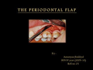
T H E P E R I O D O N T A L F L A P
By-
Aananyaa Jhaldiyal
BDS IV year (2009-10)
Roll no. 01
 A periodontal flap is a section of gingiva &/or mucosa
surgically separated from the underlying tissues to provide
visibility of and access to the bone and root surface.
 A flap also allows the gingiva to be displaced to a different
location in patients with mucogingival involvement.
 Periodontal flaps can be classified as follows –
a) Based on bone exposure after flap reflection
- mucoperiosteal or full thickness flap
- partial thickness or mucosal flap
b) Based on placement of flap after surgery
- displaced flap
- non displaced flap
c) Based on management of papilla
- conventional flap
- papilla preservation flap
1) Full thickness or mucoperiosteal flap-
 All the soft tissue, including the periosteum, is reflected to
expose the bone.
 Indication- need to view the alveolar bone
2) Partial thickness or mucosal flap-
 It includes only the epithelium and a layer of underlying
connective tissue.
 The bone remains covered by a layer of connective tissue,
including the periosteum.
 Also known as split thickness flap.
 Indication- when flap is to be positioned apically, laterally
or coronally; or when the operator does not want to expose
the bone.
A) Based on bone exposure after flap reflection
Fig :A) internal bevel incision to reflect full thickness
flap. B) internal bevel incision to reflect a partial
thickness flap.
1) Non – displaced flap
 The flap is returned and sutured in its original position.
2) Displaced flap
 The flap is placed apically, coronally, or laterally to its original
position.
1) Conventional flap
 In this the interdental papilla is split beneath the contact
point of the two approximating teeth to allow reflection of
buccal and lingual flaps.
B ) B a s e d o n f l a p p l a c e m e n t a f t e r s u r g e r y
C ) B a s e d o n m a n a g e m e n t o f p a p i l l a
 The incision is usually scalloped to maintain gingival
morphology and retain as much papilla as possible.
 Indications-
1) When the interdental spaces are too narrow, thereby
precluding the possibility of preserving the papilla.
2) When the flap is to be displaced.
 Examples- modified Widman flap, the undisplaced flap,
the apically displaced flap, & the flap for reconstructive
procedures.
2) Papilla preservation flap
 In this the entire papilla is incorporated into one of the
flaps by means of crevicular interdental incisions to sever
the connective tissue attachment and a horizontal incision
at the base of the papilla, leaving it connected to one of the
flaps.
 Indications-
1) When there are open interdental spaces
2) When esthetics is of concern
3) When bone regeneration techniques are attempted.
 Dictated by the surgical judgment of the operator.
 Depend on the objectives of the procedure.
 Factors to be considered in designing the flap are-
1) Degree of access to the underlying bone and root
surfaces
2) Final position of the flap
3) Preservation of good blood supply to the flap
 Two basic flap designs are used -
1. Conventional flap
2. Papilla preservation flap
 The incisions for the facial, and the lingual or palatal flap
reach the tip of the interdental papilla or its vicinity,
thereby splitting the papilla into facial half and a lingual
or palatal half.
Fig: flap design for
conventional flap
technique.
1 ) C o n v e n t i o n a l f l a p
 papilla is preserved ( not split).
 The entire surgical procedure should be planned in detail
before the procedure is initiated as detailed planning
allows for a better clinical result.
2 ) P a p i l l a p r e s e r v a t i o n f l a p
 There are basically two types of periodontal flap
incisions-
Horizontal
incisions
Vertical incisions
1) Internal
bevel
incision
2) Crevicular
incision
3) Interdental
incision
1) Oblique
releasing
incision
 Horizontal incisions are directed along the margin of the
gingiva in a mesial or a distal direction.
 Types of horizontal incisions recommended are-
 It is the incision from which the flap is reflected to expose
the underlying bone and root.
1 ) I n t e r n a l b e v e l i n c i s i o n
 Objectives of internal bevel incision are -
1. It removes the pocket lining
2. Conserves the relatively uninvolved outer surface of the
gingiva, which when apically positioned, becomes
attached gingiva.
3. Produces a sharp, thin flap margin for adaptation to the
bone tooth junction.
 This incision is also termed as the first incision because it
is the initial incision in the reflection of a periodontal flap.
 Also termed as reverse bevel incision because its bevel is in
reverse direction from that of the gingivectomy incision.
 Blade used for making this incision - #15C or #15
surgical blade.
Fig: Position of the knife in performing internal bevel incision.
 The internal bevel incision starts from a designated area
on the gingiva and is directed to an area at or near the
crest of the bone.
 It is made from the base of the pocket to the crest of the
bone.
 The incision together with the initial reverse bevel incision
forms a V- shaped wedge ending at or near the crest of
bone.
 This wedge of tissue contains most of the inflamed &
granulomatous areas that constitute the lateral wall of the
pocket as well as the junctional epithelium & the
connective tissue fibers that still persist between the bottom
of the pocket & the crest of the bone.
2 ) C r e v i c u l a r i n c i s i o n
Fig : Position of knife in performing crevicular incision.
 A periosteal elevator is inserted into the initial internal
bevel incision, & the flap is separated from the bone .
 The most apical end of the internal bevel incision is
exposed and visible. With this access, the surgeon is able
to make the interdental incision.
 This incision is made to separate the collar of the gingiva
that is left around the tooth.
 Knife used for this incision- Orban knife.
3 ) I n t e r d e n t a l i n c i s i o n
 The incision is made not only around the facial & the
lingual radicular area but also interdentally, connecting
the facial and the lingual segments to the free the gingiva
completely around the tooth.
Fig : Three incisions necessary for flap surgery. A) internal
bevel incision B) crevicular incision C) interdental
incision.
 Vertical or oblique releasing incisions can be used on one
or both ends of the horizontal incision, depending on the
purpose & design of the flap.
 Vertical incisions at both the ends are necessary if the flap
is to be apically displaced.
 Vertical incision must extend beyond the mucogingival
line, reaching the alveolar mucosa, to allow for the release
of the flap to be displaced.
 Vertical incisions are avoided in the lingual or palatal
areas.
 Facial vertical incisions should not be made in the centre
of an interdental papilla or over the radicular surface of a
tooth.
Fig : The incision
should be made at the
line angles.
 Incisions should be made at the line angles of a tooth
either to include the papilla in the flap or to avoid it
completely.
 Vertical incisions should also be designed to avoid short
flaps with long, apically directed incisions because this
could jeopardize the blood supply of the flap.
S.no Type of flap Reflection
accomplished by
Instrument
used
1) Full thickness
flap or
mucoperiosteal
flap
Blunt dissection Periosteal
elevator
which
separates the
mucoperioste
um from the
bone.
2) Partial thickness
flap or mucosal
flap
Sharp dissection Surgical
scalpel (#15)
Fig : Elevation of flap with
periosteal elevator to obtain full
thickness flap.
Fig: Elevation of flap with BP
knife to obtain a split thickness
flap.
 The purpose of suturing is to maintain the flap in the
desired position until healing has progressed to the point
where sutures are no longer needed.
 Suture materials for periodontal flap are –
1) Non absorbable •Silk: braided
•Nylon: monofilament
(ethilon)
•EPTfe: monofilament
(Gore- tex)
•Polyester: braided
(Ethibond)
2) Absorbable • Surgical: gut
• Plain gut ; monofilament ( 30
days)
• Chromic gut ; monofilament (
45-60 days)
3) Synthetic • Polyglycolic: braided (16 -20
days)
( vicryl; ethicon)
(Dexon; Davis & Geck)
• Polyglecaprone: monofilament
( 90- 120 days)
(Monocryl; Ethicon)
• Polyglyconate: monofilament
(Maxon)
 The resorbable sutures have gained popularity because
they enhance patient comfort & eliminate suture removal
appointments.
 The non resorbable silk braided suture was the most
commonly used in the past due to its ease of use & low
cost.
 The expanded polytetrafluoroethylene synthetic
monofilament is an excellent nonresorbable suture widely
used today.
 The most commonly used resorbable sutures are the
natural plain gut or the chromic gut. Both are mono-
filaments and are processed from purified collagen of
either sheep or cattle intestine.
 The chromic gut is a plain gut suture processed with
chromic salts to make it resistant to enzymatic resorption,
thereby increasing the resorption time.
1) The needle should enter the tissues at right angles and no
less than 2 to 3 mm from the incision.
2) The needle is then carried through the tissue, following
the needle's curvature.
T e c h n i q u e o r p r i n c i p l e s o f s u t u r i n g
3) The knot should not be placed over the incision.
4) The periodontal flap is closed either with independent
sutures or with continuous, independent sling sutures.
5) Sutures of any type in the interdental papillae should be
placed at a point located below the imaginary line that
forms the base of the triangle of the interdental papilla as
shown in the figure below.
6) The location of sutures for closure of a palatal flap depends
on the extent of flap elevation that has been performed.
The flap is divided in four quadrants as shown in the figure
below. If the elevation of the flap is slight or moderate, the
sutures can be placed in the quadrant closest to the teeth. If
the flap elevation is substantial, the sutures should be
placed in the central quadrants of the palate.
7) The clinician may or may not use periodontal dressings.
When the flaps are not apically displaced, it is not
necessary to use dressings other than for patient comfort.
L i g a t i o n
1) Interdental
ligation
Direct Loop Suture
permits a better
closure of the interdental
papilla .
should be performed
when bone grafts are
used or when close
apposition of the
scalloped incision is
required.
figure-eight
suture
there is thread
between the two flaps.
used when the flaps
are not in close
apposition because of
apical flap position or
non-scalloped
incisions.
2) Sling ligation  The sling ligation can be used for a flap
on one surface of a tooth that involves two
interdental spaces.
T y p e s O f S u t u r e s
1) Horizontal mattress
suture
often used for the
interproximal areas of diastema
or for wide interdental spaces
to adapt the interproximal
papilla properly against the
bone.
can be incorporated with the
continuous, independent sling
sutures.
2) Continuous,
independent sling
suture
is used when there is both a facial
and a lingual flap involving many
teeth.
This type of suture does not pull on
the lingual flap when this flap is
sutured.
is especially appropriate for the
maxillary arch because the palatal
gingiva is attached and fibrous
3) Anchor suture This suture closes the facial and
lingual flaps and adapts them tightly
against the tooth.
4) Closed-Anchor
Suture
Another technique to close a flap
located in an edentulous area
mesial or distal to a tooth.
 Consists of tying a direct suture
that closes the proximal flap,
carrying one of the threads around
the tooth to anchor the tissue
against the tooth, and then tying
the two threads.
5) Periosteal suture used to hold the apically
displaced partial-thickness flaps on
the periosteum.
It is of two types-
1. holding suture
2. closing suture
FIG: Periosteal sutures for an apically displaced flap. Holding
sutures, shown at the bottom, are done first, followed by the
closing sutures, shown at the coronal edge of the flap.
1. The Holding Suture - is a horizontal mattress suture placed
at the base of the displaced flap to secure it into the new
position.
2. The Closing Suture- are used to secure the flap edges to the
periosteum.
1) Immediately
after
suturing ( up
to 24 hours)
 A connection between the flap and the
tooth or bone surface is established which
contains fibrin reticulum with many PMN
leukocytes, erythrocytes, debris of injured
cells, & capillaries at the edge of the
wound.
2) 1-3 days
after flap
surgery.
 The space between the flap & the tooth or
bone is thinner & epithetlial cells migrate
over the border of the flap, usually
contacting the tooth at this time.
3) One week
after surgery.
 An epithelial attachment to the root has been
established by means of hemidesmosomes & a
basal lamina.
 Blood clot is replaced by granulation tissue
derived from the gingival connective tissue,
the bone marrow, & the PDL.
4) Two weeks
after surgery.
 Collagen fibers begin to appear parallel to the
tooth surface.
 Union of the flap to the tooth is still weak
because of presence of immature collagen
collagen fibers.
5) One month
after surgery.
 A fully epithelialized gingival crevice with a
well defined epithelial attachment is present.
 There is beginning of functional arrangement
of supra crestal fibers.
 Newman, Takei, Klokkevold, Fermin A Carranza.
CLINICAL PERIODONTOLOGY, 11th edition, WB
Saunders 2006.
The Periodontal flap

More Related Content

What's hot

Periodontal bone defects
Periodontal bone defectsPeriodontal bone defects
Periodontal bone defectsHeenal Adhyaru
 
Mucogingival surgery in periodontics
Mucogingival surgery in periodonticsMucogingival surgery in periodontics
Mucogingival surgery in periodonticsBinaya Subedi
 
General principles of Periodontal surgery
General principles of Periodontal surgeryGeneral principles of Periodontal surgery
General principles of Periodontal surgeryJignesh Patel
 
Papilla preservation flap
Papilla preservation flapPapilla preservation flap
Papilla preservation flapVidya Vishnu
 
Guided tissue regeneration
Guided tissue regenerationGuided tissue regeneration
Guided tissue regenerationParth Thakkar
 
Sonic & ultrasonic instruments
Sonic & ultrasonic instruments Sonic & ultrasonic instruments
Sonic & ultrasonic instruments Shikha Arya
 
Free gingival graft
Free gingival graftFree gingival graft
Free gingival graftMaryamAdham1
 
Aggressive periodontitis
Aggressive periodontitisAggressive periodontitis
Aggressive periodontitisBinaya Subedi
 
Periodontal plastic and esthetic surgery
Periodontal plastic and esthetic surgeryPeriodontal plastic and esthetic surgery
Periodontal plastic and esthetic surgeryjosna thankachan
 
4.furcation involvement and its treatment
4.furcation involvement and its treatment4.furcation involvement and its treatment
4.furcation involvement and its treatmentpunitnaidu07
 
furcation involvement
furcation involvementfurcation involvement
furcation involvementJignesh Patel
 
Pathologic migration
Pathologic migrationPathologic migration
Pathologic migrationsruthi K
 

What's hot (20)

Periodontal pocket
Periodontal pocketPeriodontal pocket
Periodontal pocket
 
Periodontal bone defects
Periodontal bone defectsPeriodontal bone defects
Periodontal bone defects
 
Mucogingival surgery in periodontics
Mucogingival surgery in periodonticsMucogingival surgery in periodontics
Mucogingival surgery in periodontics
 
Vestibuloplasty
VestibuloplastyVestibuloplasty
Vestibuloplasty
 
General principles of Periodontal surgery
General principles of Periodontal surgeryGeneral principles of Periodontal surgery
General principles of Periodontal surgery
 
Papilla preservation flap
Papilla preservation flapPapilla preservation flap
Papilla preservation flap
 
Guided tissue regeneration
Guided tissue regenerationGuided tissue regeneration
Guided tissue regeneration
 
Furcation ppt
Furcation pptFurcation ppt
Furcation ppt
 
Sonic & ultrasonic instruments
Sonic & ultrasonic instruments Sonic & ultrasonic instruments
Sonic & ultrasonic instruments
 
Gingivectomy
Gingivectomy Gingivectomy
Gingivectomy
 
Free gingival graft
Free gingival graftFree gingival graft
Free gingival graft
 
Aggressive periodontitis
Aggressive periodontitisAggressive periodontitis
Aggressive periodontitis
 
Frenectomy
Frenectomy Frenectomy
Frenectomy
 
Gingival crevicular fluid
Gingival crevicular fluidGingival crevicular fluid
Gingival crevicular fluid
 
Periodontal plastic and esthetic surgery
Periodontal plastic and esthetic surgeryPeriodontal plastic and esthetic surgery
Periodontal plastic and esthetic surgery
 
4.furcation involvement and its treatment
4.furcation involvement and its treatment4.furcation involvement and its treatment
4.furcation involvement and its treatment
 
furcation involvement
furcation involvementfurcation involvement
furcation involvement
 
ATTACHED GINGIVA
ATTACHED GINGIVAATTACHED GINGIVA
ATTACHED GINGIVA
 
Peridontal pocket
Peridontal pocketPeridontal pocket
Peridontal pocket
 
Pathologic migration
Pathologic migrationPathologic migration
Pathologic migration
 

Viewers also liked

Periodontal Flap Surgery
Periodontal Flap SurgeryPeriodontal Flap Surgery
Periodontal Flap SurgeryWendy Jeng
 
periodontal flap surgeries
periodontal flap surgeriesperiodontal flap surgeries
periodontal flap surgeriesSwati Gupta
 
Principles of flap surgery copy
Principles of flap surgery   copyPrinciples of flap surgery   copy
Principles of flap surgery copyNavneet Randhawa
 
Periodontal surgeries
Periodontal surgeriesPeriodontal surgeries
Periodontal surgeriesMoola Reddy
 
Principles of scaling & root planing dr alaa attia
Principles of scaling & root planing dr alaa attia Principles of scaling & root planing dr alaa attia
Principles of scaling & root planing dr alaa attia Alaa Atia
 
Periodontal flap (Carranza 57)
Periodontal flap (Carranza 57)Periodontal flap (Carranza 57)
Periodontal flap (Carranza 57)Dara Ghaznavi
 
Treatment Plan in Periodontics
Treatment Plan in PeriodonticsTreatment Plan in Periodontics
Treatment Plan in PeriodonticsDRAMITDE
 
Gingival surgical techniques
Gingival surgical techniquesGingival surgical techniques
Gingival surgical techniquesshazia26
 
JOURNAL CLUB ON CORONALLY ADVANCED FLAP vs THE POUCH TECHNIQUE COMBINED WITH ...
JOURNAL CLUB ON CORONALLY ADVANCED FLAP vs THE POUCH TECHNIQUE COMBINED WITH ...JOURNAL CLUB ON CORONALLY ADVANCED FLAP vs THE POUCH TECHNIQUE COMBINED WITH ...
JOURNAL CLUB ON CORONALLY ADVANCED FLAP vs THE POUCH TECHNIQUE COMBINED WITH ...Shilpa Shiv
 
Minimally Invasive Surgery & Acellular Dermal Matrix to Correct Gingival Rece...
Minimally Invasive Surgery & Acellular Dermal Matrix to Correct Gingival Rece...Minimally Invasive Surgery & Acellular Dermal Matrix to Correct Gingival Rece...
Minimally Invasive Surgery & Acellular Dermal Matrix to Correct Gingival Rece...Edward Gottesman
 
General principles of periodontal surgery
General principles of periodontal surgeryGeneral principles of periodontal surgery
General principles of periodontal surgeryMD Abdul Haleem
 
Gingival recession
Gingival recessionGingival recession
Gingival recessionImen Kassoma
 
Periodontal plastic surgery
Periodontal plastic surgeryPeriodontal plastic surgery
Periodontal plastic surgeryRobert Cain
 

Viewers also liked (20)

Periodontal Flap Surgery
Periodontal Flap SurgeryPeriodontal Flap Surgery
Periodontal Flap Surgery
 
periodontal flap surgeries
periodontal flap surgeriesperiodontal flap surgeries
periodontal flap surgeries
 
Periodontal Flap
Periodontal FlapPeriodontal Flap
Periodontal Flap
 
Principles of flap surgery copy
Principles of flap surgery   copyPrinciples of flap surgery   copy
Principles of flap surgery copy
 
Periodontal surgeries
Periodontal surgeriesPeriodontal surgeries
Periodontal surgeries
 
Principles of scaling & root planing dr alaa attia
Principles of scaling & root planing dr alaa attia Principles of scaling & root planing dr alaa attia
Principles of scaling & root planing dr alaa attia
 
Periodontal flap (Carranza 57)
Periodontal flap (Carranza 57)Periodontal flap (Carranza 57)
Periodontal flap (Carranza 57)
 
Gingivectomy
GingivectomyGingivectomy
Gingivectomy
 
Perio surgery
Perio surgeryPerio surgery
Perio surgery
 
Treatment Plan in Periodontics
Treatment Plan in PeriodonticsTreatment Plan in Periodontics
Treatment Plan in Periodontics
 
Gingival surgical techniques
Gingival surgical techniquesGingival surgical techniques
Gingival surgical techniques
 
JOURNAL CLUB ON CORONALLY ADVANCED FLAP vs THE POUCH TECHNIQUE COMBINED WITH ...
JOURNAL CLUB ON CORONALLY ADVANCED FLAP vs THE POUCH TECHNIQUE COMBINED WITH ...JOURNAL CLUB ON CORONALLY ADVANCED FLAP vs THE POUCH TECHNIQUE COMBINED WITH ...
JOURNAL CLUB ON CORONALLY ADVANCED FLAP vs THE POUCH TECHNIQUE COMBINED WITH ...
 
Minimally Invasive Surgery & Acellular Dermal Matrix to Correct Gingival Rece...
Minimally Invasive Surgery & Acellular Dermal Matrix to Correct Gingival Rece...Minimally Invasive Surgery & Acellular Dermal Matrix to Correct Gingival Rece...
Minimally Invasive Surgery & Acellular Dermal Matrix to Correct Gingival Rece...
 
GINGIVECTOMY AND GINGIVOPLASTY
GINGIVECTOMY AND GINGIVOPLASTYGINGIVECTOMY AND GINGIVOPLASTY
GINGIVECTOMY AND GINGIVOPLASTY
 
General principles of periodontal surgery
General principles of periodontal surgeryGeneral principles of periodontal surgery
General principles of periodontal surgery
 
Gingival recession
Gingival recession Gingival recession
Gingival recession
 
Root planing
Root planingRoot planing
Root planing
 
Gingival recession
Gingival recessionGingival recession
Gingival recession
 
Gingival Recession
Gingival RecessionGingival Recession
Gingival Recession
 
Periodontal plastic surgery
Periodontal plastic surgeryPeriodontal plastic surgery
Periodontal plastic surgery
 

Similar to The Periodontal flap

periodontal flap
periodontal flapperiodontal flap
periodontal flaps farrokhi
 
Periodontal Flap.pptx
Periodontal Flap.pptxPeriodontal Flap.pptx
Periodontal Flap.pptxChhayaDev
 
periodontal_flap_one_2-10-2013.ppt
periodontal_flap_one_2-10-2013.pptperiodontal_flap_one_2-10-2013.ppt
periodontal_flap_one_2-10-2013.pptHoeliom
 
Surgical endodontics (Apicectomy) by Dr. Amit T. Suryawanshi, Oral Surgeon, ...
Surgical endodontics (Apicectomy) by Dr. Amit T. Suryawanshi,  Oral Surgeon, ...Surgical endodontics (Apicectomy) by Dr. Amit T. Suryawanshi,  Oral Surgeon, ...
Surgical endodontics (Apicectomy) by Dr. Amit T. Suryawanshi, Oral Surgeon, ...All Good Things
 
Surgical endodontics(Apicectomy) by Dr. Amit Suryawanshi .Oral & Maxillofac...
Surgical endodontics(Apicectomy)  by  Dr. Amit Suryawanshi .Oral & Maxillofac...Surgical endodontics(Apicectomy)  by  Dr. Amit Suryawanshi .Oral & Maxillofac...
Surgical endodontics(Apicectomy) by Dr. Amit Suryawanshi .Oral & Maxillofac...All Good Things
 
Periodontal flap design for access on osseous surgery
Periodontal flap design for access on osseous surgeryPeriodontal flap design for access on osseous surgery
Periodontal flap design for access on osseous surgeryDisha Rai
 
Principles of more complex exdontia By:Cezar Edward
Principles of more complex exdontia By:Cezar EdwardPrinciples of more complex exdontia By:Cezar Edward
Principles of more complex exdontia By:Cezar EdwardCezar Edward Lahham
 
Periodontal surgery
Periodontal surgeryPeriodontal surgery
Periodontal surgeryEnas Elgendy
 
Root coverage procedures periodontics.pptx
Root coverage procedures periodontics.pptxRoot coverage procedures periodontics.pptx
Root coverage procedures periodontics.pptxwanidayim1
 
Flap Design for Minor Oral Surgery
Flap Design for Minor  Oral SurgeryFlap Design for Minor  Oral Surgery
Flap Design for Minor Oral Surgeryssuseraf61fb
 
endodontic surgery and its current concepts
endodontic surgery and its current concepts endodontic surgery and its current concepts
endodontic surgery and its current concepts boris saha
 

Similar to The Periodontal flap (20)

flap surgery.pptx
flap surgery.pptxflap surgery.pptx
flap surgery.pptx
 
Flap techniques for pocket therapy
Flap techniques for pocket therapy  Flap techniques for pocket therapy
Flap techniques for pocket therapy
 
Periodontal flaps
Periodontal flapsPeriodontal flaps
Periodontal flaps
 
Flap surgery
Flap surgeryFlap surgery
Flap surgery
 
periodontal flap
periodontal flapperiodontal flap
periodontal flap
 
Periodontal Flap.pptx
Periodontal Flap.pptxPeriodontal Flap.pptx
Periodontal Flap.pptx
 
periodontal_flap_one_2-10-2013.ppt
periodontal_flap_one_2-10-2013.pptperiodontal_flap_one_2-10-2013.ppt
periodontal_flap_one_2-10-2013.ppt
 
Surgery 2
Surgery 2Surgery 2
Surgery 2
 
General Principles of Surgical Techniques for Periodontal Regeneration
General Principles of Surgical Techniques for Periodontal RegenerationGeneral Principles of Surgical Techniques for Periodontal Regeneration
General Principles of Surgical Techniques for Periodontal Regeneration
 
Surgical endodontics (Apicectomy) by Dr. Amit T. Suryawanshi, Oral Surgeon, ...
Surgical endodontics (Apicectomy) by Dr. Amit T. Suryawanshi,  Oral Surgeon, ...Surgical endodontics (Apicectomy) by Dr. Amit T. Suryawanshi,  Oral Surgeon, ...
Surgical endodontics (Apicectomy) by Dr. Amit T. Suryawanshi, Oral Surgeon, ...
 
Surgical endodontics(Apicectomy) by Dr. Amit Suryawanshi .Oral & Maxillofac...
Surgical endodontics(Apicectomy)  by  Dr. Amit Suryawanshi .Oral & Maxillofac...Surgical endodontics(Apicectomy)  by  Dr. Amit Suryawanshi .Oral & Maxillofac...
Surgical endodontics(Apicectomy) by Dr. Amit Suryawanshi .Oral & Maxillofac...
 
Periodontal flaps
Periodontal flapsPeriodontal flaps
Periodontal flaps
 
Periodontal flap design for access on osseous surgery
Periodontal flap design for access on osseous surgeryPeriodontal flap design for access on osseous surgery
Periodontal flap design for access on osseous surgery
 
Surgical exodontia
Surgical exodontiaSurgical exodontia
Surgical exodontia
 
Principles of more complex exdontia By:Cezar Edward
Principles of more complex exdontia By:Cezar EdwardPrinciples of more complex exdontia By:Cezar Edward
Principles of more complex exdontia By:Cezar Edward
 
surgical flaps oral surgery
surgical flaps oral surgery surgical flaps oral surgery
surgical flaps oral surgery
 
Periodontal surgery
Periodontal surgeryPeriodontal surgery
Periodontal surgery
 
Root coverage procedures periodontics.pptx
Root coverage procedures periodontics.pptxRoot coverage procedures periodontics.pptx
Root coverage procedures periodontics.pptx
 
Flap Design for Minor Oral Surgery
Flap Design for Minor  Oral SurgeryFlap Design for Minor  Oral Surgery
Flap Design for Minor Oral Surgery
 
endodontic surgery and its current concepts
endodontic surgery and its current concepts endodontic surgery and its current concepts
endodontic surgery and its current concepts
 

Recently uploaded

Wessex Health Partners Wessex Integrated Care, Population Health, Research & ...
Wessex Health Partners Wessex Integrated Care, Population Health, Research & ...Wessex Health Partners Wessex Integrated Care, Population Health, Research & ...
Wessex Health Partners Wessex Integrated Care, Population Health, Research & ...Wessex Health Partners
 
Statistical modeling in pharmaceutical research and development.
Statistical modeling in pharmaceutical research and development.Statistical modeling in pharmaceutical research and development.
Statistical modeling in pharmaceutical research and development.ANJALI
 
The next social challenge to public health: the information environment.pptx
The next social challenge to public health:  the information environment.pptxThe next social challenge to public health:  the information environment.pptx
The next social challenge to public health: the information environment.pptxTina Purnat
 
SYNDESMOTIC INJURY- ANATOMICAL REPAIR.pptx
SYNDESMOTIC INJURY- ANATOMICAL REPAIR.pptxSYNDESMOTIC INJURY- ANATOMICAL REPAIR.pptx
SYNDESMOTIC INJURY- ANATOMICAL REPAIR.pptxdrashraf369
 
SWD (Short wave diathermy)- Physiotherapy.ppt
SWD (Short wave diathermy)- Physiotherapy.pptSWD (Short wave diathermy)- Physiotherapy.ppt
SWD (Short wave diathermy)- Physiotherapy.pptMumux Mirani
 
Radiation Dosimetry Parameters and Isodose Curves.pptx
Radiation Dosimetry Parameters and Isodose Curves.pptxRadiation Dosimetry Parameters and Isodose Curves.pptx
Radiation Dosimetry Parameters and Isodose Curves.pptxDr. Dheeraj Kumar
 
call girls in Dwarka Sector 21 Metro DELHI 🔝 >༒9540349809 🔝 genuine Escort Se...
call girls in Dwarka Sector 21 Metro DELHI 🔝 >༒9540349809 🔝 genuine Escort Se...call girls in Dwarka Sector 21 Metro DELHI 🔝 >༒9540349809 🔝 genuine Escort Se...
call girls in Dwarka Sector 21 Metro DELHI 🔝 >༒9540349809 🔝 genuine Escort Se...saminamagar
 
Introduction to Sports Injuries by- Dr. Anjali Rai
Introduction to Sports Injuries by- Dr. Anjali RaiIntroduction to Sports Injuries by- Dr. Anjali Rai
Introduction to Sports Injuries by- Dr. Anjali RaiGoogle
 
call girls in paharganj DELHI 🔝 >༒9540349809 🔝 genuine Escort Service 🔝✔️✔️
call girls in paharganj DELHI 🔝 >༒9540349809 🔝 genuine Escort Service 🔝✔️✔️call girls in paharganj DELHI 🔝 >༒9540349809 🔝 genuine Escort Service 🔝✔️✔️
call girls in paharganj DELHI 🔝 >༒9540349809 🔝 genuine Escort Service 🔝✔️✔️saminamagar
 
Presentació "Real-Life VR Integration for Mild Cognitive Impairment Rehabilit...
Presentació "Real-Life VR Integration for Mild Cognitive Impairment Rehabilit...Presentació "Real-Life VR Integration for Mild Cognitive Impairment Rehabilit...
Presentació "Real-Life VR Integration for Mild Cognitive Impairment Rehabilit...Badalona Serveis Assistencials
 
Measurement of Radiation and Dosimetric Procedure.pptx
Measurement of Radiation and Dosimetric Procedure.pptxMeasurement of Radiation and Dosimetric Procedure.pptx
Measurement of Radiation and Dosimetric Procedure.pptxDr. Dheeraj Kumar
 
Music Therapy's Impact in Palliative Care| IAPCON2024| Dr. Tara Rajendran
Music Therapy's Impact in Palliative Care| IAPCON2024| Dr. Tara RajendranMusic Therapy's Impact in Palliative Care| IAPCON2024| Dr. Tara Rajendran
Music Therapy's Impact in Palliative Care| IAPCON2024| Dr. Tara RajendranTara Rajendran
 
Pharmaceutical Marketting: Unit-5, Pricing
Pharmaceutical Marketting: Unit-5, PricingPharmaceutical Marketting: Unit-5, Pricing
Pharmaceutical Marketting: Unit-5, PricingArunagarwal328757
 
Case Report Peripartum Cardiomyopathy.pptx
Case Report Peripartum Cardiomyopathy.pptxCase Report Peripartum Cardiomyopathy.pptx
Case Report Peripartum Cardiomyopathy.pptxNiranjan Chavan
 
Glomerular Filtration and determinants of glomerular filtration .pptx
Glomerular Filtration and  determinants of glomerular filtration .pptxGlomerular Filtration and  determinants of glomerular filtration .pptx
Glomerular Filtration and determinants of glomerular filtration .pptxDr.Nusrat Tariq
 
Primary headache and facial pain. (2024)
Primary headache and facial pain. (2024)Primary headache and facial pain. (2024)
Primary headache and facial pain. (2024)Mohamed Rizk Khodair
 
call girls in green park DELHI 🔝 >༒9540349809 🔝 genuine Escort Service 🔝✔️✔️
call girls in green park  DELHI 🔝 >༒9540349809 🔝 genuine Escort Service 🔝✔️✔️call girls in green park  DELHI 🔝 >༒9540349809 🔝 genuine Escort Service 🔝✔️✔️
call girls in green park DELHI 🔝 >༒9540349809 🔝 genuine Escort Service 🔝✔️✔️saminamagar
 
Culture and Health Disorders Social change.pptx
Culture and Health Disorders Social change.pptxCulture and Health Disorders Social change.pptx
Culture and Health Disorders Social change.pptxDr. Dheeraj Kumar
 
world health day presentation ppt download
world health day presentation ppt downloadworld health day presentation ppt download
world health day presentation ppt downloadAnkitKumar311566
 

Recently uploaded (20)

Wessex Health Partners Wessex Integrated Care, Population Health, Research & ...
Wessex Health Partners Wessex Integrated Care, Population Health, Research & ...Wessex Health Partners Wessex Integrated Care, Population Health, Research & ...
Wessex Health Partners Wessex Integrated Care, Population Health, Research & ...
 
Statistical modeling in pharmaceutical research and development.
Statistical modeling in pharmaceutical research and development.Statistical modeling in pharmaceutical research and development.
Statistical modeling in pharmaceutical research and development.
 
The next social challenge to public health: the information environment.pptx
The next social challenge to public health:  the information environment.pptxThe next social challenge to public health:  the information environment.pptx
The next social challenge to public health: the information environment.pptx
 
SYNDESMOTIC INJURY- ANATOMICAL REPAIR.pptx
SYNDESMOTIC INJURY- ANATOMICAL REPAIR.pptxSYNDESMOTIC INJURY- ANATOMICAL REPAIR.pptx
SYNDESMOTIC INJURY- ANATOMICAL REPAIR.pptx
 
SWD (Short wave diathermy)- Physiotherapy.ppt
SWD (Short wave diathermy)- Physiotherapy.pptSWD (Short wave diathermy)- Physiotherapy.ppt
SWD (Short wave diathermy)- Physiotherapy.ppt
 
Radiation Dosimetry Parameters and Isodose Curves.pptx
Radiation Dosimetry Parameters and Isodose Curves.pptxRadiation Dosimetry Parameters and Isodose Curves.pptx
Radiation Dosimetry Parameters and Isodose Curves.pptx
 
call girls in Dwarka Sector 21 Metro DELHI 🔝 >༒9540349809 🔝 genuine Escort Se...
call girls in Dwarka Sector 21 Metro DELHI 🔝 >༒9540349809 🔝 genuine Escort Se...call girls in Dwarka Sector 21 Metro DELHI 🔝 >༒9540349809 🔝 genuine Escort Se...
call girls in Dwarka Sector 21 Metro DELHI 🔝 >༒9540349809 🔝 genuine Escort Se...
 
Introduction to Sports Injuries by- Dr. Anjali Rai
Introduction to Sports Injuries by- Dr. Anjali RaiIntroduction to Sports Injuries by- Dr. Anjali Rai
Introduction to Sports Injuries by- Dr. Anjali Rai
 
call girls in paharganj DELHI 🔝 >༒9540349809 🔝 genuine Escort Service 🔝✔️✔️
call girls in paharganj DELHI 🔝 >༒9540349809 🔝 genuine Escort Service 🔝✔️✔️call girls in paharganj DELHI 🔝 >༒9540349809 🔝 genuine Escort Service 🔝✔️✔️
call girls in paharganj DELHI 🔝 >༒9540349809 🔝 genuine Escort Service 🔝✔️✔️
 
Presentació "Real-Life VR Integration for Mild Cognitive Impairment Rehabilit...
Presentació "Real-Life VR Integration for Mild Cognitive Impairment Rehabilit...Presentació "Real-Life VR Integration for Mild Cognitive Impairment Rehabilit...
Presentació "Real-Life VR Integration for Mild Cognitive Impairment Rehabilit...
 
Measurement of Radiation and Dosimetric Procedure.pptx
Measurement of Radiation and Dosimetric Procedure.pptxMeasurement of Radiation and Dosimetric Procedure.pptx
Measurement of Radiation and Dosimetric Procedure.pptx
 
Music Therapy's Impact in Palliative Care| IAPCON2024| Dr. Tara Rajendran
Music Therapy's Impact in Palliative Care| IAPCON2024| Dr. Tara RajendranMusic Therapy's Impact in Palliative Care| IAPCON2024| Dr. Tara Rajendran
Music Therapy's Impact in Palliative Care| IAPCON2024| Dr. Tara Rajendran
 
Pharmaceutical Marketting: Unit-5, Pricing
Pharmaceutical Marketting: Unit-5, PricingPharmaceutical Marketting: Unit-5, Pricing
Pharmaceutical Marketting: Unit-5, Pricing
 
Case Report Peripartum Cardiomyopathy.pptx
Case Report Peripartum Cardiomyopathy.pptxCase Report Peripartum Cardiomyopathy.pptx
Case Report Peripartum Cardiomyopathy.pptx
 
Glomerular Filtration and determinants of glomerular filtration .pptx
Glomerular Filtration and  determinants of glomerular filtration .pptxGlomerular Filtration and  determinants of glomerular filtration .pptx
Glomerular Filtration and determinants of glomerular filtration .pptx
 
Primary headache and facial pain. (2024)
Primary headache and facial pain. (2024)Primary headache and facial pain. (2024)
Primary headache and facial pain. (2024)
 
Epilepsy
EpilepsyEpilepsy
Epilepsy
 
call girls in green park DELHI 🔝 >༒9540349809 🔝 genuine Escort Service 🔝✔️✔️
call girls in green park  DELHI 🔝 >༒9540349809 🔝 genuine Escort Service 🔝✔️✔️call girls in green park  DELHI 🔝 >༒9540349809 🔝 genuine Escort Service 🔝✔️✔️
call girls in green park DELHI 🔝 >༒9540349809 🔝 genuine Escort Service 🔝✔️✔️
 
Culture and Health Disorders Social change.pptx
Culture and Health Disorders Social change.pptxCulture and Health Disorders Social change.pptx
Culture and Health Disorders Social change.pptx
 
world health day presentation ppt download
world health day presentation ppt downloadworld health day presentation ppt download
world health day presentation ppt download
 

The Periodontal flap

  • 1. T H E P E R I O D O N T A L F L A P By- Aananyaa Jhaldiyal BDS IV year (2009-10) Roll no. 01
  • 2.  A periodontal flap is a section of gingiva &/or mucosa surgically separated from the underlying tissues to provide visibility of and access to the bone and root surface.  A flap also allows the gingiva to be displaced to a different location in patients with mucogingival involvement.
  • 3.  Periodontal flaps can be classified as follows – a) Based on bone exposure after flap reflection - mucoperiosteal or full thickness flap - partial thickness or mucosal flap b) Based on placement of flap after surgery - displaced flap - non displaced flap c) Based on management of papilla - conventional flap - papilla preservation flap
  • 4. 1) Full thickness or mucoperiosteal flap-  All the soft tissue, including the periosteum, is reflected to expose the bone.  Indication- need to view the alveolar bone 2) Partial thickness or mucosal flap-  It includes only the epithelium and a layer of underlying connective tissue.  The bone remains covered by a layer of connective tissue, including the periosteum.  Also known as split thickness flap.  Indication- when flap is to be positioned apically, laterally or coronally; or when the operator does not want to expose the bone. A) Based on bone exposure after flap reflection
  • 5. Fig :A) internal bevel incision to reflect full thickness flap. B) internal bevel incision to reflect a partial thickness flap.
  • 6. 1) Non – displaced flap  The flap is returned and sutured in its original position. 2) Displaced flap  The flap is placed apically, coronally, or laterally to its original position. 1) Conventional flap  In this the interdental papilla is split beneath the contact point of the two approximating teeth to allow reflection of buccal and lingual flaps. B ) B a s e d o n f l a p p l a c e m e n t a f t e r s u r g e r y C ) B a s e d o n m a n a g e m e n t o f p a p i l l a
  • 7.  The incision is usually scalloped to maintain gingival morphology and retain as much papilla as possible.  Indications- 1) When the interdental spaces are too narrow, thereby precluding the possibility of preserving the papilla. 2) When the flap is to be displaced.  Examples- modified Widman flap, the undisplaced flap, the apically displaced flap, & the flap for reconstructive procedures. 2) Papilla preservation flap  In this the entire papilla is incorporated into one of the flaps by means of crevicular interdental incisions to sever
  • 8. the connective tissue attachment and a horizontal incision at the base of the papilla, leaving it connected to one of the flaps.  Indications- 1) When there are open interdental spaces 2) When esthetics is of concern 3) When bone regeneration techniques are attempted.
  • 9.  Dictated by the surgical judgment of the operator.  Depend on the objectives of the procedure.  Factors to be considered in designing the flap are- 1) Degree of access to the underlying bone and root surfaces 2) Final position of the flap 3) Preservation of good blood supply to the flap  Two basic flap designs are used - 1. Conventional flap 2. Papilla preservation flap
  • 10.  The incisions for the facial, and the lingual or palatal flap reach the tip of the interdental papilla or its vicinity, thereby splitting the papilla into facial half and a lingual or palatal half. Fig: flap design for conventional flap technique. 1 ) C o n v e n t i o n a l f l a p
  • 11.  papilla is preserved ( not split).  The entire surgical procedure should be planned in detail before the procedure is initiated as detailed planning allows for a better clinical result. 2 ) P a p i l l a p r e s e r v a t i o n f l a p
  • 12.  There are basically two types of periodontal flap incisions- Horizontal incisions Vertical incisions 1) Internal bevel incision 2) Crevicular incision 3) Interdental incision 1) Oblique releasing incision
  • 13.  Horizontal incisions are directed along the margin of the gingiva in a mesial or a distal direction.  Types of horizontal incisions recommended are-  It is the incision from which the flap is reflected to expose the underlying bone and root. 1 ) I n t e r n a l b e v e l i n c i s i o n
  • 14.  Objectives of internal bevel incision are - 1. It removes the pocket lining 2. Conserves the relatively uninvolved outer surface of the gingiva, which when apically positioned, becomes attached gingiva. 3. Produces a sharp, thin flap margin for adaptation to the bone tooth junction.  This incision is also termed as the first incision because it is the initial incision in the reflection of a periodontal flap.  Also termed as reverse bevel incision because its bevel is in reverse direction from that of the gingivectomy incision.
  • 15.  Blade used for making this incision - #15C or #15 surgical blade. Fig: Position of the knife in performing internal bevel incision.  The internal bevel incision starts from a designated area on the gingiva and is directed to an area at or near the crest of the bone.
  • 16.  It is made from the base of the pocket to the crest of the bone.  The incision together with the initial reverse bevel incision forms a V- shaped wedge ending at or near the crest of bone.  This wedge of tissue contains most of the inflamed & granulomatous areas that constitute the lateral wall of the pocket as well as the junctional epithelium & the connective tissue fibers that still persist between the bottom of the pocket & the crest of the bone. 2 ) C r e v i c u l a r i n c i s i o n
  • 17. Fig : Position of knife in performing crevicular incision.
  • 18.  A periosteal elevator is inserted into the initial internal bevel incision, & the flap is separated from the bone .  The most apical end of the internal bevel incision is exposed and visible. With this access, the surgeon is able to make the interdental incision.  This incision is made to separate the collar of the gingiva that is left around the tooth.  Knife used for this incision- Orban knife. 3 ) I n t e r d e n t a l i n c i s i o n
  • 19.  The incision is made not only around the facial & the lingual radicular area but also interdentally, connecting the facial and the lingual segments to the free the gingiva completely around the tooth. Fig : Three incisions necessary for flap surgery. A) internal bevel incision B) crevicular incision C) interdental incision.
  • 20.  Vertical or oblique releasing incisions can be used on one or both ends of the horizontal incision, depending on the purpose & design of the flap.  Vertical incisions at both the ends are necessary if the flap is to be apically displaced.  Vertical incision must extend beyond the mucogingival line, reaching the alveolar mucosa, to allow for the release of the flap to be displaced.
  • 21.  Vertical incisions are avoided in the lingual or palatal areas.  Facial vertical incisions should not be made in the centre of an interdental papilla or over the radicular surface of a tooth. Fig : The incision should be made at the line angles.
  • 22.  Incisions should be made at the line angles of a tooth either to include the papilla in the flap or to avoid it completely.  Vertical incisions should also be designed to avoid short flaps with long, apically directed incisions because this could jeopardize the blood supply of the flap.
  • 23. S.no Type of flap Reflection accomplished by Instrument used 1) Full thickness flap or mucoperiosteal flap Blunt dissection Periosteal elevator which separates the mucoperioste um from the bone. 2) Partial thickness flap or mucosal flap Sharp dissection Surgical scalpel (#15)
  • 24. Fig : Elevation of flap with periosteal elevator to obtain full thickness flap. Fig: Elevation of flap with BP knife to obtain a split thickness flap.
  • 25.  The purpose of suturing is to maintain the flap in the desired position until healing has progressed to the point where sutures are no longer needed.  Suture materials for periodontal flap are – 1) Non absorbable •Silk: braided •Nylon: monofilament (ethilon) •EPTfe: monofilament (Gore- tex) •Polyester: braided (Ethibond)
  • 26. 2) Absorbable • Surgical: gut • Plain gut ; monofilament ( 30 days) • Chromic gut ; monofilament ( 45-60 days) 3) Synthetic • Polyglycolic: braided (16 -20 days) ( vicryl; ethicon) (Dexon; Davis & Geck) • Polyglecaprone: monofilament ( 90- 120 days) (Monocryl; Ethicon) • Polyglyconate: monofilament (Maxon)
  • 27.  The resorbable sutures have gained popularity because they enhance patient comfort & eliminate suture removal appointments.  The non resorbable silk braided suture was the most commonly used in the past due to its ease of use & low cost.  The expanded polytetrafluoroethylene synthetic monofilament is an excellent nonresorbable suture widely used today.  The most commonly used resorbable sutures are the natural plain gut or the chromic gut. Both are mono-
  • 28. filaments and are processed from purified collagen of either sheep or cattle intestine.  The chromic gut is a plain gut suture processed with chromic salts to make it resistant to enzymatic resorption, thereby increasing the resorption time. 1) The needle should enter the tissues at right angles and no less than 2 to 3 mm from the incision. 2) The needle is then carried through the tissue, following the needle's curvature. T e c h n i q u e o r p r i n c i p l e s o f s u t u r i n g
  • 29. 3) The knot should not be placed over the incision. 4) The periodontal flap is closed either with independent sutures or with continuous, independent sling sutures. 5) Sutures of any type in the interdental papillae should be placed at a point located below the imaginary line that forms the base of the triangle of the interdental papilla as shown in the figure below.
  • 30. 6) The location of sutures for closure of a palatal flap depends on the extent of flap elevation that has been performed. The flap is divided in four quadrants as shown in the figure below. If the elevation of the flap is slight or moderate, the sutures can be placed in the quadrant closest to the teeth. If the flap elevation is substantial, the sutures should be placed in the central quadrants of the palate.
  • 31. 7) The clinician may or may not use periodontal dressings. When the flaps are not apically displaced, it is not necessary to use dressings other than for patient comfort. L i g a t i o n 1) Interdental ligation Direct Loop Suture permits a better closure of the interdental papilla . should be performed when bone grafts are used or when close apposition of the scalloped incision is required. figure-eight suture there is thread between the two flaps. used when the flaps are not in close apposition because of apical flap position or non-scalloped incisions.
  • 32. 2) Sling ligation  The sling ligation can be used for a flap on one surface of a tooth that involves two interdental spaces. T y p e s O f S u t u r e s 1) Horizontal mattress suture often used for the interproximal areas of diastema or for wide interdental spaces to adapt the interproximal papilla properly against the bone. can be incorporated with the continuous, independent sling sutures.
  • 33. 2) Continuous, independent sling suture is used when there is both a facial and a lingual flap involving many teeth. This type of suture does not pull on the lingual flap when this flap is sutured. is especially appropriate for the maxillary arch because the palatal gingiva is attached and fibrous 3) Anchor suture This suture closes the facial and lingual flaps and adapts them tightly against the tooth.
  • 34. 4) Closed-Anchor Suture Another technique to close a flap located in an edentulous area mesial or distal to a tooth.  Consists of tying a direct suture that closes the proximal flap, carrying one of the threads around the tooth to anchor the tissue against the tooth, and then tying the two threads. 5) Periosteal suture used to hold the apically displaced partial-thickness flaps on the periosteum. It is of two types- 1. holding suture 2. closing suture
  • 35. FIG: Periosteal sutures for an apically displaced flap. Holding sutures, shown at the bottom, are done first, followed by the closing sutures, shown at the coronal edge of the flap. 1. The Holding Suture - is a horizontal mattress suture placed at the base of the displaced flap to secure it into the new position. 2. The Closing Suture- are used to secure the flap edges to the periosteum.
  • 36. 1) Immediately after suturing ( up to 24 hours)  A connection between the flap and the tooth or bone surface is established which contains fibrin reticulum with many PMN leukocytes, erythrocytes, debris of injured cells, & capillaries at the edge of the wound. 2) 1-3 days after flap surgery.  The space between the flap & the tooth or bone is thinner & epithetlial cells migrate over the border of the flap, usually contacting the tooth at this time.
  • 37. 3) One week after surgery.  An epithelial attachment to the root has been established by means of hemidesmosomes & a basal lamina.  Blood clot is replaced by granulation tissue derived from the gingival connective tissue, the bone marrow, & the PDL. 4) Two weeks after surgery.  Collagen fibers begin to appear parallel to the tooth surface.  Union of the flap to the tooth is still weak because of presence of immature collagen collagen fibers. 5) One month after surgery.  A fully epithelialized gingival crevice with a well defined epithelial attachment is present.  There is beginning of functional arrangement of supra crestal fibers.
  • 38.  Newman, Takei, Klokkevold, Fermin A Carranza. CLINICAL PERIODONTOLOGY, 11th edition, WB Saunders 2006.