7. Axial scout
Coronal T1, T2 WI & STIR
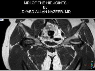
Axial T2 (gradient, T2*) , axial T1 ?!
Sagittal T2 for the diseased hip
If contrast is injected [ Axial, Sagittal ,coronal T1 WIs ].
Protocol of examination.
AP
Axial .
Lat.
10. Ball and socket joint.
Acetabulum covers 40% of the femoral head.
A fibrocartilaginous labium ↑ the depth of acetabulum.
95% of the femoral neck is intraarticular.
MR Anatomy.
18. Introduction to Avascular necrosis.
-Known also as osteonecrosis, aseptic necrosis,
ischemic necrosis , osteochondritis dissecans.
-Progressive process involving the compromise
of bone vasculature, leading to death of bone and
marrow cells and subsequently mechanical failure.
-Theory for avascular necrosis is increased intra-
osseous pressure with resultant osteonecrosis.
-Estimated 10,000-20,000 new patient diagnosed
every year, with male to female ratio as 8: 1.
Cause of approximately 10% of hip replacements.
Common site are femoral and humeral heads.
Avascular necrosis:Avascular necrosis:
19. The antrolateral aspect of the femoral head is the
commonest site, but no specific area is
protected
MR sensitive 97% specific 98%
Causes:
Traumatic
(Femoral neck fracture, Dislocation and Minor trauma).
Non-Traumatic
Chronic corticosteroids administration.
Sickle cell disease
Alcohol use/Cigarette smoking.
Gusher's disease
Radiation
Collagen disease, pancreatitis.
SLE.
HIV.
20.
21.
22.
23. Hip AVN on plain films.
Suspected AVN of the femoral head should be evaluated initially by AP
and lateral films.
Lateral films help to evaluate superior element of femoral head where
subchondral abnormality may be seen.
Plain films can remain normal months after AVN has begun.
Sclerosis,, cysts, joint space narrowing, degenerative changes in the
acetabulum.
25. Avascular necrosis.Avascular necrosis.
Hip AVN on CT:
CT scan do not demonstrate early AVN
Osteoporosis is the first visible sign of AVN on CT.
Contour irregularities and fissures
Areas of bone sclerosis .
Structural collapse
Osteoarthritic changes
26. Hip AVN on Tc-99 Bone Scan.
Technetium-99 bone scanning used for patients with suspected
disease who have negative radiographs and unilateral symptoms.
Increased bone turnover at the bridge between dead and reactive bone.
Increased uptake surrounded by a cold area lead to a radiographic
donut sign.
27.
28. Avascular necrosis.Avascular necrosis.
1-Bone marrow edema.1-Bone marrow edema.
2-Normal marrow + line2-Normal marrow + line
IIIIII Fluid signalFluid signal
VIVI Bone sclerosisBone sclerosis
29. Stage 1 - Plain radiograph and T1-weighted MRI image of the right hip.
There are no abnormal findings in the left image, although there are
already some signs of osteonecrosis on the femoral head on the MRI.
30. Stage I versus transient osteoporosisStage I versus transient osteoporosis..
32. Stage11 The line is composed of two layers [ double line sign[:
Inner layer of hyperemic granulation tissue and an outer layer of osteoblastic activity
33. Stage11. The line is composed of two layers [ double line sign[:
Inner layer of hyperemic granulation tissue and an outer layer of osteoblastic activity
36. The size and location of the lesion will affect the prognosis.
Lesions < 25% of the weight bearing area of the
femoral head responds well to core decompression
Medially and centrally located lesions have better prognosis
Contrast injection may be used to assess bone viability ?!!
Stage 11
49. Transient osteoporosis.Transient osteoporosis.
Unknown etiology
Middle aged over weight males
Male : female= 3:1
Usually unilateral [left hip in females]
Resolves spontaneously in 6-8 months
Pain & limp with no history of trauma
50. X ray Normal or ↓ bone density
Bone scan ↑ uptake in the femoral head and neck
MRI Bone marrow edema in the head and
neck
DD AVN, bone infarct, stress fracture
Septic arthritis, primary and metastatic tumors
Transient osteoporosisTransient osteoporosis
58. Subchondral fracture.
In young may be a stress fracture
In elderly may be the squeal of osteoporosis
Leads to extensive marrow edema which may progress
to femoral head collapse and secondary OA
DD include AVN , TOH .
MR shows a hypo intense line
60. Legg- Calve- Perthes diseasesLegg- Calve- Perthes diseases
Avascular necrosis of the bony femoral epiphysis
Unknown etiology
Children 4-9 years old boys: girls= 4:1
Children with knee pain must be examined for hip pathology
61. I Anterior aspect of the epiphysis.
II Anterior aspect of the epiphysis + metaphyseal reaction.
III All of the epiphysis+ metaphyseal reaction.
IV Flattening and collapse.
Legg- Calve- PerthesLegg- Calve- Perthes
diseases.diseases.Stages
67. Morphology and signal characteristics of femoral epiphysis
Normal epiphysis shows bright signal in T1 (Fat marrow)
Intra articular effusion
Legg- Calve- Perthes diseasesLegg- Calve- Perthes diseases
MR value
69. Spectrum of Perth's disease.
IVIV
Stage 1, Anterior aspect
of the epiphysis
Flattening and
collapse
70. Slipped Capital Femoral epiphysis.
Disorder of the proximal femoral physis that leads to slippage of the
epiphysis relative to the femoral neck as a result of physis fracture.
Epidemiology
incidence
most common disorder affecting adolescent hips, found in 10 per
100,000
demographics
more common in
obese children (single greatest risk factor)
males (male to female ratio is 3:2)
African Americans
Pacific islanders
during period of rapid growth
location
left hip is more common
bilateral in 17 to 50%
risk factors
femoral retroversion
obesity (single greatest risk factor for SCFE)
history of previous radiation therapy to the femoral head region
71. Classification:
Loder classification
Stable
Unstable, practically defined as when the
patient is unable to ambulate even with crutches
Temporal
Acute
Chronic
Acute-on-chronic
Radiological
Grade I = 0-33% slippage
Grade II = 34-50% slippage
Grade III = >50% slippage
77. Developmental dysplasia of the hip (DDH) denotes aberrant
development of the hip joint and results from an abnormal
relationship of the femoral head to the acetabulum. There is a
clear female predominance, and it usually occurs from
ligamentous laxity and abnormal position in utero. Therefore,
it is more common with oligohydramniotic pregnancies. This
article describes the commonly used radiographic
measurements and lines involved in DDH.
Epidemiology
The reported incidence varies between 1.5 and 20 per 1000
births, with the majority (60-80%) of abnormal hips resolving
spontaneously within 2-8 weeks (so-called immature hip).
Risk factors include :
female gender (M:F ratio ~1:8)
family history
breech presentation
oligohydramnios
metatarsus adductus
78. Plain radiograph: Assessment is looking for symmetry and defining
the relationship of the proximal femur to the developing pelvis. The
ossification of the superior femoral epiphyses should be symmetric.
Delay of ossification is a sign of DDH.
Hilgenreiner line: Hilgenreiner line is drawn horizontally through the
superior aspect of both triradiate cartilages. It should be horizontal but
is mainly used as a reference for Perkin line and measurement of the
acetabular angle.
Perkin line: Perkin line is drawn perpendicular to Hilgenreiner line,
intersecting the lateral most aspect of the acetabular roof. The upper
femoral epiphysis should be seen in the inferomedial quadrant (i.e.
below Hilgenreiner line, and medial to Perkin line)
Acetabular angle: The acetabular angle is formed by the intersection
between a line drawn tangential to the acetabular roof and Hilgenreiner
line, forming an acute angle. It should be approximately 30 degrees at
birth and progressively reduce with the maturation of the joint.
Shenton line: Shenton line is drawn along the inferior border of the
superior pubic ramus and should continue laterally along the
inferomedial aspect of the proximal femur as a smooth line. If there is a
superolateral migration of the proximal femur due to DDH then this line
will be discontinuous.
79.
80.
81.
82.
83.
84.
85.
86.
87. I Muscle edema with preserved morphology
II Disruption of up to 50% of muscle fibers with Subacute
blood at the site of tear
III Complete muscle tear ± retraction and atrophy
[ best seen in axial images with comparison to normal
side]
Muscle sprainsMuscle sprains
Grade I muscle sprain of
the obturator externus
and adductor longus
88. Coronal STIR images show tear at the hamstring muscles at ischial tuberosity.Coronal STIR images show tear at the hamstring muscles at ischial tuberosity.
94. Labral tearsLabral tears
Normal labrum is a triangular low signal structure at the superior and
inferior acetabular margins.
Surface coil
MR arthrogram.
Labral tears are part of femoro-acetabular impingement and can occur
due to trauma or secondary to degeneration.
95.
96.
97.
98.
99.
100.
101.
102.
103.
104. MR arthrogram of the left hip showing anterior paralabral
cyst(arrow) and a complex degenerative tear of the anterior labrum
105.
106.
107. Bursitis.Bursitis.
Bursae are sacs of synovial tissue
Prevent friction between bones and soft tissues.
15-20 Bursae around the hip joint
Trochanteric
Ischeo-gluteal
Iliopsoas : the largest in the body
10% - 15% communicate with the joint
108. Sagittal and coronal STIR images show Iliopsoas bursitis.Sagittal and coronal STIR images show Iliopsoas bursitis.
109. AXIAL CT Scan and axial STIR MRI images show ilioposas bursitisAXIAL CT Scan and axial STIR MRI images show ilioposas bursitis
110. Coronal STIR images show left greater trochanter bursa.Coronal STIR images show left greater trochanter bursa.
111. Axial images show left greater trochanteric bursa.Axial images show left greater trochanteric bursa.
112. Femoro-acetabular impingement (FAI)
Refers to a clinical syndrome of painful, limited hip
motion resulting from certain types of underlying
morphological abnormalities in the femoral head/neck
region and/or surrounding acetabulum. FAI can lead to
early degenerative disease.
Epidemiology
Pincer impingement is more common in middle-aged
women, occurring at an average age of 40 years, and can
occur with various disorders. It is essentially an over-
coverage of the femoral head by the acetabulum
Cam impingement is more common in young men,
occurring at an average age of 32 years . It refers to a
bony protrusion, mostly located at the anterosuperior
aspect of the femoral head-neck junction
Combined: mixture of the two occurring together.
113. Causes of Cam Lesions
Idiopathic
Developmental
Nonspherical femoral head
Coxa vara
Traumatic
Malunited femoral neck
fracture
Post-traumatic
retroversion of the femoral
head
Childhood orthopedic
condition
Perth's disease
Slipped capital femoral
epiphysis (SCFE)
Iatrogenic
Femoral osteotomy
Causes of Pincer Lesions
Idiopathic
Developmental
Retroverted acetabulum
Coxa profunda
Os acetabuli
Protrusio acetabuli
Chronic residual dysplasia
of the acetabulum
Traumatic
Post-traumatic deformity
of the acetabulum
Iatrogenic
Overcorrection of
retroversion in dysplastic
hips
114. Femro - acetabular impingementFemro - acetabular impingement..
Micro trauma from impingement of the femoral head against the acetabulum
Abnormal signal of the acetabular rim and femoral head
Labral tears and cartilage degeneration are seen
Clinically recurrent attacks of severe hip and groin pain
Pain increases by flexion and internal rotation and weight bearing
115. Plain x-ray of the hip showing bony bump (cam lesion) at the base of the ball.
130. Synovial osteochondromatosis.Synovial osteochondromatosis.
Metaplasia of subsynovial soft tissues cartilage formation
Affects any joint [ knee , hip , elbow[
Age incidence 40 years M : F = 2 : 1
Findings
Widening of the joint space
Bone erosions
Intra articular loose bodies
Secondary osteoarthritis changes
136. Femoral neck anteversion
refers to the orientation of the femoral neck
in relation to the femoral condyles at the
level of the knee. In most cases, the
femoral neck is oriented anteriorly as
compared to the femoral condyles.
Femoral anteversion averages between
30-40° at birth, and between 8-14° in adults.
Symptoms
Parents complain of an intoeing gait in
early childhood.
Child classically sits in the W position.
knee pain when associated with tibial
torsion
Awkward running style
when extreme in an older child occasional
functional limitations in sports and
activities of daily living can occur
difficulty with tripping during walking or
running activities.