SlideShare a Scribd company logo
1 of 50
Radiological imaging of chronic obstructive pulmonary disease.
Dr/ ABD ALLAH NAZEER. MD.
Chronic obstructive pulmonary disease is defined as a
preventable and treatable disease state characterized by
airflow limitation that is not fully reversible. The airflow
limitation is usually progressive and is associated with an
abnormal inflammatory response of the lungs to noxious
particles or gases, primarily caused by cigarette smoking.
Emphysema is one of its components, along with asthma
and chronic bronchitis.
Emphysema is defined pathologically as permanent
enlargement of the airspaces distal to the terminal
bronchioles, accompanied by destruction of their walls
and without obvious fibrosis.
Chronic bronchitis is defined as chronic productive cough
for 3 months in each of 2 successive years in a patient in
whom other causes of chronic cough have been excluded.
Clinical presentation
The clinical features of emphysema should be distinguished from the
signs and symptoms of chronic bronchitis. Patients with emphysema
are hypocapnoeic and are often referred to as "pink puffers". This
compares with the hypercapnoea and cyanosis of chronic bronchitis
with patients referred to as "blue bloaters". In practice, features of
these two syndromes coexist as chronic obstructive pulmonary disease.
Patients typically report dyspnea without significant sputum
production.
Signs of emphysema include:
tachypnoea
absence of cyanosis
pursed-lip breathing
chest hyperinflation
reduced breath sounds
hyper-resonant to percussion
Cor pulmonale (late)
Radiographic features
Plain film
Except in the case of very advanced disease with bulla formation, chest radiography does
not image emphysema directly, but rather infers the diagnosis due to associated features:
hyperinflation:
flattened hemidiaphragm(s): most reliable sign
increased and usually irregular radiolucency of the lungs
increased retrosternal airspace
increased antero-posterior diameter of chest
widely spaced ribs
sternal bowing
tenting of the diaphragm
saber-sheath trachea
vascular changes:
paucity of blood vessels, often distorted
pulmonary arterial hypertension
pruning of peripheral vessels
increased caliber of central arteries
right ventricular enlargement
It should be remembered, however, that the most common plain film appearance of COPD
is "normal" and the role of chest radiography is to eliminate other causes of lung
symptoms such as infection, bronchiectasis or cancer .
CT is currently the modality of choice for detecting emphysema; HRCT is
particularly effective. It should be noted, however, that there is relatively poor
correlation between autopsy-proven emphysema, pulmonary function test
abnormalities and CT with 20% of pathology-proven cases not being evident
on CT and 40% of patients with abnormal CT having normal pulmonary function
tests. CT is able to discriminate between centrilobular, panlobular, and
paraseptal emphysema.
Centrilobular emphysema
Centrilobular is by far the most common type encountered, and is a common
finding in asymptomatic elderly patients. It is predominantly located in the upper
zones of each lobe (i.e. apical and posterior segments of the upper lobes, and
superior segment of the lower lobes) and has a patchy distribution. It appears as
focal lucencies (emphysematous spaces) which measure up to 1 cm in diameter,
located centrally within the secondary pulmonary lobule, often with a central or
peripheral dot representing the central brochovascular bundle
Panlobular emphysema
Panlobular emphysema is predominantly located in the lower lobes, has a
uniform distribution across parts of the secondary pulmonary lobule, which
are homogeneously reduced in attenuation.
Paraseptal emphysema
Paraseptal emphysema is located adjacent to the pleura and
septal lines with a peripheral distribution within the secondary
pulmonary lobule. The affected lobules are almost always
subpleural, and demonstrate small focal lucencies up to 10 mm
in size.
Any lucency larger than 10 mm should be referred to as
subpleural blebs or subpleural bullae (synonymous).
In all three subtypes, the emphysematous spaces are not
bounded by any visible wall.
MRI
MRI is in the research phases for evaluation of lung parenchymal
abnormalities like emphysema. Dynamic breathing MRI may
have a future role in assessing pulmonary emphysema
Emphysema
Emphysema typically presents as areas of low attenuation without visible walls as
a result of parenchymal destruction.
Centrilobular emphysema
Most common type
Irreversible destruction of alveolar walls in the centrilobular portion of the
lobule
Upper lobe predominance and uneven distribution
Strongly associated with smoking.
Panlobular emphysema
Affects the whole secondary lobule
Lower lobe predominance
In alpha-1-antitrypsin deficiency, but also seen in smokers with advanced
emphysema
Paraseptal emphysema
Adjacent to the pleura and interlobar fissures
Can be isolated phenomenon in young adults, or in older patients with
centrilobular emphysema
In young adults often associated with spontaneous pneumothorax
Types of emphysema: line diagram shows the parts of secondary pulmonary lobule that are
affected in different types of emphysema. Respiratory bronchioles are primarily affected by
centrilobular emphysema; peripheral alveolar ducts, sacs, and alveoli in paraseptal
emphysema (PLE); all the components (i.e., respiratory bronchioles, alveolar ducts, alveolar
sacs, and alveoli) in panlobular emphysema (PLE), and any part in irregular or
paracicatricial emphysema.
Centrilobular emphysema (CLE) and edema: A, Chest radiographs, postero-
anterior and lateral views, show hyperinflation of the lungs (flattened
diaphragm), increased translucency in the upper lungs with vascular attenuation
and loss of arborization. B, Emphysematous spaces outlined by edema fluid filling
the surrounding airspaces give an appearance of reticulation in this patient with
lung edema superimposed on confluent, upper-lung predominant CLE.
Centrilobular Emphysema in a Cigarette Smoker. Axial CT image through the upper lungs
shows numerous well-defined lucencies, many of which are traversed by a central vessel.
Centrilobular emphysema due to smoking. The periphery of the lung is spared (blue
arrows). Centrilobular artery (yellow arrows) is seen in the center of the hypodense area.
Centrilobular pulmonary emphysema
Centrilobular emphysema:
A, Transverse computed
tomography images shows
centrilobular
hypoattenuation with upper
lung predominance. Note
the resemblance of the
macroscopic pathologic
image in Figure 4A. B,
Coronal minimum intensity
projection image brings out
the distribution and extent
of emphysema.
Centrilobular emphysema: Transverse computed tomography images in the first
row and minimum intensity projection images in the second row show confluent
centrilobular hypoattenuation with posterior lung predominance (arrows), Also
note para-septal emphysema in the left upper lobe (arrow heads).
Panlobular emphysema (PLE) from a-1–antitrypsin deficiency: chest radiographs
in postero-anterior and lateral projections show hyperinflation and increased
translucency in the lower lungs with vascular attenuation, indicating PLE.
Panlobular emphysema (PLE) from a-1–antitrypsin deficiency: computed tomography
images (first row) show confluent lower-lung predominant panlobular
hypoattenuation, indicating PLE. The confluence, panlobular distribution, lower-lung
predominance, and vascular attenuation are better shown by the coronal minimum
intensity projection and maximum intensity projection images (second row).
A- Chest tomography in axial cut showing areas of panlobular emphysema in predominately
lower lobes. Note cylindroids bronchiectasis and thickening of bronchial walls, most evidence
in right. B- coronal reformatted showing hyper-inflation areas in lower lobes.
Ritalin lung with panlobular emphysema: chest radiographs, postero-anterior projection,
and computed tomography (coronal reformatted image) show basal-predominant
panlobular hypoattenuation similar to that found in a-1–antitrypsin deficiency.
Panlobular Emphysema in a Patient with Alpha-1 Antitrypsin Deficiency. Axial image through
the lower lungs shows homogenous decrease in lung attenuation in both lower
lobes. Some of the interlobular septa in the right lower lobe are accentuated by the emphysema.
Panlobular emphysema
Paraseptal emphysema: computed tomography shows rectangular cysts sharing
walls in subpleural upper lobes and the superior segment of the left lower lobe.
Centrilobular emphysema is also evident in the upper lobes (arrows).
Paraseptal Emphysema in a Cigarette Smoker. Axial image through the upper
lungs shows multiple well-demarcated subpleural air-containing spaces.
Paraseptal emphysema with small bullae.
Paracicatricial emphysema (PCE) from progressive massive fibrosis caused by silicosis:
computed tomography images in lung window show conglomerate masses in the
posterior upper lobes with surrounding low attenuation (arrows) indicating PCE.
Hyperinflation of the anterior upper lungs is from traction by the conglomerate masses.
Coronal reformat demonstrating heterogeneous opacity in the right upper lobe
and areas of centrilobular emphysema and para-septal. Tuberculosis in COPD.
Bullous emphysema: postero-anterior radiograph and coronal computed tomography
multiplanar reformation and maximum intensity projection images show a large bulla
in the right upper lobe with atelectasis of the adjacent lung (arrows).
Apical bleb: computed tomography through the lung apex and multiplanar
reformation show a left apical bleb floating in small pneumothorax
(arrows). Also note unruptured blebs in the right lung apex.
Quantification of Emphysema and Air Trapping in 66 year old Individual with GOLD Stage 3 COPD (FEV1 43% predicted)
(a) Inspiratory CT: coronal image depicts voxels with CT attenuation less than -950HU indicating emphysema (14% in
this case). Color coding: Indicates lobes. (b) Inspiratory CT: coronal image depicts distribution of sizes of
emphysematous clusters, with color coding depending on cluster size. (c) Expiratory CT: coronal image depicts voxels
with CT attenuation less than -856HU indicating air trapping (38% in this case). (d) Image map derived from co-
registered inspiratory and expiratory images depicts change in voxel attenuation from inspiration to expiration.
Color coding: Red indicates voxels that were less than -950 HU on inspiration and less than -856 HU on expiration
(emphysema). Yellow indicates voxels that were greater than -950 HU on inspiration, but less than -856 HU on expiration
(air trapping). Green indicates voxels that were less than -950 HU on inspiration and less than -856 on expiration
(normal). White indicates voxels that were greater than -950 HU on inspiration but less than -856 on expiration.
Progress in Imaging COPD, 2004-2014
Follow up CT Analysis in Same Subject as Shown in Figure 6. Color coding: Same as in Figure 6. (a) %
LAA-950 has increased from 14% to 23%. (b) Cluster sizes have also increased. (c)Air trapping appears
to have increased from 38% to 45%. (d) Inspiratory-expiratory registration shows that only the amount
of emphysema increased, while the amount of gas trapping remained constant. (e) Longitudinal
registration of baseline and follow up inspiratory scans with micro mapping shows areas of stable
emphysema in red, new areas of emphysema in yellow, and stable normal lung in green.
Images in representative
patients with mild-to-
moderate or severe
chronic obstructive
pulmonary disease
(COPD).
Chronic bronchitis (CB) is often defined as the presence of
productive cough for 3 months in two successive years in a
patient in whom other causes of chronic cough, such as
tuberculosis, lung cancer and heart failure, have been
excluded. It can be an important pathological component
of chronic obstructive pulmonary disease. (often
considered as a distinct phenotype of COPD)
Pathology
Chronic bronchitis most often results from overproduction
and hypersecretion of mucus by goblet cells. This can in
turn lead to worsening airflow obstruction by luminal
obstruction of small airways, epithelial remodeling, and
alteration of airway surface tension predisposing to
collapse.
Diagnosis:
A physical examination will often reveal decreased intensity of
breath sounds, wheezing, rhonchi, and prolonged expiration. Most
physicians rely on the presence of a persistent dry or wet cough as
evidence of bronchitis.
A variety of tests may be performed in patients presenting with
cough and shortness of breath:
A chest X-ray is useful to exclude pneumonia which is more common
in those with a fever, fast heart rate, fast respiratory rate, or who
are old.
A sputum sample showing neutrophil granulocytes (inflammatory
white blood cells) and culture showing that has pathogenic
microorganisms such as Streptococcus species.
A blood test would indicate inflammation (as indicated by a raised
white blood cell count and elevated C-reactive protein).
Chronic bronchitis with bronchial wall thickening with increased brochovascular
markings, enlarged vessels and cardiomeagly (abnormal enlargement of the heart).
Chronic bronchitis: postero-anterior radiograph (A) and computed tomography
(B) show bilateral bronchial wall thickening, the so-called, ‘‘dirty lung.’’
Computed tomography (CT) scan in an elderly patient with known chronic
bronchitis. The scans demonstrate thickening of the bronchial walls (purple
arrows) and bronchi filled with mucus or phlegm (blue arrows).
Computed tomography (CT) scan in patient with chronic bronchitis showing thickening
of the bronchial walls (red arrows) and mucous within the bronchi (yellow arrows).
Thank You.

More Related Content

What's hot

Collapse consolidation
Collapse consolidationCollapse consolidation
Collapse consolidationairwave12
 
Radiological imaging of pleural diseases
Radiological imaging of pleural diseases Radiological imaging of pleural diseases
Radiological imaging of pleural diseases Pankaj Kaira
 
Diagnostic Imaging of Pulmonary infections
Diagnostic Imaging of Pulmonary infectionsDiagnostic Imaging of Pulmonary infections
Diagnostic Imaging of Pulmonary infectionsMohamed M.A. Zaitoun
 
Ascites and Pleural Effusion
 Ascites and Pleural Effusion Ascites and Pleural Effusion
Ascites and Pleural EffusionMedia Genie
 
Pulmonary embolism radiology
Pulmonary embolism radiologyPulmonary embolism radiology
Pulmonary embolism radiologyAnish Choudhary
 
Presentation1.pptx, radiological imaging of pulmonary infection.
Presentation1.pptx, radiological imaging of pulmonary infection.Presentation1.pptx, radiological imaging of pulmonary infection.
Presentation1.pptx, radiological imaging of pulmonary infection.Abdellah Nazeer
 
Interstitial lung diseases radiology
Interstitial lung diseases radiologyInterstitial lung diseases radiology
Interstitial lung diseases radiologyShrikant Nagare
 
Mediastinum-RADIOLOGY
Mediastinum-RADIOLOGYMediastinum-RADIOLOGY
Mediastinum-RADIOLOGYNavdeep Shah
 
Interpretation of chest CT
Interpretation of chest CTInterpretation of chest CT
Interpretation of chest CTSakiru Isa
 
Radiographic manifestations of pulmonary tuberculosis
Radiographic manifestations of pulmonary tuberculosisRadiographic manifestations of pulmonary tuberculosis
Radiographic manifestations of pulmonary tuberculosisDev Lakhera
 
Mediastinal lymph nodes
Mediastinal lymph nodesMediastinal lymph nodes
Mediastinal lymph nodesnipiv17
 
Presentation1, ultrasound examination of the chest.
Presentation1, ultrasound examination of the chest.Presentation1, ultrasound examination of the chest.
Presentation1, ultrasound examination of the chest.Abdellah Nazeer
 
Chest xray for evaluation of cardiovascular system
Chest xray for evaluation of cardiovascular systemChest xray for evaluation of cardiovascular system
Chest xray for evaluation of cardiovascular systemPRAVEEN GUPTA
 
Presentation1.pptx radiological imaging of mediastinal masses .
Presentation1.pptx radiological imaging of mediastinal masses .Presentation1.pptx radiological imaging of mediastinal masses .
Presentation1.pptx radiological imaging of mediastinal masses .Abdellah Nazeer
 
Radiology: Chest Imaging
Radiology: Chest ImagingRadiology: Chest Imaging
Radiology: Chest ImagingSCGH ED CME
 

What's hot (20)

Collapse consolidation
Collapse consolidationCollapse consolidation
Collapse consolidation
 
Radiological imaging of pleural diseases
Radiological imaging of pleural diseases Radiological imaging of pleural diseases
Radiological imaging of pleural diseases
 
Diagnostic Imaging of Pulmonary infections
Diagnostic Imaging of Pulmonary infectionsDiagnostic Imaging of Pulmonary infections
Diagnostic Imaging of Pulmonary infections
 
Ascites and Pleural Effusion
 Ascites and Pleural Effusion Ascites and Pleural Effusion
Ascites and Pleural Effusion
 
Pulmonary embolism radiology
Pulmonary embolism radiologyPulmonary embolism radiology
Pulmonary embolism radiology
 
Lung tumor radiology
Lung tumor radiologyLung tumor radiology
Lung tumor radiology
 
Presentation1.pptx, radiological imaging of pulmonary infection.
Presentation1.pptx, radiological imaging of pulmonary infection.Presentation1.pptx, radiological imaging of pulmonary infection.
Presentation1.pptx, radiological imaging of pulmonary infection.
 
Interstitial lung diseases radiology
Interstitial lung diseases radiologyInterstitial lung diseases radiology
Interstitial lung diseases radiology
 
Basics of CT chest
Basics of CT chestBasics of CT chest
Basics of CT chest
 
Lung cancer radiology
Lung cancer radiologyLung cancer radiology
Lung cancer radiology
 
Mediastinum-RADIOLOGY
Mediastinum-RADIOLOGYMediastinum-RADIOLOGY
Mediastinum-RADIOLOGY
 
Interpretation of chest CT
Interpretation of chest CTInterpretation of chest CT
Interpretation of chest CT
 
Radiographic manifestations of pulmonary tuberculosis
Radiographic manifestations of pulmonary tuberculosisRadiographic manifestations of pulmonary tuberculosis
Radiographic manifestations of pulmonary tuberculosis
 
HRCT Reticular pattern
HRCT Reticular pattern HRCT Reticular pattern
HRCT Reticular pattern
 
Mediastinal lymph nodes
Mediastinal lymph nodesMediastinal lymph nodes
Mediastinal lymph nodes
 
Presentation1, ultrasound examination of the chest.
Presentation1, ultrasound examination of the chest.Presentation1, ultrasound examination of the chest.
Presentation1, ultrasound examination of the chest.
 
Chest xray for evaluation of cardiovascular system
Chest xray for evaluation of cardiovascular systemChest xray for evaluation of cardiovascular system
Chest xray for evaluation of cardiovascular system
 
Presentation1.pptx radiological imaging of mediastinal masses .
Presentation1.pptx radiological imaging of mediastinal masses .Presentation1.pptx radiological imaging of mediastinal masses .
Presentation1.pptx radiological imaging of mediastinal masses .
 
Radiological features of pneumonia
Radiological features of pneumoniaRadiological features of pneumonia
Radiological features of pneumonia
 
Radiology: Chest Imaging
Radiology: Chest ImagingRadiology: Chest Imaging
Radiology: Chest Imaging
 

Viewers also liked

Presentation1.pptx, radiological imaging of the larngeal diseases.
Presentation1.pptx, radiological imaging of the larngeal diseases.Presentation1.pptx, radiological imaging of the larngeal diseases.
Presentation1.pptx, radiological imaging of the larngeal diseases.Abdellah Nazeer
 
Presentation1, mri quiz cases.
Presentation1, mri quiz cases.Presentation1, mri quiz cases.
Presentation1, mri quiz cases.Abdellah Nazeer
 
Chronic obstructive pulmonary disease
Chronic obstructive pulmonary diseaseChronic obstructive pulmonary disease
Chronic obstructive pulmonary diseaseEdson Mutandwa
 
Presentation1, radiological imaging of barium studies.
Presentation1, radiological imaging of barium studies.Presentation1, radiological imaging of barium studies.
Presentation1, radiological imaging of barium studies.Abdellah Nazeer
 
Larynx pathologies by dr avinash
Larynx pathologies  by dr avinashLarynx pathologies  by dr avinash
Larynx pathologies by dr avinashAnish Choudhary
 
Hrct in diagnosis of diffuse lung diseases
Hrct in diagnosis of  diffuse lung diseasesHrct in diagnosis of  diffuse lung diseases
Hrct in diagnosis of diffuse lung diseasesAhmed Bahnassy
 
Presentation1, film readiing for barium studies.
Presentation1, film readiing for barium studies.Presentation1, film readiing for barium studies.
Presentation1, film readiing for barium studies.Abdellah Nazeer
 
Presentation2.pptx, radiological imaging of the rectal diseases.
Presentation2.pptx, radiological imaging of the rectal diseases.Presentation2.pptx, radiological imaging of the rectal diseases.
Presentation2.pptx, radiological imaging of the rectal diseases.Abdellah Nazeer
 
Presentation1.pptx, radiological imaging of large bowel diseases
Presentation1.pptx, radiological imaging of large bowel diseasesPresentation1.pptx, radiological imaging of large bowel diseases
Presentation1.pptx, radiological imaging of large bowel diseasesAbdellah Nazeer
 
Larynx anatomy ct and mri
Larynx anatomy ct and mriLarynx anatomy ct and mri
Larynx anatomy ct and mriAnish Choudhary
 
Larynx Imaging 3rd part laryngeal neoplasm CT MRI Dr Ahmed Esawy
Larynx Imaging 3rd  part laryngeal neoplasm CT MRI Dr Ahmed EsawyLarynx Imaging 3rd  part laryngeal neoplasm CT MRI Dr Ahmed Esawy
Larynx Imaging 3rd part laryngeal neoplasm CT MRI Dr Ahmed EsawyAHMED ESAWY
 
Radiology of Tuberculosis
Radiology of TuberculosisRadiology of Tuberculosis
Radiology of TuberculosisMohit Goyal
 
Presentation1, radiological imaging of diabetic foor and charcot joint.
Presentation1, radiological imaging of diabetic foor and charcot joint.Presentation1, radiological imaging of diabetic foor and charcot joint.
Presentation1, radiological imaging of diabetic foor and charcot joint.Abdellah Nazeer
 
Presentation1, radiological imaging of adhesive capsulitis(frozen shoulder).
Presentation1, radiological imaging of adhesive capsulitis(frozen shoulder).Presentation1, radiological imaging of adhesive capsulitis(frozen shoulder).
Presentation1, radiological imaging of adhesive capsulitis(frozen shoulder).Abdellah Nazeer
 
COPD (Chronic obstructive Pulmonary Disease) PowerPoint Presentation -aslam
COPD  (Chronic obstructive Pulmonary Disease) PowerPoint Presentation -aslamCOPD  (Chronic obstructive Pulmonary Disease) PowerPoint Presentation -aslam
COPD (Chronic obstructive Pulmonary Disease) PowerPoint Presentation -aslamDr.Aslam calicut
 
How to Make Awesome SlideShares: Tips & Tricks
How to Make Awesome SlideShares: Tips & TricksHow to Make Awesome SlideShares: Tips & Tricks
How to Make Awesome SlideShares: Tips & TricksSlideShare
 

Viewers also liked (20)

Emphysema
EmphysemaEmphysema
Emphysema
 
Laryngeal Cancer
Laryngeal CancerLaryngeal Cancer
Laryngeal Cancer
 
Presentation1.pptx, radiological imaging of the larngeal diseases.
Presentation1.pptx, radiological imaging of the larngeal diseases.Presentation1.pptx, radiological imaging of the larngeal diseases.
Presentation1.pptx, radiological imaging of the larngeal diseases.
 
Presentation1, mri quiz cases.
Presentation1, mri quiz cases.Presentation1, mri quiz cases.
Presentation1, mri quiz cases.
 
Chronic obstructive pulmonary disease
Chronic obstructive pulmonary diseaseChronic obstructive pulmonary disease
Chronic obstructive pulmonary disease
 
Hepatic imaging
Hepatic imagingHepatic imaging
Hepatic imaging
 
Presentation1, radiological imaging of barium studies.
Presentation1, radiological imaging of barium studies.Presentation1, radiological imaging of barium studies.
Presentation1, radiological imaging of barium studies.
 
Larynx pathologies by dr avinash
Larynx pathologies  by dr avinashLarynx pathologies  by dr avinash
Larynx pathologies by dr avinash
 
Hrct in diagnosis of diffuse lung diseases
Hrct in diagnosis of  diffuse lung diseasesHrct in diagnosis of  diffuse lung diseases
Hrct in diagnosis of diffuse lung diseases
 
Presentation1, film readiing for barium studies.
Presentation1, film readiing for barium studies.Presentation1, film readiing for barium studies.
Presentation1, film readiing for barium studies.
 
Presentation2.pptx, radiological imaging of the rectal diseases.
Presentation2.pptx, radiological imaging of the rectal diseases.Presentation2.pptx, radiological imaging of the rectal diseases.
Presentation2.pptx, radiological imaging of the rectal diseases.
 
Presentation1.pptx, radiological imaging of large bowel diseases
Presentation1.pptx, radiological imaging of large bowel diseasesPresentation1.pptx, radiological imaging of large bowel diseases
Presentation1.pptx, radiological imaging of large bowel diseases
 
Larynx anatomy ct and mri
Larynx anatomy ct and mriLarynx anatomy ct and mri
Larynx anatomy ct and mri
 
Larynx Imaging 3rd part laryngeal neoplasm CT MRI Dr Ahmed Esawy
Larynx Imaging 3rd  part laryngeal neoplasm CT MRI Dr Ahmed EsawyLarynx Imaging 3rd  part laryngeal neoplasm CT MRI Dr Ahmed Esawy
Larynx Imaging 3rd part laryngeal neoplasm CT MRI Dr Ahmed Esawy
 
Radiology of Tuberculosis
Radiology of TuberculosisRadiology of Tuberculosis
Radiology of Tuberculosis
 
Presentation1, radiological imaging of diabetic foor and charcot joint.
Presentation1, radiological imaging of diabetic foor and charcot joint.Presentation1, radiological imaging of diabetic foor and charcot joint.
Presentation1, radiological imaging of diabetic foor and charcot joint.
 
Chest imaging
Chest imagingChest imaging
Chest imaging
 
Presentation1, radiological imaging of adhesive capsulitis(frozen shoulder).
Presentation1, radiological imaging of adhesive capsulitis(frozen shoulder).Presentation1, radiological imaging of adhesive capsulitis(frozen shoulder).
Presentation1, radiological imaging of adhesive capsulitis(frozen shoulder).
 
COPD (Chronic obstructive Pulmonary Disease) PowerPoint Presentation -aslam
COPD  (Chronic obstructive Pulmonary Disease) PowerPoint Presentation -aslamCOPD  (Chronic obstructive Pulmonary Disease) PowerPoint Presentation -aslam
COPD (Chronic obstructive Pulmonary Disease) PowerPoint Presentation -aslam
 
How to Make Awesome SlideShares: Tips & Tricks
How to Make Awesome SlideShares: Tips & TricksHow to Make Awesome SlideShares: Tips & Tricks
How to Make Awesome SlideShares: Tips & Tricks
 

Similar to Presentation1.pptx, radiological imaging of chronic obstructive airway disease.

presentation1-140523194630-phpapp01.pptx
presentation1-140523194630-phpapp01.pptxpresentation1-140523194630-phpapp01.pptx
presentation1-140523194630-phpapp01.pptxOluseyi7
 
Radiological imaging of copd
Radiological imaging of copdRadiological imaging of copd
Radiological imaging of copdharibabupedamajji
 
Interactive radiology case presentation
Interactive radiology case presentationInteractive radiology case presentation
Interactive radiology case presentationGamal Agmy
 
Radiological signs in chest medicine Part 1
Radiological signs in chest medicine Part 1Radiological signs in chest medicine Part 1
Radiological signs in chest medicine Part 1Gamal Agmy
 
Pulmonary manifestations of systemic lupus erythematosis
Pulmonary manifestations of systemic lupus erythematosisPulmonary manifestations of systemic lupus erythematosis
Pulmonary manifestations of systemic lupus erythematosisdattasrisaila
 
Pathological anatomy of emphysema, pneumosclerosis.pptx
Pathological anatomy of emphysema, pneumosclerosis.pptxPathological anatomy of emphysema, pneumosclerosis.pptx
Pathological anatomy of emphysema, pneumosclerosis.pptxabbashshah09
 
Radiological signs of chest diseases
Radiological signs of chest diseasesRadiological signs of chest diseases
Radiological signs of chest diseasesGamal Agmy
 
Chest x ray 3
Chest x ray 3Chest x ray 3
Chest x ray 3Double M
 
Bullous disease of lung.pptx
Bullous disease of lung.pptxBullous disease of lung.pptx
Bullous disease of lung.pptxTarunTiwari57
 
Presentation1.pptx. interpretation of x ray chest.
Presentation1.pptx. interpretation of x ray chest.Presentation1.pptx. interpretation of x ray chest.
Presentation1.pptx. interpretation of x ray chest.Abdellah Nazeer
 
Cxr part two
Cxr part twoCxr part two
Cxr part twoDouble M
 
Presentation1.pptx, chest film reading. lecture 1
Presentation1.pptx, chest film reading. lecture 1Presentation1.pptx, chest film reading. lecture 1
Presentation1.pptx, chest film reading. lecture 1Abdellah Nazeer
 
Imaging in opaqe hemithorax [autosaved]
Imaging in opaqe hemithorax [autosaved]Imaging in opaqe hemithorax [autosaved]
Imaging in opaqe hemithorax [autosaved]Ajay Singh
 
Radiology& Imaging in rehabilitation.pdf
Radiology& Imaging in rehabilitation.pdfRadiology& Imaging in rehabilitation.pdf
Radiology& Imaging in rehabilitation.pdfAndreiCealan1
 
Emphysema and alfa 1 antitrypsin
Emphysema and alfa 1 antitrypsinEmphysema and alfa 1 antitrypsin
Emphysema and alfa 1 antitrypsinDavid Ciulla
 
Complex clinical imaging of radiological system
Complex clinical imaging of radiological system Complex clinical imaging of radiological system
Complex clinical imaging of radiological system Sonia Iyobosa Omoregie
 
Respiratory lectures
Respiratory lecturesRespiratory lectures
Respiratory lecturesDOCTOR WHO
 

Similar to Presentation1.pptx, radiological imaging of chronic obstructive airway disease. (20)

presentation1-140523194630-phpapp01.pptx
presentation1-140523194630-phpapp01.pptxpresentation1-140523194630-phpapp01.pptx
presentation1-140523194630-phpapp01.pptx
 
Radiological imaging of copd
Radiological imaging of copdRadiological imaging of copd
Radiological imaging of copd
 
Interactive radiology case presentation
Interactive radiology case presentationInteractive radiology case presentation
Interactive radiology case presentation
 
Radiological signs in chest medicine Part 1
Radiological signs in chest medicine Part 1Radiological signs in chest medicine Part 1
Radiological signs in chest medicine Part 1
 
Pulmonary manifestations of systemic lupus erythematosis
Pulmonary manifestations of systemic lupus erythematosisPulmonary manifestations of systemic lupus erythematosis
Pulmonary manifestations of systemic lupus erythematosis
 
Pathological anatomy of emphysema, pneumosclerosis.pptx
Pathological anatomy of emphysema, pneumosclerosis.pptxPathological anatomy of emphysema, pneumosclerosis.pptx
Pathological anatomy of emphysema, pneumosclerosis.pptx
 
Chest Lungs 3
Chest Lungs 3Chest Lungs 3
Chest Lungs 3
 
Chest xrays pneumonias
Chest xrays pneumoniasChest xrays pneumonias
Chest xrays pneumonias
 
Radiological signs of chest diseases
Radiological signs of chest diseasesRadiological signs of chest diseases
Radiological signs of chest diseases
 
PNEUMOTHORAX.ppt
PNEUMOTHORAX.pptPNEUMOTHORAX.ppt
PNEUMOTHORAX.ppt
 
Chest x ray 3
Chest x ray 3Chest x ray 3
Chest x ray 3
 
Bullous disease of lung.pptx
Bullous disease of lung.pptxBullous disease of lung.pptx
Bullous disease of lung.pptx
 
Presentation1.pptx. interpretation of x ray chest.
Presentation1.pptx. interpretation of x ray chest.Presentation1.pptx. interpretation of x ray chest.
Presentation1.pptx. interpretation of x ray chest.
 
Cxr part two
Cxr part twoCxr part two
Cxr part two
 
Presentation1.pptx, chest film reading. lecture 1
Presentation1.pptx, chest film reading. lecture 1Presentation1.pptx, chest film reading. lecture 1
Presentation1.pptx, chest film reading. lecture 1
 
Imaging in opaqe hemithorax [autosaved]
Imaging in opaqe hemithorax [autosaved]Imaging in opaqe hemithorax [autosaved]
Imaging in opaqe hemithorax [autosaved]
 
Radiology& Imaging in rehabilitation.pdf
Radiology& Imaging in rehabilitation.pdfRadiology& Imaging in rehabilitation.pdf
Radiology& Imaging in rehabilitation.pdf
 
Emphysema and alfa 1 antitrypsin
Emphysema and alfa 1 antitrypsinEmphysema and alfa 1 antitrypsin
Emphysema and alfa 1 antitrypsin
 
Complex clinical imaging of radiological system
Complex clinical imaging of radiological system Complex clinical imaging of radiological system
Complex clinical imaging of radiological system
 
Respiratory lectures
Respiratory lecturesRespiratory lectures
Respiratory lectures
 

More from Abdellah Nazeer

Muculoskeletal Pediatic Imaging..pptx
Muculoskeletal Pediatic Imaging..pptxMuculoskeletal Pediatic Imaging..pptx
Muculoskeletal Pediatic Imaging..pptxAbdellah Nazeer
 
Presentation1, Ultrasound of the bowel loops and the lymph nodes..pptx
Presentation1, Ultrasound of the bowel loops and the lymph nodes..pptxPresentation1, Ultrasound of the bowel loops and the lymph nodes..pptx
Presentation1, Ultrasound of the bowel loops and the lymph nodes..pptxAbdellah Nazeer
 
Presentation1 Short cases MD..pptx
Presentation1 Short cases MD..pptxPresentation1 Short cases MD..pptx
Presentation1 Short cases MD..pptxAbdellah Nazeer
 
Presentation1, MD MCQ Cases..pptx
Presentation1, MD MCQ Cases..pptxPresentation1, MD MCQ Cases..pptx
Presentation1, MD MCQ Cases..pptxAbdellah Nazeer
 
Presentation1, Short Cases Quiz..pptx
Presentation1, Short Cases Quiz..pptxPresentation1, Short Cases Quiz..pptx
Presentation1, Short Cases Quiz..pptxAbdellah Nazeer
 
Presentation1, radiological imaging of lateral hindfoot impingement.
Presentation1, radiological imaging of lateral hindfoot impingement.Presentation1, radiological imaging of lateral hindfoot impingement.
Presentation1, radiological imaging of lateral hindfoot impingement.Abdellah Nazeer
 
Presentation2, radiological anatomy of the liver and spleen.
Presentation2, radiological anatomy of the liver and spleen.Presentation2, radiological anatomy of the liver and spleen.
Presentation2, radiological anatomy of the liver and spleen.Abdellah Nazeer
 
Presentation1, artifacts and pitfalls of the wrist and elbow joints.
Presentation1, artifacts and pitfalls of the wrist and elbow joints.Presentation1, artifacts and pitfalls of the wrist and elbow joints.
Presentation1, artifacts and pitfalls of the wrist and elbow joints.Abdellah Nazeer
 
Presentation1, artifact and pitfalls of the knee, hip and ankle joints.
Presentation1, artifact and pitfalls of the knee, hip and ankle joints.Presentation1, artifact and pitfalls of the knee, hip and ankle joints.
Presentation1, artifact and pitfalls of the knee, hip and ankle joints.Abdellah Nazeer
 
Presentation1, radiological imaging of artifact and pitfalls in shoulder join...
Presentation1, radiological imaging of artifact and pitfalls in shoulder join...Presentation1, radiological imaging of artifact and pitfalls in shoulder join...
Presentation1, radiological imaging of artifact and pitfalls in shoulder join...Abdellah Nazeer
 
Presentation1, radiological imaging of internal abdominal hernia.
Presentation1, radiological imaging of internal abdominal hernia.Presentation1, radiological imaging of internal abdominal hernia.
Presentation1, radiological imaging of internal abdominal hernia.Abdellah Nazeer
 
Presentation11, radiological imaging of ovarian torsion.
Presentation11, radiological imaging of ovarian torsion.Presentation11, radiological imaging of ovarian torsion.
Presentation11, radiological imaging of ovarian torsion.Abdellah Nazeer
 
Presentation1, musculoskeletal anatomy.
Presentation1, musculoskeletal anatomy.Presentation1, musculoskeletal anatomy.
Presentation1, musculoskeletal anatomy.Abdellah Nazeer
 
Presentation1, new mri techniques in the diagnosis and monitoring of multiple...
Presentation1, new mri techniques in the diagnosis and monitoring of multiple...Presentation1, new mri techniques in the diagnosis and monitoring of multiple...
Presentation1, new mri techniques in the diagnosis and monitoring of multiple...Abdellah Nazeer
 
Presentation1, radiological application of diffusion weighted mri in neck mas...
Presentation1, radiological application of diffusion weighted mri in neck mas...Presentation1, radiological application of diffusion weighted mri in neck mas...
Presentation1, radiological application of diffusion weighted mri in neck mas...Abdellah Nazeer
 
Presentation1, radiological application of diffusion weighted images in breas...
Presentation1, radiological application of diffusion weighted images in breas...Presentation1, radiological application of diffusion weighted images in breas...
Presentation1, radiological application of diffusion weighted images in breas...Abdellah Nazeer
 
Presentation1, radiological application of diffusion weighted images in abdom...
Presentation1, radiological application of diffusion weighted images in abdom...Presentation1, radiological application of diffusion weighted images in abdom...
Presentation1, radiological application of diffusion weighted images in abdom...Abdellah Nazeer
 
Presentation1, radiological application of diffusion weighted imges in neuror...
Presentation1, radiological application of diffusion weighted imges in neuror...Presentation1, radiological application of diffusion weighted imges in neuror...
Presentation1, radiological application of diffusion weighted imges in neuror...Abdellah Nazeer
 
Presentation1, mr physics.
Presentation1, mr physics.Presentation1, mr physics.
Presentation1, mr physics.Abdellah Nazeer
 
Presentation1. ct physics.
Presentation1. ct physics.Presentation1. ct physics.
Presentation1. ct physics.Abdellah Nazeer
 

More from Abdellah Nazeer (20)

Muculoskeletal Pediatic Imaging..pptx
Muculoskeletal Pediatic Imaging..pptxMuculoskeletal Pediatic Imaging..pptx
Muculoskeletal Pediatic Imaging..pptx
 
Presentation1, Ultrasound of the bowel loops and the lymph nodes..pptx
Presentation1, Ultrasound of the bowel loops and the lymph nodes..pptxPresentation1, Ultrasound of the bowel loops and the lymph nodes..pptx
Presentation1, Ultrasound of the bowel loops and the lymph nodes..pptx
 
Presentation1 Short cases MD..pptx
Presentation1 Short cases MD..pptxPresentation1 Short cases MD..pptx
Presentation1 Short cases MD..pptx
 
Presentation1, MD MCQ Cases..pptx
Presentation1, MD MCQ Cases..pptxPresentation1, MD MCQ Cases..pptx
Presentation1, MD MCQ Cases..pptx
 
Presentation1, Short Cases Quiz..pptx
Presentation1, Short Cases Quiz..pptxPresentation1, Short Cases Quiz..pptx
Presentation1, Short Cases Quiz..pptx
 
Presentation1, radiological imaging of lateral hindfoot impingement.
Presentation1, radiological imaging of lateral hindfoot impingement.Presentation1, radiological imaging of lateral hindfoot impingement.
Presentation1, radiological imaging of lateral hindfoot impingement.
 
Presentation2, radiological anatomy of the liver and spleen.
Presentation2, radiological anatomy of the liver and spleen.Presentation2, radiological anatomy of the liver and spleen.
Presentation2, radiological anatomy of the liver and spleen.
 
Presentation1, artifacts and pitfalls of the wrist and elbow joints.
Presentation1, artifacts and pitfalls of the wrist and elbow joints.Presentation1, artifacts and pitfalls of the wrist and elbow joints.
Presentation1, artifacts and pitfalls of the wrist and elbow joints.
 
Presentation1, artifact and pitfalls of the knee, hip and ankle joints.
Presentation1, artifact and pitfalls of the knee, hip and ankle joints.Presentation1, artifact and pitfalls of the knee, hip and ankle joints.
Presentation1, artifact and pitfalls of the knee, hip and ankle joints.
 
Presentation1, radiological imaging of artifact and pitfalls in shoulder join...
Presentation1, radiological imaging of artifact and pitfalls in shoulder join...Presentation1, radiological imaging of artifact and pitfalls in shoulder join...
Presentation1, radiological imaging of artifact and pitfalls in shoulder join...
 
Presentation1, radiological imaging of internal abdominal hernia.
Presentation1, radiological imaging of internal abdominal hernia.Presentation1, radiological imaging of internal abdominal hernia.
Presentation1, radiological imaging of internal abdominal hernia.
 
Presentation11, radiological imaging of ovarian torsion.
Presentation11, radiological imaging of ovarian torsion.Presentation11, radiological imaging of ovarian torsion.
Presentation11, radiological imaging of ovarian torsion.
 
Presentation1, musculoskeletal anatomy.
Presentation1, musculoskeletal anatomy.Presentation1, musculoskeletal anatomy.
Presentation1, musculoskeletal anatomy.
 
Presentation1, new mri techniques in the diagnosis and monitoring of multiple...
Presentation1, new mri techniques in the diagnosis and monitoring of multiple...Presentation1, new mri techniques in the diagnosis and monitoring of multiple...
Presentation1, new mri techniques in the diagnosis and monitoring of multiple...
 
Presentation1, radiological application of diffusion weighted mri in neck mas...
Presentation1, radiological application of diffusion weighted mri in neck mas...Presentation1, radiological application of diffusion weighted mri in neck mas...
Presentation1, radiological application of diffusion weighted mri in neck mas...
 
Presentation1, radiological application of diffusion weighted images in breas...
Presentation1, radiological application of diffusion weighted images in breas...Presentation1, radiological application of diffusion weighted images in breas...
Presentation1, radiological application of diffusion weighted images in breas...
 
Presentation1, radiological application of diffusion weighted images in abdom...
Presentation1, radiological application of diffusion weighted images in abdom...Presentation1, radiological application of diffusion weighted images in abdom...
Presentation1, radiological application of diffusion weighted images in abdom...
 
Presentation1, radiological application of diffusion weighted imges in neuror...
Presentation1, radiological application of diffusion weighted imges in neuror...Presentation1, radiological application of diffusion weighted imges in neuror...
Presentation1, radiological application of diffusion weighted imges in neuror...
 
Presentation1, mr physics.
Presentation1, mr physics.Presentation1, mr physics.
Presentation1, mr physics.
 
Presentation1. ct physics.
Presentation1. ct physics.Presentation1. ct physics.
Presentation1. ct physics.
 

Recently uploaded

Top Rated Bangalore Call Girls Ramamurthy Nagar ⟟ 9332606886 ⟟ Call Me For G...
Top Rated Bangalore Call Girls Ramamurthy Nagar ⟟  9332606886 ⟟ Call Me For G...Top Rated Bangalore Call Girls Ramamurthy Nagar ⟟  9332606886 ⟟ Call Me For G...
Top Rated Bangalore Call Girls Ramamurthy Nagar ⟟ 9332606886 ⟟ Call Me For G...narwatsonia7
 
Lucknow Call girls - 8800925952 - 24x7 service with hotel room
Lucknow Call girls - 8800925952 - 24x7 service with hotel roomLucknow Call girls - 8800925952 - 24x7 service with hotel room
Lucknow Call girls - 8800925952 - 24x7 service with hotel roomdiscovermytutordmt
 
Call Girls Tirupati Just Call 9907093804 Top Class Call Girl Service Available
Call Girls Tirupati Just Call 9907093804 Top Class Call Girl Service AvailableCall Girls Tirupati Just Call 9907093804 Top Class Call Girl Service Available
Call Girls Tirupati Just Call 9907093804 Top Class Call Girl Service AvailableDipal Arora
 
The Most Attractive Hyderabad Call Girls Kothapet 𖠋 6297143586 𖠋 Will You Mis...
The Most Attractive Hyderabad Call Girls Kothapet 𖠋 6297143586 𖠋 Will You Mis...The Most Attractive Hyderabad Call Girls Kothapet 𖠋 6297143586 𖠋 Will You Mis...
The Most Attractive Hyderabad Call Girls Kothapet 𖠋 6297143586 𖠋 Will You Mis...chandars293
 
Call Girls Varanasi Just Call 9907093804 Top Class Call Girl Service Available
Call Girls Varanasi Just Call 9907093804 Top Class Call Girl Service AvailableCall Girls Varanasi Just Call 9907093804 Top Class Call Girl Service Available
Call Girls Varanasi Just Call 9907093804 Top Class Call Girl Service AvailableDipal Arora
 
Call Girls Bareilly Just Call 9907093804 Top Class Call Girl Service Available
Call Girls Bareilly Just Call 9907093804 Top Class Call Girl Service AvailableCall Girls Bareilly Just Call 9907093804 Top Class Call Girl Service Available
Call Girls Bareilly Just Call 9907093804 Top Class Call Girl Service AvailableDipal Arora
 
VIP Call Girls Indore Kirti 💚😋 9256729539 🚀 Indore Escorts
VIP Call Girls Indore Kirti 💚😋  9256729539 🚀 Indore EscortsVIP Call Girls Indore Kirti 💚😋  9256729539 🚀 Indore Escorts
VIP Call Girls Indore Kirti 💚😋 9256729539 🚀 Indore Escortsaditipandeya
 
Call Girls Nagpur Just Call 9907093804 Top Class Call Girl Service Available
Call Girls Nagpur Just Call 9907093804 Top Class Call Girl Service AvailableCall Girls Nagpur Just Call 9907093804 Top Class Call Girl Service Available
Call Girls Nagpur Just Call 9907093804 Top Class Call Girl Service AvailableDipal Arora
 
Manyata Tech Park ( Call Girls ) Bangalore ✔ 6297143586 ✔ Hot Model With Sexy...
Manyata Tech Park ( Call Girls ) Bangalore ✔ 6297143586 ✔ Hot Model With Sexy...Manyata Tech Park ( Call Girls ) Bangalore ✔ 6297143586 ✔ Hot Model With Sexy...
Manyata Tech Park ( Call Girls ) Bangalore ✔ 6297143586 ✔ Hot Model With Sexy...vidya singh
 
(Rocky) Jaipur Call Girl - 09521753030 Escorts Service 50% Off with Cash ON D...
(Rocky) Jaipur Call Girl - 09521753030 Escorts Service 50% Off with Cash ON D...(Rocky) Jaipur Call Girl - 09521753030 Escorts Service 50% Off with Cash ON D...
(Rocky) Jaipur Call Girl - 09521753030 Escorts Service 50% Off with Cash ON D...indiancallgirl4rent
 
College Call Girls in Haridwar 9667172968 Short 4000 Night 10000 Best call gi...
College Call Girls in Haridwar 9667172968 Short 4000 Night 10000 Best call gi...College Call Girls in Haridwar 9667172968 Short 4000 Night 10000 Best call gi...
College Call Girls in Haridwar 9667172968 Short 4000 Night 10000 Best call gi...perfect solution
 
Call Girls Gwalior Just Call 9907093804 Top Class Call Girl Service Available
Call Girls Gwalior Just Call 9907093804 Top Class Call Girl Service AvailableCall Girls Gwalior Just Call 9907093804 Top Class Call Girl Service Available
Call Girls Gwalior Just Call 9907093804 Top Class Call Girl Service AvailableDipal Arora
 
(👑VVIP ISHAAN ) Russian Call Girls Service Navi Mumbai🖕9920874524🖕Independent...
(👑VVIP ISHAAN ) Russian Call Girls Service Navi Mumbai🖕9920874524🖕Independent...(👑VVIP ISHAAN ) Russian Call Girls Service Navi Mumbai🖕9920874524🖕Independent...
(👑VVIP ISHAAN ) Russian Call Girls Service Navi Mumbai🖕9920874524🖕Independent...Taniya Sharma
 
Pondicherry Call Girls Book Now 9630942363 Top Class Pondicherry Escort Servi...
Pondicherry Call Girls Book Now 9630942363 Top Class Pondicherry Escort Servi...Pondicherry Call Girls Book Now 9630942363 Top Class Pondicherry Escort Servi...
Pondicherry Call Girls Book Now 9630942363 Top Class Pondicherry Escort Servi...Genuine Call Girls
 
Best Rate (Guwahati ) Call Girls Guwahati ⟟ 8617370543 ⟟ High Class Call Girl...
Best Rate (Guwahati ) Call Girls Guwahati ⟟ 8617370543 ⟟ High Class Call Girl...Best Rate (Guwahati ) Call Girls Guwahati ⟟ 8617370543 ⟟ High Class Call Girl...
Best Rate (Guwahati ) Call Girls Guwahati ⟟ 8617370543 ⟟ High Class Call Girl...Dipal Arora
 
Top Rated Hyderabad Call Girls Erragadda ⟟ 6297143586 ⟟ Call Me For Genuine ...
Top Rated  Hyderabad Call Girls Erragadda ⟟ 6297143586 ⟟ Call Me For Genuine ...Top Rated  Hyderabad Call Girls Erragadda ⟟ 6297143586 ⟟ Call Me For Genuine ...
Top Rated Hyderabad Call Girls Erragadda ⟟ 6297143586 ⟟ Call Me For Genuine ...chandars293
 
Call Girls Gwalior Just Call 8617370543 Top Class Call Girl Service Available
Call Girls Gwalior Just Call 8617370543 Top Class Call Girl Service AvailableCall Girls Gwalior Just Call 8617370543 Top Class Call Girl Service Available
Call Girls Gwalior Just Call 8617370543 Top Class Call Girl Service AvailableDipal Arora
 
Best Rate (Hyderabad) Call Girls Jahanuma ⟟ 8250192130 ⟟ High Class Call Girl...
Best Rate (Hyderabad) Call Girls Jahanuma ⟟ 8250192130 ⟟ High Class Call Girl...Best Rate (Hyderabad) Call Girls Jahanuma ⟟ 8250192130 ⟟ High Class Call Girl...
Best Rate (Hyderabad) Call Girls Jahanuma ⟟ 8250192130 ⟟ High Class Call Girl...astropune
 
VIP Hyderabad Call Girls Bahadurpally 7877925207 ₹5000 To 25K With AC Room 💚😋
VIP Hyderabad Call Girls Bahadurpally 7877925207 ₹5000 To 25K With AC Room 💚😋VIP Hyderabad Call Girls Bahadurpally 7877925207 ₹5000 To 25K With AC Room 💚😋
VIP Hyderabad Call Girls Bahadurpally 7877925207 ₹5000 To 25K With AC Room 💚😋TANUJA PANDEY
 
Russian Escorts Girls Nehru Place ZINATHI 🔝9711199012 ☪ 24/7 Call Girls Delhi
Russian Escorts Girls  Nehru Place ZINATHI 🔝9711199012 ☪ 24/7 Call Girls DelhiRussian Escorts Girls  Nehru Place ZINATHI 🔝9711199012 ☪ 24/7 Call Girls Delhi
Russian Escorts Girls Nehru Place ZINATHI 🔝9711199012 ☪ 24/7 Call Girls DelhiAlinaDevecerski
 

Recently uploaded (20)

Top Rated Bangalore Call Girls Ramamurthy Nagar ⟟ 9332606886 ⟟ Call Me For G...
Top Rated Bangalore Call Girls Ramamurthy Nagar ⟟  9332606886 ⟟ Call Me For G...Top Rated Bangalore Call Girls Ramamurthy Nagar ⟟  9332606886 ⟟ Call Me For G...
Top Rated Bangalore Call Girls Ramamurthy Nagar ⟟ 9332606886 ⟟ Call Me For G...
 
Lucknow Call girls - 8800925952 - 24x7 service with hotel room
Lucknow Call girls - 8800925952 - 24x7 service with hotel roomLucknow Call girls - 8800925952 - 24x7 service with hotel room
Lucknow Call girls - 8800925952 - 24x7 service with hotel room
 
Call Girls Tirupati Just Call 9907093804 Top Class Call Girl Service Available
Call Girls Tirupati Just Call 9907093804 Top Class Call Girl Service AvailableCall Girls Tirupati Just Call 9907093804 Top Class Call Girl Service Available
Call Girls Tirupati Just Call 9907093804 Top Class Call Girl Service Available
 
The Most Attractive Hyderabad Call Girls Kothapet 𖠋 6297143586 𖠋 Will You Mis...
The Most Attractive Hyderabad Call Girls Kothapet 𖠋 6297143586 𖠋 Will You Mis...The Most Attractive Hyderabad Call Girls Kothapet 𖠋 6297143586 𖠋 Will You Mis...
The Most Attractive Hyderabad Call Girls Kothapet 𖠋 6297143586 𖠋 Will You Mis...
 
Call Girls Varanasi Just Call 9907093804 Top Class Call Girl Service Available
Call Girls Varanasi Just Call 9907093804 Top Class Call Girl Service AvailableCall Girls Varanasi Just Call 9907093804 Top Class Call Girl Service Available
Call Girls Varanasi Just Call 9907093804 Top Class Call Girl Service Available
 
Call Girls Bareilly Just Call 9907093804 Top Class Call Girl Service Available
Call Girls Bareilly Just Call 9907093804 Top Class Call Girl Service AvailableCall Girls Bareilly Just Call 9907093804 Top Class Call Girl Service Available
Call Girls Bareilly Just Call 9907093804 Top Class Call Girl Service Available
 
VIP Call Girls Indore Kirti 💚😋 9256729539 🚀 Indore Escorts
VIP Call Girls Indore Kirti 💚😋  9256729539 🚀 Indore EscortsVIP Call Girls Indore Kirti 💚😋  9256729539 🚀 Indore Escorts
VIP Call Girls Indore Kirti 💚😋 9256729539 🚀 Indore Escorts
 
Call Girls Nagpur Just Call 9907093804 Top Class Call Girl Service Available
Call Girls Nagpur Just Call 9907093804 Top Class Call Girl Service AvailableCall Girls Nagpur Just Call 9907093804 Top Class Call Girl Service Available
Call Girls Nagpur Just Call 9907093804 Top Class Call Girl Service Available
 
Manyata Tech Park ( Call Girls ) Bangalore ✔ 6297143586 ✔ Hot Model With Sexy...
Manyata Tech Park ( Call Girls ) Bangalore ✔ 6297143586 ✔ Hot Model With Sexy...Manyata Tech Park ( Call Girls ) Bangalore ✔ 6297143586 ✔ Hot Model With Sexy...
Manyata Tech Park ( Call Girls ) Bangalore ✔ 6297143586 ✔ Hot Model With Sexy...
 
(Rocky) Jaipur Call Girl - 09521753030 Escorts Service 50% Off with Cash ON D...
(Rocky) Jaipur Call Girl - 09521753030 Escorts Service 50% Off with Cash ON D...(Rocky) Jaipur Call Girl - 09521753030 Escorts Service 50% Off with Cash ON D...
(Rocky) Jaipur Call Girl - 09521753030 Escorts Service 50% Off with Cash ON D...
 
College Call Girls in Haridwar 9667172968 Short 4000 Night 10000 Best call gi...
College Call Girls in Haridwar 9667172968 Short 4000 Night 10000 Best call gi...College Call Girls in Haridwar 9667172968 Short 4000 Night 10000 Best call gi...
College Call Girls in Haridwar 9667172968 Short 4000 Night 10000 Best call gi...
 
Call Girls Gwalior Just Call 9907093804 Top Class Call Girl Service Available
Call Girls Gwalior Just Call 9907093804 Top Class Call Girl Service AvailableCall Girls Gwalior Just Call 9907093804 Top Class Call Girl Service Available
Call Girls Gwalior Just Call 9907093804 Top Class Call Girl Service Available
 
(👑VVIP ISHAAN ) Russian Call Girls Service Navi Mumbai🖕9920874524🖕Independent...
(👑VVIP ISHAAN ) Russian Call Girls Service Navi Mumbai🖕9920874524🖕Independent...(👑VVIP ISHAAN ) Russian Call Girls Service Navi Mumbai🖕9920874524🖕Independent...
(👑VVIP ISHAAN ) Russian Call Girls Service Navi Mumbai🖕9920874524🖕Independent...
 
Pondicherry Call Girls Book Now 9630942363 Top Class Pondicherry Escort Servi...
Pondicherry Call Girls Book Now 9630942363 Top Class Pondicherry Escort Servi...Pondicherry Call Girls Book Now 9630942363 Top Class Pondicherry Escort Servi...
Pondicherry Call Girls Book Now 9630942363 Top Class Pondicherry Escort Servi...
 
Best Rate (Guwahati ) Call Girls Guwahati ⟟ 8617370543 ⟟ High Class Call Girl...
Best Rate (Guwahati ) Call Girls Guwahati ⟟ 8617370543 ⟟ High Class Call Girl...Best Rate (Guwahati ) Call Girls Guwahati ⟟ 8617370543 ⟟ High Class Call Girl...
Best Rate (Guwahati ) Call Girls Guwahati ⟟ 8617370543 ⟟ High Class Call Girl...
 
Top Rated Hyderabad Call Girls Erragadda ⟟ 6297143586 ⟟ Call Me For Genuine ...
Top Rated  Hyderabad Call Girls Erragadda ⟟ 6297143586 ⟟ Call Me For Genuine ...Top Rated  Hyderabad Call Girls Erragadda ⟟ 6297143586 ⟟ Call Me For Genuine ...
Top Rated Hyderabad Call Girls Erragadda ⟟ 6297143586 ⟟ Call Me For Genuine ...
 
Call Girls Gwalior Just Call 8617370543 Top Class Call Girl Service Available
Call Girls Gwalior Just Call 8617370543 Top Class Call Girl Service AvailableCall Girls Gwalior Just Call 8617370543 Top Class Call Girl Service Available
Call Girls Gwalior Just Call 8617370543 Top Class Call Girl Service Available
 
Best Rate (Hyderabad) Call Girls Jahanuma ⟟ 8250192130 ⟟ High Class Call Girl...
Best Rate (Hyderabad) Call Girls Jahanuma ⟟ 8250192130 ⟟ High Class Call Girl...Best Rate (Hyderabad) Call Girls Jahanuma ⟟ 8250192130 ⟟ High Class Call Girl...
Best Rate (Hyderabad) Call Girls Jahanuma ⟟ 8250192130 ⟟ High Class Call Girl...
 
VIP Hyderabad Call Girls Bahadurpally 7877925207 ₹5000 To 25K With AC Room 💚😋
VIP Hyderabad Call Girls Bahadurpally 7877925207 ₹5000 To 25K With AC Room 💚😋VIP Hyderabad Call Girls Bahadurpally 7877925207 ₹5000 To 25K With AC Room 💚😋
VIP Hyderabad Call Girls Bahadurpally 7877925207 ₹5000 To 25K With AC Room 💚😋
 
Russian Escorts Girls Nehru Place ZINATHI 🔝9711199012 ☪ 24/7 Call Girls Delhi
Russian Escorts Girls  Nehru Place ZINATHI 🔝9711199012 ☪ 24/7 Call Girls DelhiRussian Escorts Girls  Nehru Place ZINATHI 🔝9711199012 ☪ 24/7 Call Girls Delhi
Russian Escorts Girls Nehru Place ZINATHI 🔝9711199012 ☪ 24/7 Call Girls Delhi
 

Presentation1.pptx, radiological imaging of chronic obstructive airway disease.

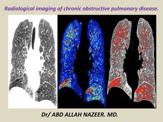
  • 1. Radiological imaging of chronic obstructive pulmonary disease. Dr/ ABD ALLAH NAZEER. MD.
  • 2. Chronic obstructive pulmonary disease is defined as a preventable and treatable disease state characterized by airflow limitation that is not fully reversible. The airflow limitation is usually progressive and is associated with an abnormal inflammatory response of the lungs to noxious particles or gases, primarily caused by cigarette smoking. Emphysema is one of its components, along with asthma and chronic bronchitis. Emphysema is defined pathologically as permanent enlargement of the airspaces distal to the terminal bronchioles, accompanied by destruction of their walls and without obvious fibrosis. Chronic bronchitis is defined as chronic productive cough for 3 months in each of 2 successive years in a patient in whom other causes of chronic cough have been excluded.
  • 3.
  • 4. Clinical presentation The clinical features of emphysema should be distinguished from the signs and symptoms of chronic bronchitis. Patients with emphysema are hypocapnoeic and are often referred to as "pink puffers". This compares with the hypercapnoea and cyanosis of chronic bronchitis with patients referred to as "blue bloaters". In practice, features of these two syndromes coexist as chronic obstructive pulmonary disease. Patients typically report dyspnea without significant sputum production. Signs of emphysema include: tachypnoea absence of cyanosis pursed-lip breathing chest hyperinflation reduced breath sounds hyper-resonant to percussion Cor pulmonale (late)
  • 5.
  • 6. Radiographic features Plain film Except in the case of very advanced disease with bulla formation, chest radiography does not image emphysema directly, but rather infers the diagnosis due to associated features: hyperinflation: flattened hemidiaphragm(s): most reliable sign increased and usually irregular radiolucency of the lungs increased retrosternal airspace increased antero-posterior diameter of chest widely spaced ribs sternal bowing tenting of the diaphragm saber-sheath trachea vascular changes: paucity of blood vessels, often distorted pulmonary arterial hypertension pruning of peripheral vessels increased caliber of central arteries right ventricular enlargement It should be remembered, however, that the most common plain film appearance of COPD is "normal" and the role of chest radiography is to eliminate other causes of lung symptoms such as infection, bronchiectasis or cancer .
  • 7. CT is currently the modality of choice for detecting emphysema; HRCT is particularly effective. It should be noted, however, that there is relatively poor correlation between autopsy-proven emphysema, pulmonary function test abnormalities and CT with 20% of pathology-proven cases not being evident on CT and 40% of patients with abnormal CT having normal pulmonary function tests. CT is able to discriminate between centrilobular, panlobular, and paraseptal emphysema. Centrilobular emphysema Centrilobular is by far the most common type encountered, and is a common finding in asymptomatic elderly patients. It is predominantly located in the upper zones of each lobe (i.e. apical and posterior segments of the upper lobes, and superior segment of the lower lobes) and has a patchy distribution. It appears as focal lucencies (emphysematous spaces) which measure up to 1 cm in diameter, located centrally within the secondary pulmonary lobule, often with a central or peripheral dot representing the central brochovascular bundle Panlobular emphysema Panlobular emphysema is predominantly located in the lower lobes, has a uniform distribution across parts of the secondary pulmonary lobule, which are homogeneously reduced in attenuation.
  • 8. Paraseptal emphysema Paraseptal emphysema is located adjacent to the pleura and septal lines with a peripheral distribution within the secondary pulmonary lobule. The affected lobules are almost always subpleural, and demonstrate small focal lucencies up to 10 mm in size. Any lucency larger than 10 mm should be referred to as subpleural blebs or subpleural bullae (synonymous). In all three subtypes, the emphysematous spaces are not bounded by any visible wall. MRI MRI is in the research phases for evaluation of lung parenchymal abnormalities like emphysema. Dynamic breathing MRI may have a future role in assessing pulmonary emphysema
  • 9. Emphysema Emphysema typically presents as areas of low attenuation without visible walls as a result of parenchymal destruction. Centrilobular emphysema Most common type Irreversible destruction of alveolar walls in the centrilobular portion of the lobule Upper lobe predominance and uneven distribution Strongly associated with smoking. Panlobular emphysema Affects the whole secondary lobule Lower lobe predominance In alpha-1-antitrypsin deficiency, but also seen in smokers with advanced emphysema Paraseptal emphysema Adjacent to the pleura and interlobar fissures Can be isolated phenomenon in young adults, or in older patients with centrilobular emphysema In young adults often associated with spontaneous pneumothorax
  • 10. Types of emphysema: line diagram shows the parts of secondary pulmonary lobule that are affected in different types of emphysema. Respiratory bronchioles are primarily affected by centrilobular emphysema; peripheral alveolar ducts, sacs, and alveoli in paraseptal emphysema (PLE); all the components (i.e., respiratory bronchioles, alveolar ducts, alveolar sacs, and alveoli) in panlobular emphysema (PLE), and any part in irregular or paracicatricial emphysema.
  • 11.
  • 12.
  • 13. Centrilobular emphysema (CLE) and edema: A, Chest radiographs, postero- anterior and lateral views, show hyperinflation of the lungs (flattened diaphragm), increased translucency in the upper lungs with vascular attenuation and loss of arborization. B, Emphysematous spaces outlined by edema fluid filling the surrounding airspaces give an appearance of reticulation in this patient with lung edema superimposed on confluent, upper-lung predominant CLE.
  • 14. Centrilobular Emphysema in a Cigarette Smoker. Axial CT image through the upper lungs shows numerous well-defined lucencies, many of which are traversed by a central vessel.
  • 15. Centrilobular emphysema due to smoking. The periphery of the lung is spared (blue arrows). Centrilobular artery (yellow arrows) is seen in the center of the hypodense area.
  • 17. Centrilobular emphysema: A, Transverse computed tomography images shows centrilobular hypoattenuation with upper lung predominance. Note the resemblance of the macroscopic pathologic image in Figure 4A. B, Coronal minimum intensity projection image brings out the distribution and extent of emphysema.
  • 18. Centrilobular emphysema: Transverse computed tomography images in the first row and minimum intensity projection images in the second row show confluent centrilobular hypoattenuation with posterior lung predominance (arrows), Also note para-septal emphysema in the left upper lobe (arrow heads).
  • 19.
  • 20. Panlobular emphysema (PLE) from a-1–antitrypsin deficiency: chest radiographs in postero-anterior and lateral projections show hyperinflation and increased translucency in the lower lungs with vascular attenuation, indicating PLE.
  • 21. Panlobular emphysema (PLE) from a-1–antitrypsin deficiency: computed tomography images (first row) show confluent lower-lung predominant panlobular hypoattenuation, indicating PLE. The confluence, panlobular distribution, lower-lung predominance, and vascular attenuation are better shown by the coronal minimum intensity projection and maximum intensity projection images (second row).
  • 22. A- Chest tomography in axial cut showing areas of panlobular emphysema in predominately lower lobes. Note cylindroids bronchiectasis and thickening of bronchial walls, most evidence in right. B- coronal reformatted showing hyper-inflation areas in lower lobes.
  • 23. Ritalin lung with panlobular emphysema: chest radiographs, postero-anterior projection, and computed tomography (coronal reformatted image) show basal-predominant panlobular hypoattenuation similar to that found in a-1–antitrypsin deficiency.
  • 24. Panlobular Emphysema in a Patient with Alpha-1 Antitrypsin Deficiency. Axial image through the lower lungs shows homogenous decrease in lung attenuation in both lower lobes. Some of the interlobular septa in the right lower lobe are accentuated by the emphysema.
  • 26.
  • 27. Paraseptal emphysema: computed tomography shows rectangular cysts sharing walls in subpleural upper lobes and the superior segment of the left lower lobe. Centrilobular emphysema is also evident in the upper lobes (arrows).
  • 28. Paraseptal Emphysema in a Cigarette Smoker. Axial image through the upper lungs shows multiple well-demarcated subpleural air-containing spaces.
  • 29. Paraseptal emphysema with small bullae.
  • 30.
  • 31. Paracicatricial emphysema (PCE) from progressive massive fibrosis caused by silicosis: computed tomography images in lung window show conglomerate masses in the posterior upper lobes with surrounding low attenuation (arrows) indicating PCE. Hyperinflation of the anterior upper lungs is from traction by the conglomerate masses.
  • 32. Coronal reformat demonstrating heterogeneous opacity in the right upper lobe and areas of centrilobular emphysema and para-septal. Tuberculosis in COPD.
  • 33. Bullous emphysema: postero-anterior radiograph and coronal computed tomography multiplanar reformation and maximum intensity projection images show a large bulla in the right upper lobe with atelectasis of the adjacent lung (arrows).
  • 34. Apical bleb: computed tomography through the lung apex and multiplanar reformation show a left apical bleb floating in small pneumothorax (arrows). Also note unruptured blebs in the right lung apex.
  • 35.
  • 36.
  • 37.
  • 38.
  • 39. Quantification of Emphysema and Air Trapping in 66 year old Individual with GOLD Stage 3 COPD (FEV1 43% predicted) (a) Inspiratory CT: coronal image depicts voxels with CT attenuation less than -950HU indicating emphysema (14% in this case). Color coding: Indicates lobes. (b) Inspiratory CT: coronal image depicts distribution of sizes of emphysematous clusters, with color coding depending on cluster size. (c) Expiratory CT: coronal image depicts voxels with CT attenuation less than -856HU indicating air trapping (38% in this case). (d) Image map derived from co- registered inspiratory and expiratory images depicts change in voxel attenuation from inspiration to expiration. Color coding: Red indicates voxels that were less than -950 HU on inspiration and less than -856 HU on expiration (emphysema). Yellow indicates voxels that were greater than -950 HU on inspiration, but less than -856 HU on expiration (air trapping). Green indicates voxels that were less than -950 HU on inspiration and less than -856 on expiration (normal). White indicates voxels that were greater than -950 HU on inspiration but less than -856 on expiration. Progress in Imaging COPD, 2004-2014
  • 40. Follow up CT Analysis in Same Subject as Shown in Figure 6. Color coding: Same as in Figure 6. (a) % LAA-950 has increased from 14% to 23%. (b) Cluster sizes have also increased. (c)Air trapping appears to have increased from 38% to 45%. (d) Inspiratory-expiratory registration shows that only the amount of emphysema increased, while the amount of gas trapping remained constant. (e) Longitudinal registration of baseline and follow up inspiratory scans with micro mapping shows areas of stable emphysema in red, new areas of emphysema in yellow, and stable normal lung in green.
  • 41. Images in representative patients with mild-to- moderate or severe chronic obstructive pulmonary disease (COPD).
  • 42. Chronic bronchitis (CB) is often defined as the presence of productive cough for 3 months in two successive years in a patient in whom other causes of chronic cough, such as tuberculosis, lung cancer and heart failure, have been excluded. It can be an important pathological component of chronic obstructive pulmonary disease. (often considered as a distinct phenotype of COPD) Pathology Chronic bronchitis most often results from overproduction and hypersecretion of mucus by goblet cells. This can in turn lead to worsening airflow obstruction by luminal obstruction of small airways, epithelial remodeling, and alteration of airway surface tension predisposing to collapse.
  • 43. Diagnosis: A physical examination will often reveal decreased intensity of breath sounds, wheezing, rhonchi, and prolonged expiration. Most physicians rely on the presence of a persistent dry or wet cough as evidence of bronchitis. A variety of tests may be performed in patients presenting with cough and shortness of breath: A chest X-ray is useful to exclude pneumonia which is more common in those with a fever, fast heart rate, fast respiratory rate, or who are old. A sputum sample showing neutrophil granulocytes (inflammatory white blood cells) and culture showing that has pathogenic microorganisms such as Streptococcus species. A blood test would indicate inflammation (as indicated by a raised white blood cell count and elevated C-reactive protein).
  • 44. Chronic bronchitis with bronchial wall thickening with increased brochovascular markings, enlarged vessels and cardiomeagly (abnormal enlargement of the heart).
  • 45. Chronic bronchitis: postero-anterior radiograph (A) and computed tomography (B) show bilateral bronchial wall thickening, the so-called, ‘‘dirty lung.’’
  • 46. Computed tomography (CT) scan in an elderly patient with known chronic bronchitis. The scans demonstrate thickening of the bronchial walls (purple arrows) and bronchi filled with mucus or phlegm (blue arrows).
  • 47. Computed tomography (CT) scan in patient with chronic bronchitis showing thickening of the bronchial walls (red arrows) and mucous within the bronchi (yellow arrows).
  • 48.
  • 49.