SlideShare a Scribd company logo
1 of 140
Radiological imaging 
of skeletal dysplasias 
Dr/ ABD ALLAH NAZEER. MD.
CLEIDOCRANIAL DYSPLASIA.
Cleidocranial dysplasia (CCD) causing respiratory distress syndrome in a newborn infant
Cleidocranial 
dysplasia. (A) AP 
view of the skull 
showing multiple 
wormian bones 
along the suture 
lines. (B) AP view of 
the upper chest 
showing an absent 
right clavicle and 
small hypoplastic 
medial segment 
(arrow) on the left.
Thanatophoric dysplasia.
Osteopoikilosis is a benign, autosomal dominant sclerosing 
dysplasia of bone characterized by the presence of numerous bone 
islands in the skeleton. 
The radiographic appearance of osteopoikilosis on an x-ray is 
characterized by a pattern of numerous white densities of similar 
size spread throughout all the bones. This is a systemic condition. It 
must be differentiated from blastic metastasis, which can also 
present radiographically as white densities interspersed 
throughout bone. Blastic metastasis tends to present with larger 
and more irregular densities in less of a uniform pattern. Another 
differentiating factor is age, with blastic metastasis mostly 
affecting older people, and osteopoikilosis being found in people 
20 years of age and younger. 
Men and women are affected in equal number. ,reflecting the fact 
that this disease attacks indiscriminately. Additionally, the disease 
is often associated with melorheostosis, despite the apparent lack 
of correlation between Melorheostosis and genetic heritability
Osteopoikilosis.
Osteopoikilosis.
Mucopolysaccharidosis type I Hurler-Scheie syndrome.
Hurler Syndrome (MPS-IH). 
(A) Lateral radiograph of 
the skull shows a large “J”- 
shaped sella and 
underdeveloped mastoids. 
Sinuses are hypoplastic. (B) 
AP radiograph of the spine 
and pelvis demonstrates 
expanded anterior ribs, 
hypoplastic ilia, and coxa 
valga. (C) Lateral 
radiograph of the spine 
shows hypoplastic 
vertebrae at the 
thoracolumbar junction 
with anterior beaking. (D) 
Hand radiograph 
demonstrates thick short 
metacarpals and phalanges 
with pointing of the 
proximal metacarpals.
Mucopolysaccharidosis, type II
Mucopolysaccharidosis type 111 .
Mucopolysaccharidosis 
type IV.
MPS IV
MPS VI.
MPS VI. Coronal MRI fast spin echo image and spoiled gradient echo image show 
irregularly enlarged growth plate, with multiple defects and/or erosions.
Multiple epiphyseal dysplasia. Radiographs of the hip (A), left knee 
(B,C), left foot and ankle (D,E), and left wrist (F) show lack of epiphyseal 
ossification centers with punctate calcifications in the knee.
Multiple epiphyseal dysplasia in an adolescent. AP and lateral radiographs of 
the knee (A,B) and ankle (C,D) showing irregular epiphysis with joint deformities.
Metaphyseal dysplasia. Standing radiographs of the knees showing 
metaphyseal irregularity and flaring with femoral bowing.
Metaphyseal dysplasia.
Metaphyseal 
chondrodysplasia
Radiographs showing abnormalities (irregularities) in different portions of the skeleton 
that aid in the diagnosis. A, A/P view of the knee showing irregular metaphyses (arrow) in 
Metaphyseal Chondrodysplasia—Schmid type. B, A/P view of the pelvis with arrow 
pointing to very small distal epiphyses in Spondyloepiphyseal Dysplasia Congenita (SEDC). 
C, A/P view of the knee demonstrating very widened diaphysis in Diaphyseal Dysplasia– 
Camurati-Hunermann type. D, A/P view of the hand showing shortened metacarpals, 
phalanges with widened metaphyses (arrow) in acrodysostosis. E, A/P and lateral views of 
the spine illustrating irregular vertebral margins in Spondyloepiphyseal Dysplasia Tarda.
Spondyloepiphyseal dysplasia congenita is an inherited disorder of bone 
growth that results in short stature (dwarfism), skeletal abnormalities, and 
problems with vision and hearing. This condition affects the bones of the spine 
(spondylo-) and the ends (epiphyses) of long bones in the arms and legs. 
Congenita indicates that the condition is present from birth. 
People with Spondyloepiphyseal dysplasia congenita have short stature from 
birth, with a very short trunk and neck and shortened limbs. Their hands and feet, 
however, are usually average-sized. Adult height ranges from 3 feet to just over 4 
feet. Abnormal curvature of the spine (kyphoscoliosis and lordosis) becomes more 
severe during childhood and can cause problems with breathing. Instability of the 
spinal bones (vertebrae) in the neck may increase the risk of spinal cord damage. 
Other skeletal features include flattened vertebrae (platyspondyly); an 
abnormality of the hip joint that causes the upper leg bones to turn inward (coxa 
vara); a broad, barrel-shaped chest; and a foot deformity called a clubfoot. 
Arthritis and decreased joint mobility often develop early in life. People with 
Spondyloepiphyseal dysplasia congenita have mild changes in their facial 
features. The cheekbones close to the nose may appear flattened. Some infants 
are born with an opening in the roof of the mouth (a cleft palate). Severe 
nearsightedness (high myopia) is common, as are other eye problems that can 
impair vision. About one quarter of people with this condition have hearing loss.
Spondyloepimetaphyseal dysplasia, Strudwick type is an inherited disorder 
of bone growth that results in short stature (dwarfism), skeletal abnormalities, 
and problems with vision. This condition affects the bones of the spine (spondylo-) 
and two regions (epiphyses and metaphyses) near the ends of long bones in the 
arms and legs. The Strudwick type was named after the first reported patient with 
the disorder. 
People with this condition have short stature from birth, with a very short trunk 
and shortened limbs. Their hands and feet, however, are usually average-sized. 
Affected individuals may have an abnormally curved lower back (lordosis) or a 
spine that curves to the side (scoliosis). This abnormal spinal curvature may be 
severe and can cause problems with breathing. Instability of the spinal bones 
(vertebrae) in the neck may increase the risk of spinal cord damage. Other skeletal 
features include flattened vertebrae (platyspondyly), severe protrusion of the 
breastbone (pectus carinatum), an abnormality of the hip joint that causes the 
upper leg bones to turn inward (coxa vara), and an inward- and upward-turning 
foot (clubfoot). Arthritis may develop early in life. 
People with spondyloepimetaphyseal dysplasia, Strudwick type have mild changes 
in their facial features. Some infants are born with an opening in the roof of the 
mouth (a cleft palate) and their cheekbones may appear flattened. Eye problems 
that can impair vision are common, such as severe nearsightedness (high myopia) 
and tearing of the lining of the eye (retinal detachment).
Chondroectodermal dysplasia (also known as the Ellis-van Creveld syndrome is 
a rare type of skeletal dysplasia. It is classified as a type of mesomelic limb 
shortening . 
Clinical spectrum 
Clinical features include: 
cleft lip and / or palate 
epispadias 
cryptorchidism 
polydactyly : tends to be post axial 
limb anomalies 
short limbs : especially forearm and lower leg 
short stature 
sparse, absent, or fine textured hair 
dental anomalies. 
peg teeth 
widely spaced teeth 
natal teeth 
delayed teeth 
missing teeth 
Associated congenital cardiac anomalies is present in 50 % of cases.
CHONDROECTODERMAL DYSPLASIA
Achondroplasia.
Achondroplasia.
Radiographs demonstrating abnormalities in the skeleton (pseudoachondroplasia). A, 
Lateral view of vertebral bodies showing rounded bodies with anterior beaking (arrow) 
and wide intervertebral disc spaces. B, A/P view of the knee showing irregular 
metaphyses (small arrow) and irregular, small for age epiphyses (large arrow). C, A/P 
view of the pelvis. Arrow points to small to almost absent, irregular epiphyses with mild 
metaphyseal abnormalities at the acetabulum surface and proximal femoral region.
Polyostotic fibrous dysplasia.
Polyostotic fibrous dysplasia
Fibrous dysplasia.
Fibrous dysplasia.
Multiple epiphyseal dysplasia. 
Fairbanks disease or multiple epiphyseal dysplasia (MED) is a rare genetic disorder 
(dominant form—1 in 10,000 births) which affects the growing ends of bones. Bones 
usually elongate by a process that involves the depositing of cartilage at the ends of the 
bones, called ossification. This cartilage then mineralizes and hardens to become bone. In 
MED, this process is defective. 
Multiple epiphyseal dysplasia (MED) encompasses a spectrum of skeletal disorders, most of 
which are inherited in an autosomal dominant form. However, there is also an autosomal 
recessive form. 
Signs and Symptoms: 
Children with autosomal dominant MED experience joint pain and fatigue after 
exercising. Their x-rays show small and irregular ossifications centers, most 
apparent in the hips and knees. A waddling gait may develop. Flat feet are very 
common. The spine is normal but may have a few irregularities, such as scoliosis. 
There are very small capital femoral epiphyses and hypoplastic, poorly formed 
acetabular roofs. Knees have metaphyseal widening and irregularity while hands 
have brachydactyly (short fingers) and proximal metacarpal rounding. By 
adulthood, people with MED are of short stature or in the low range of normal 
and have short limbs relative to their trunks. Frequently, movement becomes 
limited at the major joints, especially at the elbows and hips. However, loose knee 
and finger joints can occur. Signs of osteoarthritis usually begin in early adulthood.
Multiple epiphyseal dysplasia.
Dysplasia epiphysealis hemimelica is a rare 
skeletal developmental disorder characterized by asymmetric 
overgrowth of cartilage in the epiphyses. Due to the unusual 
and variable clinical picture, there is no standardized 
treatment and evolution is variable. We report the case of an 
8 year-old boy, who was referred for the gradual appearance 
of a mass in the anterior region of the right knee. Plain films 
matched with dysplasia epiphysealis hemimelica. The 
histological findings confirmed the diagnosis. Treatment of 
dysplasia epiphysealis hemimelica is not clearly defined in the 
literature. However, only surgically symptomatic lesions or 
those that interfere with the function should be treated. 
Prognosis is variable and depends on the location and size of 
the lesion. Due to the risk of recurrence, patients with this 
unusual dysplasia should be monitored on a regular basis.
Dysplasia epiphysealis hemimelica (Trevor-Fairbank disease).
Dysplasia Epiphysealis Hemimelica.
Du Pan Syndrome causes skeletal abnormalities and short stature. 
In this disease, the GDF5 gene responsible for regulating cell growth 
and differentiation is defective. Individuals with Du Pan Syndrome 
have typical trunk size but shortened limbs. Fibulae, the smaller of 
the two long bones that join the knee to the ankle, are rudimentary 
or absent. Knees and ankles are contorted. In addition, fingers are 
stumpy and bent in atypical directions. Thumbs and toes are small, 
button-like, and non-functional. Affected individuals are of typical 
intelligence. 
Du Pan syndrome is very rare, with less than 30 cases reported. 
Therefore, incidence of this condition is thought to be less than 1 in 
1,000,000. Almost all the affected individuals reported have been 
from Middle Eastern countries. 
Prognosis is generally favorable. Skeletal symptoms may be 
progressive with age and growth. However, affected individuals are 
of typical intelligence and do not have a shortened lifespan.
An Egyptian patient with du Pan syndrome. Note absence of both fibulae (A), X-ray of both 
hands and feet showing severe brachydactyly and radial deviation of fingers (B, C).
Diastrophic dysplasia is a disorder of cartilage and bone 
development. Affected individuals have short stature with very short 
arms and legs. Most also have early-onset joint pain (osteoarthritis) 
and joint deformities called contractures, which restrict movement. 
These joint problems often make it difficult to walk and tend to 
worsen with age. Additional features of diastrophic dysplasia 
include an inward- and upward-turning foot (clubfoot), progressive 
abnormal curvature of the spine, and unusually positioned thumbs 
(hitchhiker thumbs). About half of infants with diastrophic dysplasia 
are born with an opening in the roof of the mouth (a cleft palate). 
Swelling of the external ears is also common in newborns and can 
lead to thickened, deformed ears. 
The signs and symptoms of diastrophic dysplasia are similar to those 
of another skeletal disorder called atelosteogenesis type 2; however, 
diastrophic dysplasia tends to be less severe. Although some 
affected infants have breathing problems, most people with 
diastrophic dysplasia live into adulthood
Diastrophic Dysplasia
Diastrophic dysplasia.
Proximal femoral focal deficiency (PFFD) is a rare, non-hereditary 
birth defect that affects the pelvis, particularly the hip bone, and the 
proximal femur. The disorder may affect one side or both, with the hip 
being deformed and the leg shortened. 
It is commonly linked with the absence or shortening of a leg bone (fibular 
hemimelia) and the absence of a kneecap. Other linked birth defects 
include the dislocation or instability of the joint between the femur and 
the kneecap, a shortened tibia or fibula, and foot deformities. 
Classifications: 
There are typically four classes (or types) of PFFD, ranging from class A to 
class D, as detailed by Aitken 
Pathology: 
The etiology of this disorder is uncertain. The diagnosis and classification 
have been based mainly on plain film findings. This method does not 
permit definite classification during the 1st year of life. 
Associations: 
asbent patella.
Proximal focal femoral deficiency.
Down syndrome (DS) or Down's syndrome, also known as trisomy 21, is a 
genetic disorder caused by the presence of all or part of a third copy of 
chromosome 21. It is typically associated with physical growth delays, 
characteristic facial features, and mild to moderate intellectual disability. 
Axial CT scan at the level of the occiput/C1 demonstrating the odontoid peg 
migrating into the foramen magnum; (B) sagittal CT scan demonstrating C1 
subluxation on C2 with migration of the odontoid peg into the brainstem; and (C) 
sagittal T2-weighted MRI demonstrating anterior subluxation of the occiput on C1 
by 5 mm, anterior subluxation of C1 on C2 by 11 mm, and severe canal stenosis.
Turner syndrome, a condition that affects only girls and women, 
results when a sex chromosome (the X chromosome) is missing or partially 
missing. Turner syndrome can cause a variety of medical and developmental 
problems, including short height, failure to start puberty, infertility, heart 
defects, certain learning disabilities and social adjustment problems. 
Both hands show small fourth finger in a case of Turner syndrome.
Thank You.

More Related Content

What's hot

Skeletal dysplasia musculoskeletal radiology
Skeletal dysplasia musculoskeletal radiologySkeletal dysplasia musculoskeletal radiology
Skeletal dysplasia musculoskeletal radiologyDr pradeep Kumar
 
Ascites and Pleural Effusion
 Ascites and Pleural Effusion Ascites and Pleural Effusion
Ascites and Pleural EffusionMedia Genie
 
Presentation2.pptx wrist joint.
Presentation2.pptx wrist joint.Presentation2.pptx wrist joint.
Presentation2.pptx wrist joint.Abdellah Nazeer
 
Mri anatomy of knee Dr. Muhammad Bin Zulfiqar
Mri anatomy of knee Dr. Muhammad Bin ZulfiqarMri anatomy of knee Dr. Muhammad Bin Zulfiqar
Mri anatomy of knee Dr. Muhammad Bin ZulfiqarDr. Muhammad Bin Zulfiqar
 
Presentation1.pptx, radiological imaging of metabolic bone diseases.
Presentation1.pptx, radiological imaging of metabolic bone diseases.Presentation1.pptx, radiological imaging of metabolic bone diseases.
Presentation1.pptx, radiological imaging of metabolic bone diseases.Abdellah Nazeer
 
54 DAVID SUTTON PICTURES NEURORADIOLOGY OF SPINE
54 DAVID SUTTON PICTURES NEURORADIOLOGY OF SPINE54 DAVID SUTTON PICTURES NEURORADIOLOGY OF SPINE
54 DAVID SUTTON PICTURES NEURORADIOLOGY OF SPINEDr. Muhammad Bin Zulfiqar
 
Presentation1.pptx, radiological imaging of spinal dysraphism.
Presentation1.pptx, radiological imaging of spinal dysraphism.Presentation1.pptx, radiological imaging of spinal dysraphism.
Presentation1.pptx, radiological imaging of spinal dysraphism.Abdellah Nazeer
 
Musculoskeletal radiology revision notes
Musculoskeletal radiology revision notesMusculoskeletal radiology revision notes
Musculoskeletal radiology revision notesTONY SCARIA
 
Imaging of white matter diseases
Imaging of white matter diseasesImaging of white matter diseases
Imaging of white matter diseasesNavni Garg
 
Presentation1.pptx, radiological anatomy of the thigh and leg.
Presentation1.pptx, radiological anatomy of the thigh and leg.Presentation1.pptx, radiological anatomy of the thigh and leg.
Presentation1.pptx, radiological anatomy of the thigh and leg.Abdellah Nazeer
 
Presentation2, radiological imaging of phakomatosis.
Presentation2, radiological imaging of phakomatosis.Presentation2, radiological imaging of phakomatosis.
Presentation2, radiological imaging of phakomatosis.Abdellah Nazeer
 
Presentation1, radiological imaging of hyperparathyroidism.
Presentation1, radiological imaging of hyperparathyroidism.Presentation1, radiological imaging of hyperparathyroidism.
Presentation1, radiological imaging of hyperparathyroidism.Abdellah Nazeer
 
KEYS OF RADIOLOGY SPOTTERS GIT
KEYS OF RADIOLOGY SPOTTERS GITKEYS OF RADIOLOGY SPOTTERS GIT
KEYS OF RADIOLOGY SPOTTERS GITAnish Choudhary
 
Radiology Spotters mixed Bag Collection for post graduates student .PPT
Radiology Spotters mixed Bag Collection for post graduates student .PPTRadiology Spotters mixed Bag Collection for post graduates student .PPT
Radiology Spotters mixed Bag Collection for post graduates student .PPTDr pradeep Kumar
 
radiology Spotters mixed bag
radiology Spotters mixed bagradiology Spotters mixed bag
radiology Spotters mixed bagAnish Choudhary
 

What's hot (20)

Skeletal dysplasia musculoskeletal radiology
Skeletal dysplasia musculoskeletal radiologySkeletal dysplasia musculoskeletal radiology
Skeletal dysplasia musculoskeletal radiology
 
Ascites and Pleural Effusion
 Ascites and Pleural Effusion Ascites and Pleural Effusion
Ascites and Pleural Effusion
 
Presentation2.pptx wrist joint.
Presentation2.pptx wrist joint.Presentation2.pptx wrist joint.
Presentation2.pptx wrist joint.
 
Spotters ppt 1
Spotters ppt 1Spotters ppt 1
Spotters ppt 1
 
Mri anatomy of knee Dr. Muhammad Bin Zulfiqar
Mri anatomy of knee Dr. Muhammad Bin ZulfiqarMri anatomy of knee Dr. Muhammad Bin Zulfiqar
Mri anatomy of knee Dr. Muhammad Bin Zulfiqar
 
Spots with keys
Spots with keysSpots with keys
Spots with keys
 
Renal doppler
Renal dopplerRenal doppler
Renal doppler
 
Presentation1.pptx, radiological imaging of metabolic bone diseases.
Presentation1.pptx, radiological imaging of metabolic bone diseases.Presentation1.pptx, radiological imaging of metabolic bone diseases.
Presentation1.pptx, radiological imaging of metabolic bone diseases.
 
54 DAVID SUTTON PICTURES NEURORADIOLOGY OF SPINE
54 DAVID SUTTON PICTURES NEURORADIOLOGY OF SPINE54 DAVID SUTTON PICTURES NEURORADIOLOGY OF SPINE
54 DAVID SUTTON PICTURES NEURORADIOLOGY OF SPINE
 
Presentation1.pptx, radiological imaging of spinal dysraphism.
Presentation1.pptx, radiological imaging of spinal dysraphism.Presentation1.pptx, radiological imaging of spinal dysraphism.
Presentation1.pptx, radiological imaging of spinal dysraphism.
 
Musculoskeletal radiology revision notes
Musculoskeletal radiology revision notesMusculoskeletal radiology revision notes
Musculoskeletal radiology revision notes
 
Hip joint
Hip jointHip joint
Hip joint
 
Imaging of white matter diseases
Imaging of white matter diseasesImaging of white matter diseases
Imaging of white matter diseases
 
Diagnostic Imaging of Arthritis
Diagnostic Imaging of ArthritisDiagnostic Imaging of Arthritis
Diagnostic Imaging of Arthritis
 
Presentation1.pptx, radiological anatomy of the thigh and leg.
Presentation1.pptx, radiological anatomy of the thigh and leg.Presentation1.pptx, radiological anatomy of the thigh and leg.
Presentation1.pptx, radiological anatomy of the thigh and leg.
 
Presentation2, radiological imaging of phakomatosis.
Presentation2, radiological imaging of phakomatosis.Presentation2, radiological imaging of phakomatosis.
Presentation2, radiological imaging of phakomatosis.
 
Presentation1, radiological imaging of hyperparathyroidism.
Presentation1, radiological imaging of hyperparathyroidism.Presentation1, radiological imaging of hyperparathyroidism.
Presentation1, radiological imaging of hyperparathyroidism.
 
KEYS OF RADIOLOGY SPOTTERS GIT
KEYS OF RADIOLOGY SPOTTERS GITKEYS OF RADIOLOGY SPOTTERS GIT
KEYS OF RADIOLOGY SPOTTERS GIT
 
Radiology Spotters mixed Bag Collection for post graduates student .PPT
Radiology Spotters mixed Bag Collection for post graduates student .PPTRadiology Spotters mixed Bag Collection for post graduates student .PPT
Radiology Spotters mixed Bag Collection for post graduates student .PPT
 
radiology Spotters mixed bag
radiology Spotters mixed bagradiology Spotters mixed bag
radiology Spotters mixed bag
 

Similar to Presentation1.pptx, radiological imaging of skeletal dysplasia

Skeletal dysplasia
Skeletal dysplasiaSkeletal dysplasia
Skeletal dysplasiaRMLIMS
 
Dysplasias of bone
Dysplasias of boneDysplasias of bone
Dysplasias of boneYassera Awan
 
Orthopedic Aspects Of Metabolic Bone Disease By Xiu
Orthopedic Aspects Of Metabolic Bone Disease By XiuOrthopedic Aspects Of Metabolic Bone Disease By Xiu
Orthopedic Aspects Of Metabolic Bone Disease By XiuXiu Srithammasit
 
Achondroplasia, pseudoachondroplasia, hypochondroplasia
Achondroplasia, pseudoachondroplasia, hypochondroplasiaAchondroplasia, pseudoachondroplasia, hypochondroplasia
Achondroplasia, pseudoachondroplasia, hypochondroplasiaNikhil Murkey
 
Skeletal dysplasia grand round
Skeletal dysplasia   grand round Skeletal dysplasia   grand round
Skeletal dysplasia grand round Amr Mansour Hassan
 
Approach to skeletal dysplasia
Approach to skeletal dysplasiaApproach to skeletal dysplasia
Approach to skeletal dysplasiaNavni Garg
 
SKELETAL DYSPLASIA Radiodiagnosis BY ABHIJIT R SINGH
SKELETAL DYSPLASIA Radiodiagnosis BY ABHIJIT R SINGHSKELETAL DYSPLASIA Radiodiagnosis BY ABHIJIT R SINGH
SKELETAL DYSPLASIA Radiodiagnosis BY ABHIJIT R SINGHDrABHIJITRSINGH
 
FETAL SKELETAL ANOMALIES GROUP 3.pptx
FETAL SKELETAL ANOMALIES GROUP 3.pptxFETAL SKELETAL ANOMALIES GROUP 3.pptx
FETAL SKELETAL ANOMALIES GROUP 3.pptxDeogratiusGivenOkodi
 
SKELTAL DYSPLASIA.pptx
SKELTAL DYSPLASIA.pptxSKELTAL DYSPLASIA.pptx
SKELTAL DYSPLASIA.pptxssuserf420c11
 
Radiological approach to Congenital skeletal dysplasia
Radiological approach to Congenital skeletal dysplasiaRadiological approach to Congenital skeletal dysplasia
Radiological approach to Congenital skeletal dysplasiaSachin Balutkar
 
Dwarfism, bonydysplasia
Dwarfism, bonydysplasiaDwarfism, bonydysplasia
Dwarfism, bonydysplasiaDr.Nikhil. S.U
 
Skeletal dysplasia final
Skeletal dysplasia finalSkeletal dysplasia final
Skeletal dysplasia finalNihit Jain
 
Skeletal dysplasia
Skeletal dysplasiaSkeletal dysplasia
Skeletal dysplasiaPankaj Kaira
 
Malignant connective tissue tumors
Malignant connective tissue tumorsMalignant connective tissue tumors
Malignant connective tissue tumorsanila namboodiripad
 

Similar to Presentation1.pptx, radiological imaging of skeletal dysplasia (20)

Skeletal dysplasia
Skeletal dysplasiaSkeletal dysplasia
Skeletal dysplasia
 
Dysplasias of bone
Dysplasias of boneDysplasias of bone
Dysplasias of bone
 
Orthopedic Aspects Of Metabolic Bone Disease By Xiu
Orthopedic Aspects Of Metabolic Bone Disease By XiuOrthopedic Aspects Of Metabolic Bone Disease By Xiu
Orthopedic Aspects Of Metabolic Bone Disease By Xiu
 
Achondroplasia, pseudoachondroplasia, hypochondroplasia
Achondroplasia, pseudoachondroplasia, hypochondroplasiaAchondroplasia, pseudoachondroplasia, hypochondroplasia
Achondroplasia, pseudoachondroplasia, hypochondroplasia
 
Skeletal dysplasia grand round
Skeletal dysplasia   grand round Skeletal dysplasia   grand round
Skeletal dysplasia grand round
 
Approach to skeletal dysplasia
Approach to skeletal dysplasiaApproach to skeletal dysplasia
Approach to skeletal dysplasia
 
SKELETAL DYSPLASIA Radiodiagnosis BY ABHIJIT R SINGH
SKELETAL DYSPLASIA Radiodiagnosis BY ABHIJIT R SINGHSKELETAL DYSPLASIA Radiodiagnosis BY ABHIJIT R SINGH
SKELETAL DYSPLASIA Radiodiagnosis BY ABHIJIT R SINGH
 
FETAL SKELETAL ANOMALIES GROUP 3.pptx
FETAL SKELETAL ANOMALIES GROUP 3.pptxFETAL SKELETAL ANOMALIES GROUP 3.pptx
FETAL SKELETAL ANOMALIES GROUP 3.pptx
 
SKELTAL DYSPLASIA.pptx
SKELTAL DYSPLASIA.pptxSKELTAL DYSPLASIA.pptx
SKELTAL DYSPLASIA.pptx
 
skeletal dysplasia ppt
skeletal dysplasia pptskeletal dysplasia ppt
skeletal dysplasia ppt
 
Osteopetrosis
OsteopetrosisOsteopetrosis
Osteopetrosis
 
Osteopetrosis
OsteopetrosisOsteopetrosis
Osteopetrosis
 
Radiological approach to Congenital skeletal dysplasia
Radiological approach to Congenital skeletal dysplasiaRadiological approach to Congenital skeletal dysplasia
Radiological approach to Congenital skeletal dysplasia
 
Dwarfism, bonydysplasia
Dwarfism, bonydysplasiaDwarfism, bonydysplasia
Dwarfism, bonydysplasia
 
Skeletal dysplasia final
Skeletal dysplasia finalSkeletal dysplasia final
Skeletal dysplasia final
 
Vol 22 congenital 3
Vol 22 congenital 3Vol 22 congenital 3
Vol 22 congenital 3
 
Vol 21 congenital 2
Vol 21 congenital 2Vol 21 congenital 2
Vol 21 congenital 2
 
Skeletal dysplasia
Skeletal dysplasiaSkeletal dysplasia
Skeletal dysplasia
 
Malignant connective tissue tumors
Malignant connective tissue tumorsMalignant connective tissue tumors
Malignant connective tissue tumors
 
Radiology 5th year, 4th lecture (Dr. Salah Mohammad Fatih)
Radiology 5th year, 4th lecture (Dr. Salah Mohammad Fatih)Radiology 5th year, 4th lecture (Dr. Salah Mohammad Fatih)
Radiology 5th year, 4th lecture (Dr. Salah Mohammad Fatih)
 

More from Abdellah Nazeer

Muculoskeletal Pediatic Imaging..pptx
Muculoskeletal Pediatic Imaging..pptxMuculoskeletal Pediatic Imaging..pptx
Muculoskeletal Pediatic Imaging..pptxAbdellah Nazeer
 
Presentation1, Ultrasound of the bowel loops and the lymph nodes..pptx
Presentation1, Ultrasound of the bowel loops and the lymph nodes..pptxPresentation1, Ultrasound of the bowel loops and the lymph nodes..pptx
Presentation1, Ultrasound of the bowel loops and the lymph nodes..pptxAbdellah Nazeer
 
Presentation1 Short cases MD..pptx
Presentation1 Short cases MD..pptxPresentation1 Short cases MD..pptx
Presentation1 Short cases MD..pptxAbdellah Nazeer
 
Presentation1, MD MCQ Cases..pptx
Presentation1, MD MCQ Cases..pptxPresentation1, MD MCQ Cases..pptx
Presentation1, MD MCQ Cases..pptxAbdellah Nazeer
 
Presentation1, Short Cases Quiz..pptx
Presentation1, Short Cases Quiz..pptxPresentation1, Short Cases Quiz..pptx
Presentation1, Short Cases Quiz..pptxAbdellah Nazeer
 
Presentation1, radiological imaging of lateral hindfoot impingement.
Presentation1, radiological imaging of lateral hindfoot impingement.Presentation1, radiological imaging of lateral hindfoot impingement.
Presentation1, radiological imaging of lateral hindfoot impingement.Abdellah Nazeer
 
Presentation2, radiological anatomy of the liver and spleen.
Presentation2, radiological anatomy of the liver and spleen.Presentation2, radiological anatomy of the liver and spleen.
Presentation2, radiological anatomy of the liver and spleen.Abdellah Nazeer
 
Presentation1, artifacts and pitfalls of the wrist and elbow joints.
Presentation1, artifacts and pitfalls of the wrist and elbow joints.Presentation1, artifacts and pitfalls of the wrist and elbow joints.
Presentation1, artifacts and pitfalls of the wrist and elbow joints.Abdellah Nazeer
 
Presentation1, artifact and pitfalls of the knee, hip and ankle joints.
Presentation1, artifact and pitfalls of the knee, hip and ankle joints.Presentation1, artifact and pitfalls of the knee, hip and ankle joints.
Presentation1, artifact and pitfalls of the knee, hip and ankle joints.Abdellah Nazeer
 
Presentation1, radiological imaging of artifact and pitfalls in shoulder join...
Presentation1, radiological imaging of artifact and pitfalls in shoulder join...Presentation1, radiological imaging of artifact and pitfalls in shoulder join...
Presentation1, radiological imaging of artifact and pitfalls in shoulder join...Abdellah Nazeer
 
Presentation1, radiological imaging of internal abdominal hernia.
Presentation1, radiological imaging of internal abdominal hernia.Presentation1, radiological imaging of internal abdominal hernia.
Presentation1, radiological imaging of internal abdominal hernia.Abdellah Nazeer
 
Presentation11, radiological imaging of ovarian torsion.
Presentation11, radiological imaging of ovarian torsion.Presentation11, radiological imaging of ovarian torsion.
Presentation11, radiological imaging of ovarian torsion.Abdellah Nazeer
 
Presentation1, musculoskeletal anatomy.
Presentation1, musculoskeletal anatomy.Presentation1, musculoskeletal anatomy.
Presentation1, musculoskeletal anatomy.Abdellah Nazeer
 
Presentation1, new mri techniques in the diagnosis and monitoring of multiple...
Presentation1, new mri techniques in the diagnosis and monitoring of multiple...Presentation1, new mri techniques in the diagnosis and monitoring of multiple...
Presentation1, new mri techniques in the diagnosis and monitoring of multiple...Abdellah Nazeer
 
Presentation1, radiological application of diffusion weighted mri in neck mas...
Presentation1, radiological application of diffusion weighted mri in neck mas...Presentation1, radiological application of diffusion weighted mri in neck mas...
Presentation1, radiological application of diffusion weighted mri in neck mas...Abdellah Nazeer
 
Presentation1, radiological application of diffusion weighted images in breas...
Presentation1, radiological application of diffusion weighted images in breas...Presentation1, radiological application of diffusion weighted images in breas...
Presentation1, radiological application of diffusion weighted images in breas...Abdellah Nazeer
 
Presentation1, radiological application of diffusion weighted images in abdom...
Presentation1, radiological application of diffusion weighted images in abdom...Presentation1, radiological application of diffusion weighted images in abdom...
Presentation1, radiological application of diffusion weighted images in abdom...Abdellah Nazeer
 
Presentation1, radiological application of diffusion weighted imges in neuror...
Presentation1, radiological application of diffusion weighted imges in neuror...Presentation1, radiological application of diffusion weighted imges in neuror...
Presentation1, radiological application of diffusion weighted imges in neuror...Abdellah Nazeer
 
Presentation1, mr physics.
Presentation1, mr physics.Presentation1, mr physics.
Presentation1, mr physics.Abdellah Nazeer
 
Presentation1. ct physics.
Presentation1. ct physics.Presentation1. ct physics.
Presentation1. ct physics.Abdellah Nazeer
 

More from Abdellah Nazeer (20)

Muculoskeletal Pediatic Imaging..pptx
Muculoskeletal Pediatic Imaging..pptxMuculoskeletal Pediatic Imaging..pptx
Muculoskeletal Pediatic Imaging..pptx
 
Presentation1, Ultrasound of the bowel loops and the lymph nodes..pptx
Presentation1, Ultrasound of the bowel loops and the lymph nodes..pptxPresentation1, Ultrasound of the bowel loops and the lymph nodes..pptx
Presentation1, Ultrasound of the bowel loops and the lymph nodes..pptx
 
Presentation1 Short cases MD..pptx
Presentation1 Short cases MD..pptxPresentation1 Short cases MD..pptx
Presentation1 Short cases MD..pptx
 
Presentation1, MD MCQ Cases..pptx
Presentation1, MD MCQ Cases..pptxPresentation1, MD MCQ Cases..pptx
Presentation1, MD MCQ Cases..pptx
 
Presentation1, Short Cases Quiz..pptx
Presentation1, Short Cases Quiz..pptxPresentation1, Short Cases Quiz..pptx
Presentation1, Short Cases Quiz..pptx
 
Presentation1, radiological imaging of lateral hindfoot impingement.
Presentation1, radiological imaging of lateral hindfoot impingement.Presentation1, radiological imaging of lateral hindfoot impingement.
Presentation1, radiological imaging of lateral hindfoot impingement.
 
Presentation2, radiological anatomy of the liver and spleen.
Presentation2, radiological anatomy of the liver and spleen.Presentation2, radiological anatomy of the liver and spleen.
Presentation2, radiological anatomy of the liver and spleen.
 
Presentation1, artifacts and pitfalls of the wrist and elbow joints.
Presentation1, artifacts and pitfalls of the wrist and elbow joints.Presentation1, artifacts and pitfalls of the wrist and elbow joints.
Presentation1, artifacts and pitfalls of the wrist and elbow joints.
 
Presentation1, artifact and pitfalls of the knee, hip and ankle joints.
Presentation1, artifact and pitfalls of the knee, hip and ankle joints.Presentation1, artifact and pitfalls of the knee, hip and ankle joints.
Presentation1, artifact and pitfalls of the knee, hip and ankle joints.
 
Presentation1, radiological imaging of artifact and pitfalls in shoulder join...
Presentation1, radiological imaging of artifact and pitfalls in shoulder join...Presentation1, radiological imaging of artifact and pitfalls in shoulder join...
Presentation1, radiological imaging of artifact and pitfalls in shoulder join...
 
Presentation1, radiological imaging of internal abdominal hernia.
Presentation1, radiological imaging of internal abdominal hernia.Presentation1, radiological imaging of internal abdominal hernia.
Presentation1, radiological imaging of internal abdominal hernia.
 
Presentation11, radiological imaging of ovarian torsion.
Presentation11, radiological imaging of ovarian torsion.Presentation11, radiological imaging of ovarian torsion.
Presentation11, radiological imaging of ovarian torsion.
 
Presentation1, musculoskeletal anatomy.
Presentation1, musculoskeletal anatomy.Presentation1, musculoskeletal anatomy.
Presentation1, musculoskeletal anatomy.
 
Presentation1, new mri techniques in the diagnosis and monitoring of multiple...
Presentation1, new mri techniques in the diagnosis and monitoring of multiple...Presentation1, new mri techniques in the diagnosis and monitoring of multiple...
Presentation1, new mri techniques in the diagnosis and monitoring of multiple...
 
Presentation1, radiological application of diffusion weighted mri in neck mas...
Presentation1, radiological application of diffusion weighted mri in neck mas...Presentation1, radiological application of diffusion weighted mri in neck mas...
Presentation1, radiological application of diffusion weighted mri in neck mas...
 
Presentation1, radiological application of diffusion weighted images in breas...
Presentation1, radiological application of diffusion weighted images in breas...Presentation1, radiological application of diffusion weighted images in breas...
Presentation1, radiological application of diffusion weighted images in breas...
 
Presentation1, radiological application of diffusion weighted images in abdom...
Presentation1, radiological application of diffusion weighted images in abdom...Presentation1, radiological application of diffusion weighted images in abdom...
Presentation1, radiological application of diffusion weighted images in abdom...
 
Presentation1, radiological application of diffusion weighted imges in neuror...
Presentation1, radiological application of diffusion weighted imges in neuror...Presentation1, radiological application of diffusion weighted imges in neuror...
Presentation1, radiological application of diffusion weighted imges in neuror...
 
Presentation1, mr physics.
Presentation1, mr physics.Presentation1, mr physics.
Presentation1, mr physics.
 
Presentation1. ct physics.
Presentation1. ct physics.Presentation1. ct physics.
Presentation1. ct physics.
 

Presentation1.pptx, radiological imaging of skeletal dysplasia

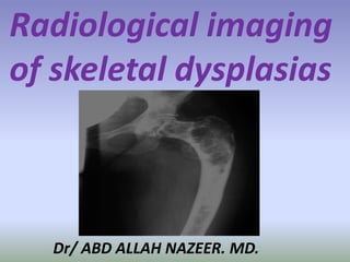
  • 1. Radiological imaging of skeletal dysplasias Dr/ ABD ALLAH NAZEER. MD.
  • 2.
  • 3.
  • 4.
  • 5.
  • 6.
  • 7.
  • 8.
  • 10.
  • 13. Cleidocranial dysplasia (CCD) causing respiratory distress syndrome in a newborn infant
  • 14. Cleidocranial dysplasia. (A) AP view of the skull showing multiple wormian bones along the suture lines. (B) AP view of the upper chest showing an absent right clavicle and small hypoplastic medial segment (arrow) on the left.
  • 15.
  • 16.
  • 17.
  • 18.
  • 19.
  • 20.
  • 21.
  • 22.
  • 24.
  • 25.
  • 26.
  • 27.
  • 28.
  • 29.
  • 30.
  • 31.
  • 32.
  • 33.
  • 34.
  • 35.
  • 36.
  • 37.
  • 38.
  • 39.
  • 40.
  • 41.
  • 42.
  • 43.
  • 44. Osteopoikilosis is a benign, autosomal dominant sclerosing dysplasia of bone characterized by the presence of numerous bone islands in the skeleton. The radiographic appearance of osteopoikilosis on an x-ray is characterized by a pattern of numerous white densities of similar size spread throughout all the bones. This is a systemic condition. It must be differentiated from blastic metastasis, which can also present radiographically as white densities interspersed throughout bone. Blastic metastasis tends to present with larger and more irregular densities in less of a uniform pattern. Another differentiating factor is age, with blastic metastasis mostly affecting older people, and osteopoikilosis being found in people 20 years of age and younger. Men and women are affected in equal number. ,reflecting the fact that this disease attacks indiscriminately. Additionally, the disease is often associated with melorheostosis, despite the apparent lack of correlation between Melorheostosis and genetic heritability
  • 45.
  • 46.
  • 49.
  • 50.
  • 51.
  • 52.
  • 53.
  • 54.
  • 55.
  • 56.
  • 57.
  • 58.
  • 59.
  • 60.
  • 61.
  • 62. Mucopolysaccharidosis type I Hurler-Scheie syndrome.
  • 63. Hurler Syndrome (MPS-IH). (A) Lateral radiograph of the skull shows a large “J”- shaped sella and underdeveloped mastoids. Sinuses are hypoplastic. (B) AP radiograph of the spine and pelvis demonstrates expanded anterior ribs, hypoplastic ilia, and coxa valga. (C) Lateral radiograph of the spine shows hypoplastic vertebrae at the thoracolumbar junction with anterior beaking. (D) Hand radiograph demonstrates thick short metacarpals and phalanges with pointing of the proximal metacarpals.
  • 64.
  • 65.
  • 67.
  • 68.
  • 70.
  • 71.
  • 72.
  • 73.
  • 74.
  • 78. MPS VI. Coronal MRI fast spin echo image and spoiled gradient echo image show irregularly enlarged growth plate, with multiple defects and/or erosions.
  • 79.
  • 80.
  • 81. Multiple epiphyseal dysplasia. Radiographs of the hip (A), left knee (B,C), left foot and ankle (D,E), and left wrist (F) show lack of epiphyseal ossification centers with punctate calcifications in the knee.
  • 82. Multiple epiphyseal dysplasia in an adolescent. AP and lateral radiographs of the knee (A,B) and ankle (C,D) showing irregular epiphysis with joint deformities.
  • 83.
  • 84.
  • 85.
  • 86.
  • 87.
  • 88. Metaphyseal dysplasia. Standing radiographs of the knees showing metaphyseal irregularity and flaring with femoral bowing.
  • 91. Radiographs showing abnormalities (irregularities) in different portions of the skeleton that aid in the diagnosis. A, A/P view of the knee showing irregular metaphyses (arrow) in Metaphyseal Chondrodysplasia—Schmid type. B, A/P view of the pelvis with arrow pointing to very small distal epiphyses in Spondyloepiphyseal Dysplasia Congenita (SEDC). C, A/P view of the knee demonstrating very widened diaphysis in Diaphyseal Dysplasia– Camurati-Hunermann type. D, A/P view of the hand showing shortened metacarpals, phalanges with widened metaphyses (arrow) in acrodysostosis. E, A/P and lateral views of the spine illustrating irregular vertebral margins in Spondyloepiphyseal Dysplasia Tarda.
  • 92.
  • 93.
  • 94.
  • 95.
  • 96. Spondyloepiphyseal dysplasia congenita is an inherited disorder of bone growth that results in short stature (dwarfism), skeletal abnormalities, and problems with vision and hearing. This condition affects the bones of the spine (spondylo-) and the ends (epiphyses) of long bones in the arms and legs. Congenita indicates that the condition is present from birth. People with Spondyloepiphyseal dysplasia congenita have short stature from birth, with a very short trunk and neck and shortened limbs. Their hands and feet, however, are usually average-sized. Adult height ranges from 3 feet to just over 4 feet. Abnormal curvature of the spine (kyphoscoliosis and lordosis) becomes more severe during childhood and can cause problems with breathing. Instability of the spinal bones (vertebrae) in the neck may increase the risk of spinal cord damage. Other skeletal features include flattened vertebrae (platyspondyly); an abnormality of the hip joint that causes the upper leg bones to turn inward (coxa vara); a broad, barrel-shaped chest; and a foot deformity called a clubfoot. Arthritis and decreased joint mobility often develop early in life. People with Spondyloepiphyseal dysplasia congenita have mild changes in their facial features. The cheekbones close to the nose may appear flattened. Some infants are born with an opening in the roof of the mouth (a cleft palate). Severe nearsightedness (high myopia) is common, as are other eye problems that can impair vision. About one quarter of people with this condition have hearing loss.
  • 97.
  • 98.
  • 99.
  • 100. Spondyloepimetaphyseal dysplasia, Strudwick type is an inherited disorder of bone growth that results in short stature (dwarfism), skeletal abnormalities, and problems with vision. This condition affects the bones of the spine (spondylo-) and two regions (epiphyses and metaphyses) near the ends of long bones in the arms and legs. The Strudwick type was named after the first reported patient with the disorder. People with this condition have short stature from birth, with a very short trunk and shortened limbs. Their hands and feet, however, are usually average-sized. Affected individuals may have an abnormally curved lower back (lordosis) or a spine that curves to the side (scoliosis). This abnormal spinal curvature may be severe and can cause problems with breathing. Instability of the spinal bones (vertebrae) in the neck may increase the risk of spinal cord damage. Other skeletal features include flattened vertebrae (platyspondyly), severe protrusion of the breastbone (pectus carinatum), an abnormality of the hip joint that causes the upper leg bones to turn inward (coxa vara), and an inward- and upward-turning foot (clubfoot). Arthritis may develop early in life. People with spondyloepimetaphyseal dysplasia, Strudwick type have mild changes in their facial features. Some infants are born with an opening in the roof of the mouth (a cleft palate) and their cheekbones may appear flattened. Eye problems that can impair vision are common, such as severe nearsightedness (high myopia) and tearing of the lining of the eye (retinal detachment).
  • 101.
  • 102.
  • 103.
  • 104. Chondroectodermal dysplasia (also known as the Ellis-van Creveld syndrome is a rare type of skeletal dysplasia. It is classified as a type of mesomelic limb shortening . Clinical spectrum Clinical features include: cleft lip and / or palate epispadias cryptorchidism polydactyly : tends to be post axial limb anomalies short limbs : especially forearm and lower leg short stature sparse, absent, or fine textured hair dental anomalies. peg teeth widely spaced teeth natal teeth delayed teeth missing teeth Associated congenital cardiac anomalies is present in 50 % of cases.
  • 105.
  • 106.
  • 108.
  • 109.
  • 110.
  • 113. Radiographs demonstrating abnormalities in the skeleton (pseudoachondroplasia). A, Lateral view of vertebral bodies showing rounded bodies with anterior beaking (arrow) and wide intervertebral disc spaces. B, A/P view of the knee showing irregular metaphyses (small arrow) and irregular, small for age epiphyses (large arrow). C, A/P view of the pelvis. Arrow points to small to almost absent, irregular epiphyses with mild metaphyseal abnormalities at the acetabulum surface and proximal femoral region.
  • 114.
  • 115.
  • 116.
  • 117.
  • 122. Multiple epiphyseal dysplasia. Fairbanks disease or multiple epiphyseal dysplasia (MED) is a rare genetic disorder (dominant form—1 in 10,000 births) which affects the growing ends of bones. Bones usually elongate by a process that involves the depositing of cartilage at the ends of the bones, called ossification. This cartilage then mineralizes and hardens to become bone. In MED, this process is defective. Multiple epiphyseal dysplasia (MED) encompasses a spectrum of skeletal disorders, most of which are inherited in an autosomal dominant form. However, there is also an autosomal recessive form. Signs and Symptoms: Children with autosomal dominant MED experience joint pain and fatigue after exercising. Their x-rays show small and irregular ossifications centers, most apparent in the hips and knees. A waddling gait may develop. Flat feet are very common. The spine is normal but may have a few irregularities, such as scoliosis. There are very small capital femoral epiphyses and hypoplastic, poorly formed acetabular roofs. Knees have metaphyseal widening and irregularity while hands have brachydactyly (short fingers) and proximal metacarpal rounding. By adulthood, people with MED are of short stature or in the low range of normal and have short limbs relative to their trunks. Frequently, movement becomes limited at the major joints, especially at the elbows and hips. However, loose knee and finger joints can occur. Signs of osteoarthritis usually begin in early adulthood.
  • 127. Dysplasia epiphysealis hemimelica is a rare skeletal developmental disorder characterized by asymmetric overgrowth of cartilage in the epiphyses. Due to the unusual and variable clinical picture, there is no standardized treatment and evolution is variable. We report the case of an 8 year-old boy, who was referred for the gradual appearance of a mass in the anterior region of the right knee. Plain films matched with dysplasia epiphysealis hemimelica. The histological findings confirmed the diagnosis. Treatment of dysplasia epiphysealis hemimelica is not clearly defined in the literature. However, only surgically symptomatic lesions or those that interfere with the function should be treated. Prognosis is variable and depends on the location and size of the lesion. Due to the risk of recurrence, patients with this unusual dysplasia should be monitored on a regular basis.
  • 128. Dysplasia epiphysealis hemimelica (Trevor-Fairbank disease).
  • 130. Du Pan Syndrome causes skeletal abnormalities and short stature. In this disease, the GDF5 gene responsible for regulating cell growth and differentiation is defective. Individuals with Du Pan Syndrome have typical trunk size but shortened limbs. Fibulae, the smaller of the two long bones that join the knee to the ankle, are rudimentary or absent. Knees and ankles are contorted. In addition, fingers are stumpy and bent in atypical directions. Thumbs and toes are small, button-like, and non-functional. Affected individuals are of typical intelligence. Du Pan syndrome is very rare, with less than 30 cases reported. Therefore, incidence of this condition is thought to be less than 1 in 1,000,000. Almost all the affected individuals reported have been from Middle Eastern countries. Prognosis is generally favorable. Skeletal symptoms may be progressive with age and growth. However, affected individuals are of typical intelligence and do not have a shortened lifespan.
  • 131. An Egyptian patient with du Pan syndrome. Note absence of both fibulae (A), X-ray of both hands and feet showing severe brachydactyly and radial deviation of fingers (B, C).
  • 132. Diastrophic dysplasia is a disorder of cartilage and bone development. Affected individuals have short stature with very short arms and legs. Most also have early-onset joint pain (osteoarthritis) and joint deformities called contractures, which restrict movement. These joint problems often make it difficult to walk and tend to worsen with age. Additional features of diastrophic dysplasia include an inward- and upward-turning foot (clubfoot), progressive abnormal curvature of the spine, and unusually positioned thumbs (hitchhiker thumbs). About half of infants with diastrophic dysplasia are born with an opening in the roof of the mouth (a cleft palate). Swelling of the external ears is also common in newborns and can lead to thickened, deformed ears. The signs and symptoms of diastrophic dysplasia are similar to those of another skeletal disorder called atelosteogenesis type 2; however, diastrophic dysplasia tends to be less severe. Although some affected infants have breathing problems, most people with diastrophic dysplasia live into adulthood
  • 135. Proximal femoral focal deficiency (PFFD) is a rare, non-hereditary birth defect that affects the pelvis, particularly the hip bone, and the proximal femur. The disorder may affect one side or both, with the hip being deformed and the leg shortened. It is commonly linked with the absence or shortening of a leg bone (fibular hemimelia) and the absence of a kneecap. Other linked birth defects include the dislocation or instability of the joint between the femur and the kneecap, a shortened tibia or fibula, and foot deformities. Classifications: There are typically four classes (or types) of PFFD, ranging from class A to class D, as detailed by Aitken Pathology: The etiology of this disorder is uncertain. The diagnosis and classification have been based mainly on plain film findings. This method does not permit definite classification during the 1st year of life. Associations: asbent patella.
  • 136. Proximal focal femoral deficiency.
  • 137. Proximal focal femoral deficiency.
  • 138. Down syndrome (DS) or Down's syndrome, also known as trisomy 21, is a genetic disorder caused by the presence of all or part of a third copy of chromosome 21. It is typically associated with physical growth delays, characteristic facial features, and mild to moderate intellectual disability. Axial CT scan at the level of the occiput/C1 demonstrating the odontoid peg migrating into the foramen magnum; (B) sagittal CT scan demonstrating C1 subluxation on C2 with migration of the odontoid peg into the brainstem; and (C) sagittal T2-weighted MRI demonstrating anterior subluxation of the occiput on C1 by 5 mm, anterior subluxation of C1 on C2 by 11 mm, and severe canal stenosis.
  • 139. Turner syndrome, a condition that affects only girls and women, results when a sex chromosome (the X chromosome) is missing or partially missing. Turner syndrome can cause a variety of medical and developmental problems, including short height, failure to start puberty, infertility, heart defects, certain learning disabilities and social adjustment problems. Both hands show small fourth finger in a case of Turner syndrome.