Presentation1.pptx, radiological vascular anatomy of the upper and lower limbs.
•Download as PPTX, PDF•
160 likes•22,647 views


Recommended
Recommended
More Related Content
What's hot
What's hot (20)
Viewers also liked
Similar to Presentation1.pptx, radiological vascular anatomy of the upper and lower limbs.
Similar to Presentation1.pptx, radiological vascular anatomy of the upper and lower limbs. (20)
More from Abdellah Nazeer
More from Abdellah Nazeer (20)
Presentation1.pptx, radiological vascular anatomy of the upper and lower limbs.
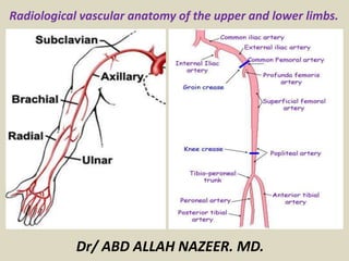
- 1. Radiological vascular anatomy of the upper and lower limbs. Dr/ ABD ALLAH NAZEER. MD.
- 2. The arterial supply to the upper limb begins in the chest as the subclavian artery. The right subclavian artery arises from the brachiocephalic trunk, while the left subclavian branches directly off the arch of aorta. When the subclavian arteries cross the lateral edge of the 1st rib, they enter the axilla, and are called axillary arteries.
- 3. The axillary artery passes through the axilla, just underneath the pectoralis minor muscle, enclosed in the axillary sheath. At the level of the humeral surgical neck, the posterior and anterior circumflex humeral arteries arise. They circle posteriorly round the humerus to supply the shoulder region. The largest branch of the humerus also arises here; the subscapular artery. The axillary artery becomes the brachial artery at the level of the teres major muscle
- 4. In the Upper Arm When the axillary artery reaches the lower border of the teres major, it becomes the brachial artery. The brachial artery is the main source of blood for the arm. Immediately distal to the teres major, the brachial artery gives rise to the profunda brachii – the deep artery of the arm. It travels along the posterior surface of the humerus, running in the radial groove. It supplies structures in the posterior aspect of the arm (e.g the triceps brachii, and terminates by contributing to a network of vessels at the elbow joint. The brachial artery descends down the arm immediately posterior to the median nerve. As it crosses the cubital fossa, underneath the brachialis muscle, the brachial artery terminates by bifurcating into the radial and ulnar arteries.
- 5. In the Forearm In the distal region of the cubital fossa, the brachial artery bifurcates into the radial artery and the ulnar artery. The radial artery supplies the posterior aspect of the forearm and the ulnar artery supplies the anterior aspect. The two arteries anastamose in the hand, by forming two arches, the superficial palmar arch, and the deep palmar arch.
- 6. In the Hand The hand has a very good blood supply, with many anastomosing arteries, allowing the hand to be perfused when grasping or applying pressure. A good majority of these arteries are superficial, allowing for heat loss when needed. In the hand, the ulnar and radial arteries interconnect to form two arches, from which branches to the digits emerge. Radial artery – contributes mainly to supply of the thumb and the lateral side of the index finger Ulnar artery – contributes mainly to the supply of the rest of the digits, and the medial side of the index finger.
- 7. CTA of the left upper limb – CPR reconstruction. No contrast enhancement in the distal part of the brachial artery, for approx. 40 mm. Contrast enhancement of the forearm arteries.
- 8. CT angiography showing bones and vessels of the chest, neck and upper extremities.
- 9. MDCT angiography of the upper right limb. 1. Axillary artery; 2. Common trunk of circumflex humeral arteries; 3. Circumflex scapular artery; 4. Brachial artery; 5. Superficial radial artery (at the level of the arm); 5. Superficial radial artery (at the level of the forearm); 6. Profunda brachii artery; 7. Inferior recurrent radial artery; 8. Inferior recurrent ulnar artery; 9. Posterior interosseous artery; 10. Anterior interosseous artery.
- 10. Normal upper extremity CT angiogram.
- 11. MRA images of the upper limb arteries.
- 12. Upper extremity CE-MRA: A) Arterial acquisition, and B) Venous acquisition
- 13. CE-MRA acquisition in arterial phase (A) and venous phase (B) with corresponding cross-sectional reformations
- 15. Magnetic resonance angiography (MRA) of the hand.
- 17. Selective angiogram of right upper extremity.
- 18. Angiogra m of the radial artery.
- 19. Superficial Veins The major superficial veins of the upper limb are the cephalic and basilic veins. As their name suggests, they are located within the subcutaneous tissue of the upper limb. The basilic vein originates from the dorsal venous network of the hand. It ascends the medial aspect of the upper limb. At the border of the teres major, the vein moves deep into the arm. Here, it combines with the brachial veins to form the axillary vein. The cephalic vein arises from the dorsal venous network of the hand. It ascends the antero-lateral aspect of the upper limb, passing anteriorly at the elbow. At the shoulder, the cephalic vein travels between the deltoid and pectoralis major muscles (known as the deltopectoral groove), and enters the axilla region via the clavipectoral triangle. Within the axilla, the cephalic vein terminates by joining the axillary vein. At the elbow, the cephalic and basilic veins are connected by the median cubital vein.
- 20. Deep Veins The deep veins of the upper limb are situated underneath the deep fascia. They are paired veins that accompany and lie either side of an artery. The brachial veins are the largest in size, and are situated either side of the brachial artery. The pulsations of the brachial artery aids the venous return. Veins that are structured in this way are known as vena comitantes. Perforating veins run between the deep and superficial veins of the upper limb, connecting the two systems.
- 21. The superficial veins of the upper extremity are shown in blue. The deep veins are shown in blue.
- 22. Basic deep venous anatomy of the arm. Basic superficial venous anatomy of the arm.
- 23. The red line shows the subclavian vein origin scan plane. Doppler of the Subclavian vein origin.
- 29. lower limb vessels. Femoral Artery. The main artery of the lower limb is femoral artery. It is a continuation of the external iliac artery (terminal branch of the abdominal aorta). The external iliac becomes the femoral artery when it crosses under the inguinal ligament and enters the femoral triangle. In the femoral triangle, the profunda femoris artery arises from the posterolateral aspect of the femoral artery. It travels posteriorly and distally, giving off three main branches: Perforating branches – Consists of three or four arteries that perforate the adductor magnus, contributing to the supply of the muscles in the medial and posterior thigh. Lateral femoral circumflex artery – Wraps round the anterior, lateral side of the femur, supplying some of the muscles in the lateral side of the thigh. Medial femoral circumflex artery – Wraps round the posterior side of the femur, supplying the neck and head of the femur. In a fracture of the femoral neck, this artery can easily be damaged, and avascular necrosis of the femur head can occur.
- 34. The course and branching of the common iliac artery into the external iliac artery becoming femoral artery. Of particular interest is the course of the femoral artery at the hip and knee joints. The sagittal maximum intensity projection (MIP) CT image (middle) and coronal (right) better demonstrate its course
- 36. CTA of the lower limb arteries.
- 37. Computed tomography angiography curved multiplanar reformat of the left lower extremity arterial vessels.
- 38. Axial images obtained at the level of abdominal aorta (a), aortic carrefour (b), common iliac arteries (c), and external and internal iliac arteries (d)
- 39. Axial images obtained at the level of common femoral artery (e), femoral bifurcation (f-h).
- 40. Axial images obtained at the level of proximal popliteal artery (i), popliteal artery (j), distal popliteal artery (k), and anterior tibial artery and tibio-peroneal trunk (l)
- 41. Axial images obtained at the level of anterior tibial, posterior tibial and peroneal arteries (m, n) and pedideal artery and plantar arch (o)
- 42. MRA images.
- 45. Magnetic resonance angiogram (MRA) obtained by using the bolus-chase technique shows the normal anatomy of the lower extremity arterial vasculature, including the aorta (a), the common iliac artery (b), the external iliac artery (c), the internal iliac artery (d), and the common femoral artery (e).
- 47. Magnetic resonance angiogram (MRA) obtained by using the bolus-chase technique shows the normal anatomy of the lower-extremity arterial vasculature, including the deep femoral artery (a) and the superficial femoral artery (b).
- 48. Superficial and deep femoral arteries of the thigh.
- 50. Magnetic resonance angiogram (MRA) obtained by using the bolus-chase technique shows the normal anatomy of the lower-extremity arterial vasculature, including the popliteal artery (a), the anterior tibial artery (b), the tibioperoneal trunk (c), the peroneal artery (d), and the posterior tibial artery (e).
- 51. Normal lower extremity CTA using maximum intensity projection (MIP) display and curved planar reformation (CPR). (A) Complete topographic MIP display of aortoiliac inflow and femoropopliteal and tibioperoneal outflow segments. (B) MIP display of normal aortoiliac inflow segment. (C) MIP display of femoral and above knee popliteal segment. (D) MIP display of the distal popliteal and tibioperoneal segments. (E) CPR in lateral projection of the right aortoiliac arterial inflow segment. (F) CPR in lateral projection of left aortoiliac arterial inflow segment.
- 52. The obturator artery arises from internal iliac artery in the pelvic region. It descends via the obturator canal to enter the medial thigh, bifurcating into two branches: Anterior branch – This supplies the pectineus, obturator externus, adductor muscles and gracilis. Posterior branch – This supplies some of the deep gluteal muscles. The gluteal region is largely supplied by the superior and inferior gluteal arteries. These arteries also arise from the internal iliac artery, entering the gluteal region via the greater sciatic foramen. The superior gluteal artery leaves the foramen above the piriformis muscle, the inferior below the muscle. In addition to the gluteal muscles, the inferior gluteal artery also contributes towards the vasculature of the posterior thigh.
- 54. The popliteal artery descends down the posterior thigh, giving off genicular branches that supply the knee joint. It moves through the popliteal fossa, exiting sandwiched between the gastrocnemius and popliteus muscles. At the lower border of the popliteus, the popliteal artery terminates by dividing into anterior and posterior tibial arteries. The posterior tibial artery continues inferiorly, along the surface of the deep muscles (such as tibialis posterior). It accompanies the tibial nerve in entering the sole of the foot via the tarsal tunnel. During the descent of the posterior tibial artery in the leg, the fibular artery arises. This artery moves laterally, penetrating the lateral compartment of the leg. It supplies muscles in the lateral compartment, and adjacent muscles in the posterior compartment. The other division of the popliteal artery, the anterior tibial artery, passes anteriorly between the tibia and fibula, through a gap in the interosseous membrane. It then moves inferiorly down the leg. It runs down the entire length of the leg, and into the foot, where it becomes the dorsalis pedis artery.
- 57. Dorsalis pedis (a continuation of the anterior tibial artery) Posterior tibial The dorsalis pedis artery begins as the anterior tibial artery enters the foot. It passes over the dorsal aspect of the tarsal bones, then moves inferiorly, towards the sole of the foot. It then anastamoses with the lateral plantar artery to form the deep plantar arch. The dorsalis pedis artery supplies the tarsal bones and the dorsal aspect of the metatarsals. Via the deep plantar arch, it also contributes to the supply of the toes. The posterior tibial artery enters the sole of the foot through the tarsal tunnel. It then splits into the lateral and medial plantar arteries. These arteries supply the plantar side of the foot, and contributes to the supply of the toes via the deep plantar arch.
- 61. Angiography with bilateral external iliac artery (EIA), common femoral artery (CFA) and superficial femoral artery (SFA). Bilateral internal iliac artery (IIA) continues as the inferior gluteal artery and persistent sciatic artery (PSA).
- 62. Angiogram of the superficial and deep femoral arteries.
- 63. Angiography of the popliteal artery (A) and posterior tibial artery .
- 66. The Deep Veins of the Lower Limb The deep venous drainage system of the lower limb is located beneath the deep fascia of the lower limb. As a general rule, the deep veins accompany and share the name of the major arteries in the lower limb. Often, the artery and vein are located within the same vascular sheath – so that the arterial pulsations aid the venous return. The Foot and Leg The main venous structure of the foot is the dorsal venous arch, which mostly drains into the superficial veins. Some veins from the arch penetrate deep into the leg, forming the anterior tibial vein. On the plantar aspect of the foot, medial and lateral plantar veins arise. These veins combine to form the posterior tibial and fibular veins. The posterior tibial vein accompanies the posterior tibial artery, entering the leg posteriorly to the medial malleolus. On the posterior surface of the knee, the anterior tibial, posterior tibial and fibular veins unite to form the popliteal vein. The popliteal vein enters the thigh via the adductor canal
- 67. The Thigh Once the popliteal vein has entered the thigh, it is known as the femoral vein. It is situated anteriorly, accompanying the femoral artery. The deep vein of the thigh is the other major venous structure. Via perforating veins, it drains blood from the thigh muscles. It then empties into the distal section of the femoral vein. The femoral vein leaves the thigh by running underneath the inguinal ligament, at which point it is known as the external iliac vein. The Gluteal Region The gluteal region is drained by inferior and superior gluteal veins. These empty into the internal iliac vein
- 70. The Superficial Veins of the Lower Limb The superficial veins of the lower limb run in the subcutaneous tissue. There are two major superficial veins – the great saphenous vein, and the small saphenous vein. The Great Saphenous Vein The great saphenous vein is formed by the dorsal venous arch of the foot, and the dorsal vein of the great toe. It ascends up the medial side of the leg, passing anteriorly to the medial malleolus at the ankle, and posteriorly to the medial condyle at the knee. As the vein moves up the leg, it receives tributaries from other small superficial veins. The great saphenous vein terminates by draining into the femoral vein immediately inferior to the inguinal ligament. Surgically, the great saphenous vein can be harvested and used as a vessel in coronary artery bypasses. The Small Saphenous Vein The small saphenous vein is formed by the dorsal venous arch of the foot, and the dorsal vein of the little toe. It moves up the posterior side of the leg, passing posteriorly to the lateral malleolus, along the lateral border of the calcaneal tendon. It moves between the two heads of the gastrocnemius muscle and empties into the popliteal vein in the popliteal fossa.
- 73. Basic deep venous anatomy of the leg Patient position
- 74. The blue line shows the CFV scan plane. The CFV, pre and post compression.
- 75. The blue line shows the distal FV scan plane. (Superficial) Femoral Vein.
- 76. The blue arrow shows the POPV scan direction. POPV.
- 77. Transverse mid calf. Transverse view of the posterior tibial and peroneal veins. Pre & Post compression.
- 78. A sagittal scan plane for the calf veins. Longitudinal calf veins. Split screen, color Doppler and B-mode.
- 80. Thank You.