SlideShare a Scribd company logo
1 of 120
Download to read offline
Dr/ ABD ALLAH NAZEER. MD.
Ultrasound examination of the uterus and ovaries.
ULTRASOUND OF THE UTERUS – Normal.
Uterus TA probe positioning
for longitudinal scan. Uterus sagittal US image.
Uterus TA probe positioning
for transverse scan.
Trans abdominal view of the uterus:
transverse. Both ovaries are visible
Transvaginal Technique
Anteverted uterus. Normal TV image anteverted sagittal.
Retroverted transvaginal technique Retroverted uterus transvaginal scan.
Sagittal US image of the uterus
obtained during the proliferative
phase of the menstrual cycle
demonstrates the endometrium
with a multilayered appearance.
Normal premenopausal endometrium.
Sagittal US image of the uterus
obtained during the secretory phase of
the menstrual cycle shows a thickened,
echogenic endometrium.
ENDOMETRIAL MEASUREMENT.
Coronal 3D image of a Mirena IUD shows the
expected location of the shaft and crossbars
simultaneously in the body and fundus of the
uterus. The endometrium is also seen well
without the normal shadowing of the mirena
always seen in the sagittal plane.
The string can be difficult to
identify on the 3D in the cervix
but a 2D sagittal scan should
easily show its placement.
ROLE OF ULTRASOUND:
To examine the uterus, ovaries, cervix, vagina and adnexae.
Classification of a mass identified on other modalities e.g solid, cystic, mixed.
Post surgical complications e.g abscess, edema.
Guidance of injections, aspiration or biopsy.
Assistance with IVF.
To identify the relationship of normal anatomy and pathology to each other.
INDICATIONS:
P/V bleeding/discharge, Menorrhagia, Metrorrhagia (irregular uterine
bleeding), Polymenorrhea, Menometrorrhagia (excessive irregular bleeding)
Amenorrhea, Oligoamenorrhea, Pelvic pain, Dysmenorrhea (Painful Menses)
F/H uterine or ovarian Cancer, Palpable lump, Infertility- primary or secondary
(evaluation, monitoring and/or treatment), Anomalies/evaluation, Follow-up
of previous abnormality, Precocious Puberty, delayed menses or vaginal
bleeding in a prepubertal child. postmenopausal bleeding, Signs/symptoms of
pelvic infection, IUCD Localization (intrauterine contraceptive Device)
Guidance for interventional or surgical procedures.
urinary incontinence or pelvic organ prolapse.
COMMON PATHOLOGIES
VAGINAL
Gartners duct cyst, Epidermal inclusion cysts, Bartholin Gland Cysts, Vaginal
carcinoma, Hydro/hematocolpos (secondary to imperforate hymen or vaginal
stenosis), Foreign body, leiomyoma and endometriosis.
CERVICAL
Nabothian (retention) cysts, Polyps, Cervical fibroids, Cervical carcinoma, Cervical stenosis
UTERINE
Fibroids (leiomyoma), submucosal, intramural, subserosal, pedunculated
Leiomyosarcoma
Adenomyosis
Lipoleiomyoma
ENDOMETRIAL
Endometrial Polyps, Endometrial Carcinoma, Endometrial hyperplasia
Endometritis, endometriosis.
Cystic hyperplasia secondary to Tamoxifen
Adhesions- Ashermans Syndrome
Submucosal fibroids
Arterio-venous malformation (AVM)
Hydro/hematometra
Blood/fluid/infection or retained products of conception (RPOC)
Common cystic lesions of the vagina and their origins.
Gartner duct cysts are embryologic secretory
retention cysts that arise from the residual wolffian
(mesonephric) duct remnant usually solitary and less
than 2 cm, most commonly develop in the
anterolateral wall of the proximal one-third of the
vagina; may contain septa; when located at the level
of the urethra, they can cause mass effect on the
urethra, giving rise to urinary tract symptoms; often
associated with other wolffian abnormalities
(unilateral renal agenesis, renal hypoplasia, and
ectopic ureteral insertion); although usually
asymptomatic, cyst aspiration, tetracycline
sclerotherapy, or surgical excision may be used to
treat larger lesions.
Gartner’s cyst (*) in a young woman with dysuria imaged with (a) suprapubic, (b) trans-
labial approach and (c) reconstructed images from translabial volume acquisition.
Posterior urethral diverticula (*) creating mass effect on the anterior wall
of the vagina in a woman with dysuria, and severe dyspareunia imaged
during voiding. The inhomogeneous appearance is due to turbulent flow.
u, urethra; b, bladder; ant and post, anterior and posterior vaginal walls.
Oblique sagittal transabdominal US (a) and coronal T1-weighted MR (b) showing
hematocolpos (*) and hematosalpinges (s) in bicornuate-unicollis uterus with associated
hypoplasia of the cervix and imperforate hymen in a young woman with cyclic pelvic pain
and primary amenorrhea. Dotted line represents the orientation of the US scan.
Imperforate hymen is the most common congenital obstructive abnormality of
the female genital tract, with a frequency that can vary from 1 case per 1000
population to 1 case per 10,000 population; most commonly occurs in
isolation; occurs due to a failure of recanalization of this membranous vestige;
Axial and sagittal scans at different levels (schemes on the right) show the presence
of a didelphys uterus and vaginal duplication with noncanalized right emisystem. A:
axial section of the pelvis at the level of uterine body. On the right: hematometra
(*); on the left, hypoplastic body without hematometra. bl, bladder; B: axial plane of
the pelvis at the level of the vagina. Right vaginal lumen distended by hematic
material; left vagina is undilated and opened. C: right paramedian sagittal plane on
the right uterus and vagina showing hematometra (hm) and hematocolpos (hc). D:
left paramedian sagittal plane on the left hypoplastic uterus and vagina.
Reconstructed axial image from translabial volumetric acquisition showing a common
asymptomatic cyst (calipers) in the posterior vaginal wall. U, urethra; R, rectum.
Epidermal inclusion cysts (also termed “sebaceous cysts”) are the most
common type of non-embryological cyst; vary in size from a few millimeters to
several centimeters; mostly asymptomatic; their location almost always
correlates with a site of previous surgery and represents a traumatic inclusion
of normal vaginal mucosa following vaginal laceration or episiotomy.
Leiomyomas
Leiomyomas are an uncommon entity in the vagina with
only about 300 reported cases in literature; commonly
seen in the age group ranging from 35 to 50 years; may
originate in the smooth muscle of the vagina, local arterial
musculature, or smooth muscle of the bladder or urethra;
however, midline of the anterior vaginal wall is the most
common location; at US, their characteristics are similar
to uterine leiomyomas, showing a heterogeneous but
predominately hypoechoic and well circumscribed
appearance.
at CEUS examination, the enhancement of leiomyomas is
uniform, as opposed to the heterogeneous enhancement
of leiomyosarcoma.
Leiomyoma of the vaginal wall. TVUS axial (A) and sagittal (B) scans demonstrate a solid,
inhomogeneously hypoechoic, well-defined lesion (*) deforming the outer vaginal wall. At
CEUS (C-F) a slow enhancement followed by a relative hypo enhancement is observed.
Translabial sagittal US scan (a) showing a myoma (*) of the anterior vaginal
wall (V, vagina, b, bladder) . Contrast-enhanced US sequences (b-e)
demonstrate high but homogenous enhancement of the lesion (arrow).
Sagittal US scan with a linear probe of a slow-growth inhomogeneous solid mass of the right
major labrum. Dynamic CEUS (D, E and F) shows a ready enhancement of the lesion. The lesion
appeared hyperintense in T2-fatsat MR sequences (G) and iso-hypointense on T1 (H). At
microscopical examination the lesion was demonstrated to be an angiomixoma of the vulva.
Primary vaginal malignancies are relatively rare, representing about 1%–2% of all
gynecologic malignancies and less than 20% of vaginal malignancies. primary cancers are
defined as having no involvement of the external os of the cervix or the vulva inferiorly
Cancer vagina with translabial sagittal US (a) of the vagina in a patient with
vaginal bleeding and dyspareunia. A large, solid, inhomogeneous vaginal
mass (arrowheads) is demonstrated with compression of the urethra (u).
Sagittal (b) and coronal (c) T2-weighted MR images show inhomogeneous
signal intensity of the mass and micro-infiltrative behavior.
Bartholin Gland Cysts
Bartholin’s glands secretions provide to lubricate vulvar vestibule with
ducts that drain at the 4 and 8 o’ clock positions of the vaginal vestibule.
Bartholin gland cysts are the most common vulvar cysts, developing in 2%
of women during their lifetime; located at the posterolateral vaginal
introits, medial to the labia minora; their location at or below the level of
the pubic symphysis helps differentiate them from Gartner duct cysts;
range in size from 1 to 4 cm (but can increase with repeated sexual
stimulation develop as a result of an obstruction of the gland’s duct by a
stone or a stenosis related to prior infection or trauma); patients are often
asymptomatic but can present with mild dyspareunia, typically in the 2nd
to 3rd decade of life; superimposed infection or abscess formation may
require drainage; a rare complication is development of squamous car-
cinoma or adenocarcinoma in a Bartholin's gland duct or cyst,
respectively; at US, simple cysts are anechoic, thin-walled and unilocular,
but septations can be seen. the cyst wall may be thickened and, if infected,
enhancement can be observed at CEUS examination; visualization of any
solid component within the cyst should raise concern for malignancy.
US coronal scan of a Bartholin’s cyst in a patient with discomfort and dyspareunia. A cystic
mass (*) in (a) is demonstrated in the right major labrum. Color-Doppler evaluation (b) shows
an implementation of vascular signs related to hyperemia and inflammation. V, vagina.
Sagittal translabial US (a). Endometriotic implant (*) in the rectovaginal
septum in a young woman presenting with elective pain at palpation.
Real-time elastography (b) demonstrates hard stiffness (red) due to
perilesional fibrotic reaction. U, uterus; PF, posterior vaginal fornix.
Vaginal endometriosis consists in the presence of ectopic endometrial glandular
and stromal tissue within the vaginal walls. May occur during surgical procedures
or from the spread of a recto-vaginal pouch endometriosis; often associated with
dysmenorrheal, postcoital spotting, dyspareunia and infertility; the condition is
almost always associated with endometriosis in other pelvic locations.
Vaginal cuff represents the apex of the vagina where the apposed upper walls are
sutured together; is a site of recurrent gynecologic malignancy after hysterectomy
(seen most often with cervical carcinoma, but it also occurs with endometrial
cancer and rarely with ovarian cancer); postoperative anteroposterior vaginal cuff
size quoted in the literature is up to 2.1 cm; at pelvic US, normal vaginal cuff is
generally small, symmetric, and homogeneously hypoechoic.
Transvaginal US and
color-Doppler scan of a
recurrent gynecologic
malignancy (arrows)
on the vaginal cuff
after hysterectomy.
Vaginal varices. Patient with severe dyspareunia and dysuria.
Color Doppler evaluation with translabial approach shows a high
flow within the uterine-vaginal planes with venous ectasia.
Vagina is an unlikely location to develop varices, because of the presence of a
bilateral draining system (similar to uterus), which largely communicates with
the uterine, vesical and hemorroidal plexuses; vaginal varices are usually
found in cirrhotic patients or after hysterectomy; color-Doppler US reveals
venous flow inside these hypoechoic, serpiginous lesions.
Nabothian cysts (also known as a retention cysts of the cervix) are non-
neoplastic cystic lesion that occurs in relation to the uterine cervix.
Pelvic ultrasound
Typically seen an anechoic well defined cystic lesions near the
endocervical canal. If the cysts are large, the cervical region can appear
enlarged Colour Doppler interrogation shows no associated colour flow.
Cervical Nabothian cyst.
Cervical polyp in ultrasound image.
A cervical polyp is a common benign polyp or tumour on the surface of the cervical
canal. They can cause irregular menstrual bleeding but often show no symptoms.
Cervical fibroid.
Cervical myoma are benign tumors composed partly of muscle tissue. They
seldom develop in the cervix, the lower part of the uterus. When they do, they
are usually accompanied by myoma in the larger upper part of the uterus.
Sagittal transabdominal US (a) and color-Doppler (b) demonstrating a large
intracavitary pedunculated myoma (*) protruding in vagina from cervical os.
The vascular pedicle is depicted at color-Doppler US. b, bladder; c, cervix.
Carcinoma of the cervix is a malignancy arising from the cervix and is
considered the third most common gynecologic malignancy.
Ultrasound
hypoechoic, heterogeneous mass involving the cervix
may show increased vascularity on colour Doppler
although cervical cancer is staged clinically, ultrasound can be a useful
adjunct by showing
size (<4 cm or >4 cm)
parametrial invasion
tumour invasion into the vagina
tumor invasion into adjacent organs
hydronephrosis: implies stage IIIB tumour.
This sagittal transvaginal color Doppler sonogram
shows prominent vascular flow in the cervical tumor.
Transvaginal and Doppler study of cancer cervix.
Sagittal and transverse transvaginal sonogram
shows a circumscribed hypoechoic cancer cervix.
Uterine leiomyomas (uterine fibroids) are benign tumours of
myometrial origin and are the most common solid benign uterine
neoplasm. Commonly an incidental finding on imaging, they rarely
cause a diagnostic dilemma. They occur in ~25% of women of
reproductive age and are particularly common in the African
population.
Fibroids may have a number of locations within or external to the
uterus:
intra-uterine
intramural leiomyoma: most common
subserosal leiomyoma
submucosal leiomyoma: least common (10-15%)
extra-uterine
broad ligament leiomyoma
cervical leiomyoma
parasitic leiomyoma
diffuse uterine leiomyomatosis
Pelvic ultrasound
ultrasound is used to diagnose the presence and monitor the growth of fibroids
uncomplicated leiomyomas are usually hypoechoic, but can be isoechoic, or even
hyperechoic compared to normal myometrium, calcification is seen as echogenic
foci with shadowing, cystic areas of necrosis or degeneration may be seen
Subserosal leiomyoma.
Subserosal Leiomyoma.
Intra-mural uterine leiomyoma.
TA USG image (A) shows a 7-cm intramural fibroid containing cystic areas
(arrows). Axial T2W MRI image (B) shows an 11 × 8 cm fibroid (arrows)
containing central high signal, consistent with cystic degeneration (arrowhead)
Transabdominal (TA) USG image (A) shows a bulky uterus showing a 10-cm
submucosal fibroid (between cursors). Sagittal T2W MRI image (B) in the same
patient shows that the submucosal fibroid (arrowhead) is heterogeneous
indicating degeneration. There is also a 2.5-cm cervical fibroid (arrow)
Ultrasound image shows sub mucosal fibroid Picture
B. MRI image shows multiple intramural fibroids.
Multiple uterine fibroid.
Endometrial polyps are benign nodular protrusions of the endometrial surface, and one
of the entities included in a differential of endometrial thickening. Endometrial polyps
can either be sessile or pedunculated. They can often be suggested on ultrasound or MRI
studies, but may require sonohysterography or direct visualization for confirmation.
Ultrasound.
Although endometrial polyps may be visualized at transvaginal ultrasound as nonspecific
endometrial thickening, they may also be identified as focal masses within the
endometrial canal. a stalk to the polyp may either be thin or broad based a feeding
vessel may be seen extending to the polyp on colour Doppler imaging cystic spaces
corresponding to dilated glands filled with proteinaceous fluid may be seen within the
polyp and is considered a relatively characteristic feature may appear as just diffusely
thickened endometrium, without visualization of a discrete mass (mimicking endometrial
hyperplasia)
3D ultrasound may be useful to help delineate the borders of a polyp.
Sonohysterography .
Polyps are best characterized on sonohysterography and appear as echogenic, smooth,
intra-cavitary masses outlined by fluid. The typical appearance of an endometrial polyp
at sonohysterography is as a well-defined, homogeneous, polypoid lesion that is
isoechoic to the endometrium with preservation of the endometrial-myometrial interface
There is usually a well-defined vascular pedicle within the stalk.
Colour Doppler interrogation may show flow within the stalk.
Endometrial polyp.
TVS of an endometrial polyp hidden
within a secretorial endometrium
After transcervical instillation of saline
(SISH), applying power Doppler, a polyp with
single vessel arrangement can be seen.
Images show endometrial polyp within the uterine cavity
with its vascular pedicle visible on color Doppler scans.
Multiple endometrial polyp.
Endometrial hyperplasia (EH) refers to an abnormal proliferation
of endometrial stromal and glandular components. It represents a
spectrum of endometrial changes ranging from cystically dilated
glands to cellular atypia.
Ultrasound
premenopausal
normal endometrial thickness depends on the stage of the
menstrual cycle, but a thickness of >10 mm is top normal or
abnormal
hyperplasia can be reliably excluded in patients only when the
endometrium measures less than 6 mm
postmenopausal
a thickness of >5 mm is considered abnormal
The appearance can be non-specific and cannot reliably allow
differentiation between hyperplasia and carcinoma. Usually there is
a homogeneous increase in endometrial thickness, but endometrial
hyperplasia may also cause asymmetric/focal thickening with surface
irregularity, an appearance that is suspicious for carcinoma.
Endometrial hyperplasia.
Endometrial carcinoma is generally considered the most common
gynecological malignancy. It frequently present with vaginal
bleeding and both ultrasound and pelvic MRI are useful modalities
for evaluation.
Transvaginal ultrasound
Endometrial carcinoma usually appears as thickening of the
endometrium though may appear as a polypoid mass
premenopausal: normal endometrial thickness varies through the
menstrual cycle
diagnosing abnormally thickened endometrium depends on
knowing what the patient's point in the menstrual cycle
postmenopausal: >5 mm is thickened (if not on Tamoxifen)
Sonographic features are non specific and endometrial thickening
can also be due to benign proliferation, endometrial hyperplasia or
polyps.
Disruption of a subendometrial halo on ultrasound may be
suggestive of myometrial involvement.
Endometrial carcinoma.
Endometrial Carcinoma.
Multiple vessels in women
with endometrial cancer.
Stage IC endometrial carcinoma. Sagittal TVUS
image of the uterus shows marked, inhomogeneous
endometrial thickening(thick arrows ).
Endometritis refers to inflammation of the endometrium, the inner
lining of the uterus. Pathologists have traditionally classified endometritis
as either acute or chronic.
Ultrasound
While the sonographic appearance of the uterus and endometrium may be
normal in early stages, findings may include a thickened, heterogeneous
endometrium, intracavitary fluid +/- intrauterine air.
Sagittal image of the uterus showing complex fluid within the endometrial cavity. Endometritis.
Endometriosis is a hormonal and immune system disease in which
cells similar to that which line the uterus (endometrium) grow
outside the uterine cavity, most commonly on the membrane which
lines the abdominal cavity, the peritoneum.
Ovarian endometrioma.
Endometriosis.
Uterine arteriovenous malformations (UAVM)
result from formation of multiple arteriovenous fistulous
communications within the uterus without an intervening
capillary network.
Ultrasound
Gray-scale sonographic appearances can be non-specific and
can have a range of manifestations including areas of subtle
myometrial inhomogeneity, tubular spaces within the
myometrium, a intramural uterine, endometrial or cervical
mass like region or sometimes as prominent parametrial
vessels. The extent of mass is effect is however minimal.
Colour Doppler
Typically shows serpiginous/tubular anechoic structures within
the myometrium with a low resistance (RI ~0.2-0.5), high
velocity flow pattern on colour Doppler interrogation.
Uterine AVM.
Uterine arterio-venous malformation. In addition
a large disproportionately dilated vascular.
ULTRASOUND OF THE OVARIES – Normal.
Use the full urinary bladder
as an acoustic window to
angle across to the ovary.
Axial trans-abdominal image with
the ovary lateral to the uterus.
Axial trans-vaginal scan plane. Axial trans-vaginal image.
Normal transvaginal ovary demonstration
normal peripheral follicles.
The uterus may be oblique and squash the
ovary giving it a flattened ovoid shape.
ROLE OF ULTRASOUND
To examine the uterus, ovaries cervix vagina and adnexae.
Classification of a mass identified on other modalities e.g solid, cystic, mixed.
Post surgical complications e.g abscess, edema.
Guidance of injections, aspiration or biopsy.
Assistance with IVF.
To identify the relationship of normal anatomy and pathology to each other.
INDICATIONS
P/V bleeding/discharge
Menorrhagia
Polymenorrhea
Amenorrhea
Irregular periods
Pelvic pain
F/H uterine or ovarian Cancer
Palpable lump
Infertility- primary or secondary
Anomalies
COMMON PATHOLOGIES
OVARIAN
Ovarian cysts
simple Vs complex (hemorrhagic, corpus luteal, ruptured, septated).
any mural nodules
Dermoid
Ovarian tumours:
Cystadenoma (serous/mucinous)-Benign
Cystadenocarcinoma (serous/mucinous)-Malignant
Polycystic Ovarian Disease
Endometrioma
Torsion
Hyper stimulation syndrome
Ectopic pregnancy
POUCH OF DOUGLAS (POD) & ADNEXAE
Fluid, Pus, Blood
Pelvic inflammatory disease-PID (may be indicated by above conditions)
Cysts (Mesenteric)
Ectopic pregnancy
Endometriosis
Pelvic venous congestion
FALLOPIAN TUBES
PID, Pyosalpinx, Hydrosalpinx, Ectopic pregnancy, Cyst, Ectopic pregnancy, Endometriosis
An ovarian cyst is any collection of fluid, surrounded by a very thin wall, within
an ovary . Any ovarian follicle that is larger than about two centimeters is termed
an ovarian cyst. The vast majority of ovarian cysts are harmless (benign ), most
are functional cysts. Ovarian cysts occur in women of all ages including neonatal
period and infancy. They are most prevalent during infancy, adolescence and
during the childbearing years.
Types of cysts
physiological cysts: mean diameter ≤3.0 cm( ovarian follicle)( corpus luteum).
functional cysts (can produce hormones):
follicular cysts of the ovary (estrogen): >3.0 cm
corpus luteum cysts (progesterone)
theca lutein cyst: gestational trophoblastic disease
complications in functional cysts:
Hemorrhage, enlargement, rupture and torsion
other cysts:
multiple large ovarian cysts in ovarian hyperstimulation syndrome
post-menopausal cyst: serous inclusion cysts of the ovary
polycystic ovaries
ovarian torsion
ovarian cystic neoplasms
Left ovarian cyst simple cyst.
Radiographic features
Simple ovarian follicular cysts
anechoic
intraovarian or exophytic
imperceptible wall
posterior acoustic enhancement (may not be as obvious with harmonic or
compound imaging)
visible far wall
Simple ovarian cyst ultrasound.
Simple looking cyst measuring 8.7cm in maximal diameter.
Hemorrhagic ovarian cyst.
Hemorrhagic ovarian cysts (HOCs) usually result from hemorrhage into a corpus luteum
or other functional cyst. Radiographic features are variable depending on the age of the
hemorrhage. They typically resolve within eight weeks.
HOCs can have a variety of appearances depending on the stage of evolution of the blood
products and clot. lace-like reticular echoes or an intracystic solid clot, a fluid-fluid level is
possible. thin wall, clot may adhere to cyst wall mimicking a nodule, but has no blood flow
on Doppler imaging, retracting clot may have sharp or concave borders, mural nodularity
does not, posterior acoustic enhancement, may be less noticeable if harmonics or
compounding is used, there should not be any internal blood flow, circumferential blood
flow in the cyst wall is typical.
US and power Doppler mode in a hemorrhagic
cyst demonstrates the absence of blood vessels.
Hemorrhagic ovarian cyst.
A corpus luteal (CL) cyst is a type of functional ovarian cyst which results when a
corpus luteum fails to regress following the release of an ovum. When associated
with pregnancy, it is the most common pelvic mass encountered within the 1st
trimester. They can have a range of sonographic appearances depending on the
stage of evolution and age of the associated intracystic hemorrhage. General
characteristics include: diffusely thick wall, peripheral vascularity, <3 cm
possible crenulated contour.
Hemorrhagic corpus luteal cyst.
Corpus luteum ovarian cyst.
Corpus luteum
ovarian cyst
Corpus luteum ovarian cyst.
Septated ovarian cyst.
A dermoid cyst is a cystic teratoma that contains an array of developmentally
mature, solid tissues. It frequently consists of skin, complete with hair follicles,
and sweat glands. Other commonly found components include: clumps of long
hair, pockets of sebum, blood, fat, bone, nails, teeth, eyes, cartilage, and thyroid
tissue. Ultrasound images show diffusely or partially echogenic mass with
posterior sound attenuation owing to sebaceous material and hair within the cyst
cavity, echogenic interface at edge of mass that obscures deep structures: the tip
of the iceberg sign, mural hyperechoic Rokitansky nodule: dermoid plug, echogenic,
shadowing calcific or dental (tooth) components, presence of fluid-fluid levels.
Dermoid cyst.
Dermoid ovarian cyst.
Ovarian dermoid cyst with Rokitansky nodule.
Complicated septated ovarian cyst.
Endometrioma right ovary with homogenous distribution of low to medium fine echoes in a
cystic mass with only one septation, no solid components. No blood flow within the mass,
Ovarian serous cyst adenomas (OSC) are a type of benign ovarian
epithelial tumour.
Ultrasound
usually seen as a unilocular cystic adnexal lesion
papillary projections are absent
if there is any wall irregularity, it is thin, with an acute angle with the cyst
wall and have a regular surface
some lesions may contain sonographically detectable septations.
Bilateral ovarian serous cystadenoma.
Left ovarian papillary serous cystoma in a patient of 55 years, RI above
0.42, sharp margins, thin internal septations, moderate vascularization
Mucinous cystadenoma images.
Ovarian cystadenocarcinoma.
Ovarian serous cystadenocarcinoma is an ovarian epithelial tumor
at the malignant end of the spectrum of ovarian serous tumours.
Ultrasound: More heterogeneous in appearance than a serous cystadenoma
papillary projections, thick septations, and/or solid components
presence of ascites concerning for peritoneal metastatic spread, discrete peritoneal
deposits may be seen.
Colour Doppler is useful to confirm vascularity of the solid components quantitative
parameters (resistive index and pulsatility index) do not reliably predict malignancy.
Ovarian serous cystadenocarcinoma. A. Coronal TVUS image with
superimposed color Doppler of a left ovarian mass shows a cystic mass
with a large vascular solid component. B. Spectral Doppler image of the
same ovary shows high diastolic flow, indicating neovascularization.
Malignant cystic ovarian neoplasm.
TVS image left ovary. bilateral serous
papillary ovarian carcinoma. She had a
normal gynecological checkup without
ultrasound one year prior to diagnosis.
Randomly dispersed vessels in the
echogenic area of the adnexal mass
TVS of the other ovary, same
patient. B-mode features
typical ovarian malignancy.
Polycystic ovarian syndrome (PCOS) is a chronic anovulation
syndrome. Sonographic findings alone are not specific, and the
diagnosis is made on the combined clinical, biochemical and
sonographic grounds.
Ultrasound
Ovaries
may show sonographic features of polycystic ovaries
bilateral enlarged ovaries with multiple small follicles: 50%
increased ovarian size (>10 cc)
12 or more follicles measuring 2-9 mm
follicles of similar size
peripheral location of follicles: which can give a string of
pearl appearance
hyperechoic central stroma
the ovarian outline may be slightly irregular
hypo-echoic ovary without individual cysts: 25%
normal ovaries: 25%.
Polycystic ovary.
Polycystic ovary.
The ovarian hyper-stimulation syndrome (OHSS) is a complication of
ovarian stimulation treatment (ovarian induction therapy).
Ultrasound
typically shows bilateral symmetric enlargement of ovaries (often >12 cm in size)
multiple cysts of varying sizes, giving the classic spoke-wheel appearance
associated ascites and pleural +/- pericardial effusion (which is due to capillary
leak) may also be present.
Ovarian hyper-stimulation syndrome.
Pelvic inflammatory disease (PID) is an broad term that
encompasses a spectrum of infection and inflammation of the upper
female genital tract, resulting in a spectrum of abnormalities.
Ultrasound
Ultrasound often only demonstrates ascitic fluid in the peritoneal
cavity or nonspecific thickening and increased vascularity of the
endometrium.
In the most severe cases, may show adnexal masses with a
heterogeneous echo-pattern.
Some sonographic signs associated with tubal inflammation include:
thickened/dilated fallopian tubes
incomplete septa in the tube
increased vascularity around the tube
echogenic fluid in the tube (pyosalpinx )
cogwheel sign
beads on a string sign
Pelvic inflammatory disease and tuboovarian complex. A, Sagittal
endovaginal sonogram reveals complex free fluid (FF). U = uterus.
Pelvic inflammatory disease with fluid at the Cul De Sac and pyosalpinx.
Pelvic congestion syndrome is a condition that results from retrograde
flow through incompetent valves in ovarian veins. It is one of commonly
missed and potentially treatable cause of chronic abdominal or pelvic pain.
Ultrasound
ovarian vein >5-6 mm (positive predictive value of 71-83%)
may show multiple dilated veins in the adnexae with reversed venous flow on
colour Doppler, especially after Valsalva maneuver
the venous calibers may increase on real time during Valsalva
Pelvic congestion syndrome
US images for Pelvic congestion syndrome.
Fallopian Tube Disease.
There is a wide range of acute and chronic diseases affecting the fallopian tubes.
Knowledge of the imaging appearances of these entities is important for correct
diagnosis and differentiation from other disease entities. Ultrasonography (US) is
the first-line imaging modality for the evaluation of signs and symptoms referable
to the female pelvis.
PID is infection of the upper female genital tract, resulting in a spectrum of
abnormalities affecting the fallopian tube, including salpingitis, pyosalpinx, and
tubo-ovarian abscess (TOA).
Salpingitis.—In salpingitis, the fallopian tubes become edematous with ascending
infection and inflammation. Although not dilated, the swollen fallopian tubes may
be more conspicuous at imaging due to wall thickening, enhancement, and
surrounding inflammation.
Pyosalpinx.—Infection with superimposed obstruction of the fallopian tube leads to
pyosalpinx. Inflammation results in tubal and peritubal adhesions with obstruction
of the fimbrial end. Unable to drain, the fallopian tube distends with pus.
US demonstrates a tubular structure distended with echogenic fluid and debris.
Thick hypervascular walls and a fluid-debris level may be seen. There may be
accompanying peri-oophoritis with ovarian enlargement, indistinct contours, and
periovarian fluid or endometritis with poor definition of the endometrial echo
complex.
Fallopian tube torsion with gradual onset of lower abdominal pain (worse in the left
lower quadrant). (a, b) Transverse (a) and longitudinal (b) US images of the pelvis
show a dilated tubular cystic structure (arrow) with thickened edematous walls.
Hydrosalpinx. US image shows a cystic adnexal mass, whose tubular
shape and incomplete septa (*) are characteristic of hydrosalpinx.
Right adnexal mass 7 over 3 cm, histology chlamydia salpingitis.
A–C. Grayscale images showing thick walls, low-level echoic fluid (pus?) and the
“cogwheel sign” (CW). D. Subserosal vascularization typical of an inflammatory
response in hollow abdominal viscera. E,F. Edematous fimbrial end (arrow) of the
inflamed tubes, floating in a small amount of free pelvic fluid. G. Low-level echoic,
fluid-filled, thick-walled tubes with incomplete septae (arrow) are the hallmarks of
hydrosalpinx. H–J. Bilateral hydrosalpinx. Note the thin walls and anechoic fluid-
filled forms (sausage-shaped) (CL = corpus luteum; OV = ovary; UT = uterus).
A–D. Various cases of tubal ectopic
gestation (arrows point to each
gestation). E–G. Power Doppler localizes
the ectopic pregnancy (arrows), side by
side with the corpus luteum (CL).
A–C. Ovarian torsion. Hyperechoic, large ovary with
follicles pushed toward the surface. Power Doppler
reveals no blood flow in the ovary. D–F. Tubal torsion.
Cystic dilatation with a small beak and a normal ovary.
G. Intraoperative view of the tube (twisted three times;
yellow arrows) and the normal ovary (white arrows).
Fallopian tube cancer: A. Fluid-filled uterine cavity. B. Large cystic dilatation
of the tube. C. A thickened tubal wall (arrow). D. Doppler interrogation
reveals high diastolic flow (arrows). E. Macroscopic gross appearance.
Thank You.

More Related Content

What's hot

Breast ultrasound
Breast ultrasoundBreast ultrasound
Breast ultrasoundairwave12
 
Ultrasound Imaging of Placenta
Ultrasound Imaging of PlacentaUltrasound Imaging of Placenta
Ultrasound Imaging of PlacentaVishwanath R S
 
Presentation1.pptx, radiological imaging of endometriosis.
Presentation1.pptx, radiological imaging of endometriosis.Presentation1.pptx, radiological imaging of endometriosis.
Presentation1.pptx, radiological imaging of endometriosis.Abdellah Nazeer
 
First trimester ultrasound
First trimester ultrasoundFirst trimester ultrasound
First trimester ultrasoundRoshan Valentine
 
Placenta ultrasound
Placenta ultrasoundPlacenta ultrasound
Placenta ultrasoundDoaa Gadalla
 
Imaging in obstetrics & gynaecology (part 1- Gynaecological scans in benign c...
Imaging in obstetrics & gynaecology (part 1- Gynaecological scans in benign c...Imaging in obstetrics & gynaecology (part 1- Gynaecological scans in benign c...
Imaging in obstetrics & gynaecology (part 1- Gynaecological scans in benign c...drmcbansal
 
Retained products of conception dr.mohamed Soliman
Retained products of conception dr.mohamed SolimanRetained products of conception dr.mohamed Soliman
Retained products of conception dr.mohamed SolimanMohamed Soliman
 
Transvaginal ultrasound presentation by Dr. Taila Amber
Transvaginal ultrasound presentation by Dr. Taila AmberTransvaginal ultrasound presentation by Dr. Taila Amber
Transvaginal ultrasound presentation by Dr. Taila AmberTaila Amber
 
radiology & imaging in OB/GYN
radiology & imaging in OB/GYNradiology & imaging in OB/GYN
radiology & imaging in OB/GYNtariggally
 
Ultrasound of breast
Ultrasound of  breastUltrasound of  breast
Ultrasound of breastLALIT KARKI
 
First trimester ultrasound Dr. Muhammad Bin Zulfiqar
First trimester ultrasound Dr. Muhammad Bin ZulfiqarFirst trimester ultrasound Dr. Muhammad Bin Zulfiqar
First trimester ultrasound Dr. Muhammad Bin ZulfiqarDr. Muhammad Bin Zulfiqar
 
Obstetrics doppler ultrasound
Obstetrics doppler ultrasoundObstetrics doppler ultrasound
Obstetrics doppler ultrasoundBharti Gahtori
 
Sonographic evaluation of breast Dr. Muhammad Bin Zulfiqar
Sonographic evaluation of breast Dr. Muhammad Bin ZulfiqarSonographic evaluation of breast Dr. Muhammad Bin Zulfiqar
Sonographic evaluation of breast Dr. Muhammad Bin ZulfiqarDr. Muhammad Bin Zulfiqar
 
Liver Ultrasound
Liver UltrasoundLiver Ultrasound
Liver UltrasoundSafi. Khan
 
Sonographic Evaluation of Pelvic Masses
Sonographic Evaluation of Pelvic MassesSonographic Evaluation of Pelvic Masses
Sonographic Evaluation of Pelvic MassesAboubakr Elnashar
 

What's hot (20)

Breast ultrasound
Breast ultrasoundBreast ultrasound
Breast ultrasound
 
Ultrasound Imaging of Placenta
Ultrasound Imaging of PlacentaUltrasound Imaging of Placenta
Ultrasound Imaging of Placenta
 
Presentation1.pptx, radiological imaging of endometriosis.
Presentation1.pptx, radiological imaging of endometriosis.Presentation1.pptx, radiological imaging of endometriosis.
Presentation1.pptx, radiological imaging of endometriosis.
 
First trimester ultrasound
First trimester ultrasoundFirst trimester ultrasound
First trimester ultrasound
 
Placenta ultrasound
Placenta ultrasoundPlacenta ultrasound
Placenta ultrasound
 
Fetal brain usg 1
Fetal brain usg   1Fetal brain usg   1
Fetal brain usg 1
 
Imaging in obstetrics & gynaecology (part 1- Gynaecological scans in benign c...
Imaging in obstetrics & gynaecology (part 1- Gynaecological scans in benign c...Imaging in obstetrics & gynaecology (part 1- Gynaecological scans in benign c...
Imaging in obstetrics & gynaecology (part 1- Gynaecological scans in benign c...
 
Retained products of conception dr.mohamed Soliman
Retained products of conception dr.mohamed SolimanRetained products of conception dr.mohamed Soliman
Retained products of conception dr.mohamed Soliman
 
Transvaginal ultrasound presentation by Dr. Taila Amber
Transvaginal ultrasound presentation by Dr. Taila AmberTransvaginal ultrasound presentation by Dr. Taila Amber
Transvaginal ultrasound presentation by Dr. Taila Amber
 
Antenatal doppler
Antenatal dopplerAntenatal doppler
Antenatal doppler
 
Early pregnancy ultrasonographic evaluation
Early pregnancy ultrasonographic evaluationEarly pregnancy ultrasonographic evaluation
Early pregnancy ultrasonographic evaluation
 
SCROTAL ULTRASOUND
SCROTAL ULTRASOUNDSCROTAL ULTRASOUND
SCROTAL ULTRASOUND
 
radiology & imaging in OB/GYN
radiology & imaging in OB/GYNradiology & imaging in OB/GYN
radiology & imaging in OB/GYN
 
Ultrasound of breast
Ultrasound of  breastUltrasound of  breast
Ultrasound of breast
 
1 ultrasound diagnosis of fetal anomalies
1 ultrasound diagnosis of fetal anomalies1 ultrasound diagnosis of fetal anomalies
1 ultrasound diagnosis of fetal anomalies
 
First trimester ultrasound Dr. Muhammad Bin Zulfiqar
First trimester ultrasound Dr. Muhammad Bin ZulfiqarFirst trimester ultrasound Dr. Muhammad Bin Zulfiqar
First trimester ultrasound Dr. Muhammad Bin Zulfiqar
 
Obstetrics doppler ultrasound
Obstetrics doppler ultrasoundObstetrics doppler ultrasound
Obstetrics doppler ultrasound
 
Sonographic evaluation of breast Dr. Muhammad Bin Zulfiqar
Sonographic evaluation of breast Dr. Muhammad Bin ZulfiqarSonographic evaluation of breast Dr. Muhammad Bin Zulfiqar
Sonographic evaluation of breast Dr. Muhammad Bin Zulfiqar
 
Liver Ultrasound
Liver UltrasoundLiver Ultrasound
Liver Ultrasound
 
Sonographic Evaluation of Pelvic Masses
Sonographic Evaluation of Pelvic MassesSonographic Evaluation of Pelvic Masses
Sonographic Evaluation of Pelvic Masses
 

Viewers also liked

Atlas of opthalmology_tanta_university
Atlas of opthalmology_tanta_universityAtlas of opthalmology_tanta_university
Atlas of opthalmology_tanta_universityDrAfiqahMF
 
Eye ultrasound
Eye ultrasoundEye ultrasound
Eye ultrasoundairwave12
 
Presentation1.pptx, ultrasound examination of the 1st trimester pregnancy.
Presentation1.pptx, ultrasound examination of the 1st trimester pregnancy.Presentation1.pptx, ultrasound examination of the 1st trimester pregnancy.
Presentation1.pptx, ultrasound examination of the 1st trimester pregnancy.Abdellah Nazeer
 
Ultrasonography of eye
Ultrasonography of eyeUltrasonography of eye
Ultrasonography of eyeNikita Jaiswal
 
Presentation1.pptx, ultrasound examination of the 2nd & 3rd trimester pregnancy.
Presentation1.pptx, ultrasound examination of the 2nd & 3rd trimester pregnancy.Presentation1.pptx, ultrasound examination of the 2nd & 3rd trimester pregnancy.
Presentation1.pptx, ultrasound examination of the 2nd & 3rd trimester pregnancy.Abdellah Nazeer
 
Presentation1.pptx, ultrasound examination of the orbit.
Presentation1.pptx, ultrasound examination of the orbit.Presentation1.pptx, ultrasound examination of the orbit.
Presentation1.pptx, ultrasound examination of the orbit.Abdellah Nazeer
 
Ultrasonography in ophthalmology
Ultrasonography in ophthalmologyUltrasonography in ophthalmology
Ultrasonography in ophthalmologyBarun Garg
 
Presentation1.pptx, ultrasound examination of the adrenal glands and kidneys.
Presentation1.pptx, ultrasound examination of the adrenal glands and kidneys.Presentation1.pptx, ultrasound examination of the adrenal glands and kidneys.
Presentation1.pptx, ultrasound examination of the adrenal glands and kidneys.Abdellah Nazeer
 

Viewers also liked (11)

Ct orbit
Ct orbitCt orbit
Ct orbit
 
Atlas of opthalmology_tanta_university
Atlas of opthalmology_tanta_universityAtlas of opthalmology_tanta_university
Atlas of opthalmology_tanta_university
 
Eye ultrasound
Eye ultrasoundEye ultrasound
Eye ultrasound
 
Presentation1.pptx, ultrasound examination of the 1st trimester pregnancy.
Presentation1.pptx, ultrasound examination of the 1st trimester pregnancy.Presentation1.pptx, ultrasound examination of the 1st trimester pregnancy.
Presentation1.pptx, ultrasound examination of the 1st trimester pregnancy.
 
Ultrasonography of eye
Ultrasonography of eyeUltrasonography of eye
Ultrasonography of eye
 
Presentation1.pptx, ultrasound examination of the 2nd & 3rd trimester pregnancy.
Presentation1.pptx, ultrasound examination of the 2nd & 3rd trimester pregnancy.Presentation1.pptx, ultrasound examination of the 2nd & 3rd trimester pregnancy.
Presentation1.pptx, ultrasound examination of the 2nd & 3rd trimester pregnancy.
 
Presentation1.pptx, ultrasound examination of the orbit.
Presentation1.pptx, ultrasound examination of the orbit.Presentation1.pptx, ultrasound examination of the orbit.
Presentation1.pptx, ultrasound examination of the orbit.
 
Ultrasonography in ophthalmology
Ultrasonography in ophthalmologyUltrasonography in ophthalmology
Ultrasonography in ophthalmology
 
Presentation1.pptx, ultrasound examination of the adrenal glands and kidneys.
Presentation1.pptx, ultrasound examination of the adrenal glands and kidneys.Presentation1.pptx, ultrasound examination of the adrenal glands and kidneys.
Presentation1.pptx, ultrasound examination of the adrenal glands and kidneys.
 
USG B Scan
USG B ScanUSG B Scan
USG B Scan
 
Ophthalmic ultrasound
Ophthalmic ultrasoundOphthalmic ultrasound
Ophthalmic ultrasound
 

Similar to Presentation1.pptx, ultrasound examination of the uterus and ovaries.

ULTRASOUND EXAMINATION OF Uterine and ovarian pathology by DR ABHIJIT R SINGH
ULTRASOUND EXAMINATION OF Uterine and ovarian pathology by DR ABHIJIT R SINGHULTRASOUND EXAMINATION OF Uterine and ovarian pathology by DR ABHIJIT R SINGH
ULTRASOUND EXAMINATION OF Uterine and ovarian pathology by DR ABHIJIT R SINGHDrABHIJITRSINGH
 
Benign pelvic diseases in females 2
Benign pelvic diseases in females 2Benign pelvic diseases in females 2
Benign pelvic diseases in females 2Sangeeta Jha
 
Cancer of the Female Genital Tract
Cancer of the Female Genital TractCancer of the Female Genital Tract
Cancer of the Female Genital TractEneutron
 
Presentation1.pptx, radiological imaging of uterine lesions.
Presentation1.pptx, radiological imaging of uterine lesions.Presentation1.pptx, radiological imaging of uterine lesions.
Presentation1.pptx, radiological imaging of uterine lesions.Abdellah Nazeer
 
23205065
2320506523205065
23205065radgirl
 
Presentation1.pptx, radiological imaging of female infertility.
Presentation1.pptx, radiological imaging of female infertility.Presentation1.pptx, radiological imaging of female infertility.
Presentation1.pptx, radiological imaging of female infertility.Abdellah Nazeer
 
Advanced usg lounge gynaecology imaging series
Advanced usg lounge gynaecology imaging seriesAdvanced usg lounge gynaecology imaging series
Advanced usg lounge gynaecology imaging seriesRitesh Mahajan
 
testis presentation.pptx by dr. shahariar hossain
testis presentation.pptx by dr. shahariar hossaintestis presentation.pptx by dr. shahariar hossain
testis presentation.pptx by dr. shahariar hossainshahariarhossainshaw
 
role of Imaging in female infertility
role of Imaging in  female infertilityrole of Imaging in  female infertility
role of Imaging in female infertilitycharusmita chaudhary
 
Endometrial Polyps 2021 dr.ajami
Endometrial Polyps 2021 dr.ajamiEndometrial Polyps 2021 dr.ajami
Endometrial Polyps 2021 dr.ajami’Mohamed Alajami
 
Endometriosis & Adenomyosis
Endometriosis & AdenomyosisEndometriosis & Adenomyosis
Endometriosis & AdenomyosisBahgat Yassin
 
Imaging of the placenta
Imaging of the placentaImaging of the placenta
Imaging of the placentathambimbbs
 
radiology.Gynecology.(dr.nasreen)
radiology.Gynecology.(dr.nasreen)radiology.Gynecology.(dr.nasreen)
radiology.Gynecology.(dr.nasreen)student
 
Reproductive tract anomalies
Reproductive tract anomaliesReproductive tract anomalies
Reproductive tract anomaliesSantosh Kumari
 
Dismenorrea art
Dismenorrea artDismenorrea art
Dismenorrea artMar
 

Similar to Presentation1.pptx, ultrasound examination of the uterus and ovaries. (20)

ULTRASOUND EXAMINATION OF Uterine and ovarian pathology by DR ABHIJIT R SINGH
ULTRASOUND EXAMINATION OF Uterine and ovarian pathology by DR ABHIJIT R SINGHULTRASOUND EXAMINATION OF Uterine and ovarian pathology by DR ABHIJIT R SINGH
ULTRASOUND EXAMINATION OF Uterine and ovarian pathology by DR ABHIJIT R SINGH
 
Benign pelvic diseases in females 2
Benign pelvic diseases in females 2Benign pelvic diseases in females 2
Benign pelvic diseases in females 2
 
Cancer of the Female Genital Tract
Cancer of the Female Genital TractCancer of the Female Genital Tract
Cancer of the Female Genital Tract
 
Presentation1.pptx, radiological imaging of uterine lesions.
Presentation1.pptx, radiological imaging of uterine lesions.Presentation1.pptx, radiological imaging of uterine lesions.
Presentation1.pptx, radiological imaging of uterine lesions.
 
23205065
2320506523205065
23205065
 
Presentation1.pptx, radiological imaging of female infertility.
Presentation1.pptx, radiological imaging of female infertility.Presentation1.pptx, radiological imaging of female infertility.
Presentation1.pptx, radiological imaging of female infertility.
 
Advanced usg lounge gynaecology imaging series
Advanced usg lounge gynaecology imaging seriesAdvanced usg lounge gynaecology imaging series
Advanced usg lounge gynaecology imaging series
 
testis presentation.pptx by dr. shahariar hossain
testis presentation.pptx by dr. shahariar hossaintestis presentation.pptx by dr. shahariar hossain
testis presentation.pptx by dr. shahariar hossain
 
Placental evaluation
Placental evaluationPlacental evaluation
Placental evaluation
 
Congenital abnormaleties of the uterus
Congenital abnormaleties of the uterusCongenital abnormaleties of the uterus
Congenital abnormaleties of the uterus
 
role of Imaging in female infertility
role of Imaging in  female infertilityrole of Imaging in  female infertility
role of Imaging in female infertility
 
Endometrial Polyps 2021 dr.ajami
Endometrial Polyps 2021 dr.ajamiEndometrial Polyps 2021 dr.ajami
Endometrial Polyps 2021 dr.ajami
 
Endometriosis & Adenomyosis
Endometriosis & AdenomyosisEndometriosis & Adenomyosis
Endometriosis & Adenomyosis
 
Imaging of the placenta
Imaging of the placentaImaging of the placenta
Imaging of the placenta
 
radiology.Gynecology.(dr.nasreen)
radiology.Gynecology.(dr.nasreen)radiology.Gynecology.(dr.nasreen)
radiology.Gynecology.(dr.nasreen)
 
Radiology 5th year, 7th lecture (Dr. Nasrin Alatrushi)
Radiology 5th year, 7th lecture (Dr. Nasrin Alatrushi)Radiology 5th year, 7th lecture (Dr. Nasrin Alatrushi)
Radiology 5th year, 7th lecture (Dr. Nasrin Alatrushi)
 
Endometriosis by Dr syeda komal
Endometriosis by Dr syeda komalEndometriosis by Dr syeda komal
Endometriosis by Dr syeda komal
 
Reproductive tract anomalies
Reproductive tract anomaliesReproductive tract anomalies
Reproductive tract anomalies
 
Uterine malformations
Uterine malformationsUterine malformations
Uterine malformations
 
Dismenorrea art
Dismenorrea artDismenorrea art
Dismenorrea art
 

More from Abdellah Nazeer

Muculoskeletal Pediatic Imaging..pptx
Muculoskeletal Pediatic Imaging..pptxMuculoskeletal Pediatic Imaging..pptx
Muculoskeletal Pediatic Imaging..pptxAbdellah Nazeer
 
Presentation1, Ultrasound of the bowel loops and the lymph nodes..pptx
Presentation1, Ultrasound of the bowel loops and the lymph nodes..pptxPresentation1, Ultrasound of the bowel loops and the lymph nodes..pptx
Presentation1, Ultrasound of the bowel loops and the lymph nodes..pptxAbdellah Nazeer
 
Presentation1 Short cases MD..pptx
Presentation1 Short cases MD..pptxPresentation1 Short cases MD..pptx
Presentation1 Short cases MD..pptxAbdellah Nazeer
 
Presentation1, MD MCQ Cases..pptx
Presentation1, MD MCQ Cases..pptxPresentation1, MD MCQ Cases..pptx
Presentation1, MD MCQ Cases..pptxAbdellah Nazeer
 
Presentation1, Short Cases Quiz..pptx
Presentation1, Short Cases Quiz..pptxPresentation1, Short Cases Quiz..pptx
Presentation1, Short Cases Quiz..pptxAbdellah Nazeer
 
Presentation1, radiological imaging of lateral hindfoot impingement.
Presentation1, radiological imaging of lateral hindfoot impingement.Presentation1, radiological imaging of lateral hindfoot impingement.
Presentation1, radiological imaging of lateral hindfoot impingement.Abdellah Nazeer
 
Presentation2, radiological anatomy of the liver and spleen.
Presentation2, radiological anatomy of the liver and spleen.Presentation2, radiological anatomy of the liver and spleen.
Presentation2, radiological anatomy of the liver and spleen.Abdellah Nazeer
 
Presentation1, artifacts and pitfalls of the wrist and elbow joints.
Presentation1, artifacts and pitfalls of the wrist and elbow joints.Presentation1, artifacts and pitfalls of the wrist and elbow joints.
Presentation1, artifacts and pitfalls of the wrist and elbow joints.Abdellah Nazeer
 
Presentation1, artifact and pitfalls of the knee, hip and ankle joints.
Presentation1, artifact and pitfalls of the knee, hip and ankle joints.Presentation1, artifact and pitfalls of the knee, hip and ankle joints.
Presentation1, artifact and pitfalls of the knee, hip and ankle joints.Abdellah Nazeer
 
Presentation1, radiological imaging of artifact and pitfalls in shoulder join...
Presentation1, radiological imaging of artifact and pitfalls in shoulder join...Presentation1, radiological imaging of artifact and pitfalls in shoulder join...
Presentation1, radiological imaging of artifact and pitfalls in shoulder join...Abdellah Nazeer
 
Presentation1, radiological imaging of internal abdominal hernia.
Presentation1, radiological imaging of internal abdominal hernia.Presentation1, radiological imaging of internal abdominal hernia.
Presentation1, radiological imaging of internal abdominal hernia.Abdellah Nazeer
 
Presentation1, musculoskeletal anatomy.
Presentation1, musculoskeletal anatomy.Presentation1, musculoskeletal anatomy.
Presentation1, musculoskeletal anatomy.Abdellah Nazeer
 
Presentation1, new mri techniques in the diagnosis and monitoring of multiple...
Presentation1, new mri techniques in the diagnosis and monitoring of multiple...Presentation1, new mri techniques in the diagnosis and monitoring of multiple...
Presentation1, new mri techniques in the diagnosis and monitoring of multiple...Abdellah Nazeer
 
Presentation1, radiological application of diffusion weighted mri in neck mas...
Presentation1, radiological application of diffusion weighted mri in neck mas...Presentation1, radiological application of diffusion weighted mri in neck mas...
Presentation1, radiological application of diffusion weighted mri in neck mas...Abdellah Nazeer
 
Presentation1, radiological application of diffusion weighted images in breas...
Presentation1, radiological application of diffusion weighted images in breas...Presentation1, radiological application of diffusion weighted images in breas...
Presentation1, radiological application of diffusion weighted images in breas...Abdellah Nazeer
 
Presentation1, radiological application of diffusion weighted images in abdom...
Presentation1, radiological application of diffusion weighted images in abdom...Presentation1, radiological application of diffusion weighted images in abdom...
Presentation1, radiological application of diffusion weighted images in abdom...Abdellah Nazeer
 
Presentation1, radiological application of diffusion weighted imges in neuror...
Presentation1, radiological application of diffusion weighted imges in neuror...Presentation1, radiological application of diffusion weighted imges in neuror...
Presentation1, radiological application of diffusion weighted imges in neuror...Abdellah Nazeer
 
Presentation1, mr physics.
Presentation1, mr physics.Presentation1, mr physics.
Presentation1, mr physics.Abdellah Nazeer
 
Presentation1. ct physics.
Presentation1. ct physics.Presentation1. ct physics.
Presentation1. ct physics.Abdellah Nazeer
 
Presentation1, ultrasound physics.
Presentation1, ultrasound physics.Presentation1, ultrasound physics.
Presentation1, ultrasound physics.Abdellah Nazeer
 

More from Abdellah Nazeer (20)

Muculoskeletal Pediatic Imaging..pptx
Muculoskeletal Pediatic Imaging..pptxMuculoskeletal Pediatic Imaging..pptx
Muculoskeletal Pediatic Imaging..pptx
 
Presentation1, Ultrasound of the bowel loops and the lymph nodes..pptx
Presentation1, Ultrasound of the bowel loops and the lymph nodes..pptxPresentation1, Ultrasound of the bowel loops and the lymph nodes..pptx
Presentation1, Ultrasound of the bowel loops and the lymph nodes..pptx
 
Presentation1 Short cases MD..pptx
Presentation1 Short cases MD..pptxPresentation1 Short cases MD..pptx
Presentation1 Short cases MD..pptx
 
Presentation1, MD MCQ Cases..pptx
Presentation1, MD MCQ Cases..pptxPresentation1, MD MCQ Cases..pptx
Presentation1, MD MCQ Cases..pptx
 
Presentation1, Short Cases Quiz..pptx
Presentation1, Short Cases Quiz..pptxPresentation1, Short Cases Quiz..pptx
Presentation1, Short Cases Quiz..pptx
 
Presentation1, radiological imaging of lateral hindfoot impingement.
Presentation1, radiological imaging of lateral hindfoot impingement.Presentation1, radiological imaging of lateral hindfoot impingement.
Presentation1, radiological imaging of lateral hindfoot impingement.
 
Presentation2, radiological anatomy of the liver and spleen.
Presentation2, radiological anatomy of the liver and spleen.Presentation2, radiological anatomy of the liver and spleen.
Presentation2, radiological anatomy of the liver and spleen.
 
Presentation1, artifacts and pitfalls of the wrist and elbow joints.
Presentation1, artifacts and pitfalls of the wrist and elbow joints.Presentation1, artifacts and pitfalls of the wrist and elbow joints.
Presentation1, artifacts and pitfalls of the wrist and elbow joints.
 
Presentation1, artifact and pitfalls of the knee, hip and ankle joints.
Presentation1, artifact and pitfalls of the knee, hip and ankle joints.Presentation1, artifact and pitfalls of the knee, hip and ankle joints.
Presentation1, artifact and pitfalls of the knee, hip and ankle joints.
 
Presentation1, radiological imaging of artifact and pitfalls in shoulder join...
Presentation1, radiological imaging of artifact and pitfalls in shoulder join...Presentation1, radiological imaging of artifact and pitfalls in shoulder join...
Presentation1, radiological imaging of artifact and pitfalls in shoulder join...
 
Presentation1, radiological imaging of internal abdominal hernia.
Presentation1, radiological imaging of internal abdominal hernia.Presentation1, radiological imaging of internal abdominal hernia.
Presentation1, radiological imaging of internal abdominal hernia.
 
Presentation1, musculoskeletal anatomy.
Presentation1, musculoskeletal anatomy.Presentation1, musculoskeletal anatomy.
Presentation1, musculoskeletal anatomy.
 
Presentation1, new mri techniques in the diagnosis and monitoring of multiple...
Presentation1, new mri techniques in the diagnosis and monitoring of multiple...Presentation1, new mri techniques in the diagnosis and monitoring of multiple...
Presentation1, new mri techniques in the diagnosis and monitoring of multiple...
 
Presentation1, radiological application of diffusion weighted mri in neck mas...
Presentation1, radiological application of diffusion weighted mri in neck mas...Presentation1, radiological application of diffusion weighted mri in neck mas...
Presentation1, radiological application of diffusion weighted mri in neck mas...
 
Presentation1, radiological application of diffusion weighted images in breas...
Presentation1, radiological application of diffusion weighted images in breas...Presentation1, radiological application of diffusion weighted images in breas...
Presentation1, radiological application of diffusion weighted images in breas...
 
Presentation1, radiological application of diffusion weighted images in abdom...
Presentation1, radiological application of diffusion weighted images in abdom...Presentation1, radiological application of diffusion weighted images in abdom...
Presentation1, radiological application of diffusion weighted images in abdom...
 
Presentation1, radiological application of diffusion weighted imges in neuror...
Presentation1, radiological application of diffusion weighted imges in neuror...Presentation1, radiological application of diffusion weighted imges in neuror...
Presentation1, radiological application of diffusion weighted imges in neuror...
 
Presentation1, mr physics.
Presentation1, mr physics.Presentation1, mr physics.
Presentation1, mr physics.
 
Presentation1. ct physics.
Presentation1. ct physics.Presentation1. ct physics.
Presentation1. ct physics.
 
Presentation1, ultrasound physics.
Presentation1, ultrasound physics.Presentation1, ultrasound physics.
Presentation1, ultrasound physics.
 

Presentation1.pptx, ultrasound examination of the uterus and ovaries.

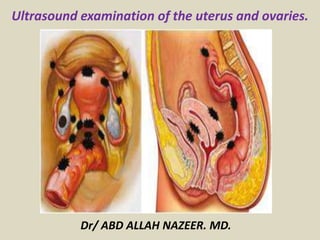
  • 1. Dr/ ABD ALLAH NAZEER. MD. Ultrasound examination of the uterus and ovaries.
  • 2. ULTRASOUND OF THE UTERUS – Normal. Uterus TA probe positioning for longitudinal scan. Uterus sagittal US image.
  • 3. Uterus TA probe positioning for transverse scan. Trans abdominal view of the uterus: transverse. Both ovaries are visible
  • 4. Transvaginal Technique Anteverted uterus. Normal TV image anteverted sagittal.
  • 5. Retroverted transvaginal technique Retroverted uterus transvaginal scan.
  • 6. Sagittal US image of the uterus obtained during the proliferative phase of the menstrual cycle demonstrates the endometrium with a multilayered appearance. Normal premenopausal endometrium. Sagittal US image of the uterus obtained during the secretory phase of the menstrual cycle shows a thickened, echogenic endometrium. ENDOMETRIAL MEASUREMENT.
  • 7. Coronal 3D image of a Mirena IUD shows the expected location of the shaft and crossbars simultaneously in the body and fundus of the uterus. The endometrium is also seen well without the normal shadowing of the mirena always seen in the sagittal plane. The string can be difficult to identify on the 3D in the cervix but a 2D sagittal scan should easily show its placement.
  • 8. ROLE OF ULTRASOUND: To examine the uterus, ovaries, cervix, vagina and adnexae. Classification of a mass identified on other modalities e.g solid, cystic, mixed. Post surgical complications e.g abscess, edema. Guidance of injections, aspiration or biopsy. Assistance with IVF. To identify the relationship of normal anatomy and pathology to each other. INDICATIONS: P/V bleeding/discharge, Menorrhagia, Metrorrhagia (irregular uterine bleeding), Polymenorrhea, Menometrorrhagia (excessive irregular bleeding) Amenorrhea, Oligoamenorrhea, Pelvic pain, Dysmenorrhea (Painful Menses) F/H uterine or ovarian Cancer, Palpable lump, Infertility- primary or secondary (evaluation, monitoring and/or treatment), Anomalies/evaluation, Follow-up of previous abnormality, Precocious Puberty, delayed menses or vaginal bleeding in a prepubertal child. postmenopausal bleeding, Signs/symptoms of pelvic infection, IUCD Localization (intrauterine contraceptive Device) Guidance for interventional or surgical procedures. urinary incontinence or pelvic organ prolapse.
  • 9. COMMON PATHOLOGIES VAGINAL Gartners duct cyst, Epidermal inclusion cysts, Bartholin Gland Cysts, Vaginal carcinoma, Hydro/hematocolpos (secondary to imperforate hymen or vaginal stenosis), Foreign body, leiomyoma and endometriosis. CERVICAL Nabothian (retention) cysts, Polyps, Cervical fibroids, Cervical carcinoma, Cervical stenosis UTERINE Fibroids (leiomyoma), submucosal, intramural, subserosal, pedunculated Leiomyosarcoma Adenomyosis Lipoleiomyoma ENDOMETRIAL Endometrial Polyps, Endometrial Carcinoma, Endometrial hyperplasia Endometritis, endometriosis. Cystic hyperplasia secondary to Tamoxifen Adhesions- Ashermans Syndrome Submucosal fibroids Arterio-venous malformation (AVM) Hydro/hematometra Blood/fluid/infection or retained products of conception (RPOC)
  • 10. Common cystic lesions of the vagina and their origins.
  • 11. Gartner duct cysts are embryologic secretory retention cysts that arise from the residual wolffian (mesonephric) duct remnant usually solitary and less than 2 cm, most commonly develop in the anterolateral wall of the proximal one-third of the vagina; may contain septa; when located at the level of the urethra, they can cause mass effect on the urethra, giving rise to urinary tract symptoms; often associated with other wolffian abnormalities (unilateral renal agenesis, renal hypoplasia, and ectopic ureteral insertion); although usually asymptomatic, cyst aspiration, tetracycline sclerotherapy, or surgical excision may be used to treat larger lesions.
  • 12. Gartner’s cyst (*) in a young woman with dysuria imaged with (a) suprapubic, (b) trans- labial approach and (c) reconstructed images from translabial volume acquisition.
  • 13. Posterior urethral diverticula (*) creating mass effect on the anterior wall of the vagina in a woman with dysuria, and severe dyspareunia imaged during voiding. The inhomogeneous appearance is due to turbulent flow. u, urethra; b, bladder; ant and post, anterior and posterior vaginal walls.
  • 14. Oblique sagittal transabdominal US (a) and coronal T1-weighted MR (b) showing hematocolpos (*) and hematosalpinges (s) in bicornuate-unicollis uterus with associated hypoplasia of the cervix and imperforate hymen in a young woman with cyclic pelvic pain and primary amenorrhea. Dotted line represents the orientation of the US scan. Imperforate hymen is the most common congenital obstructive abnormality of the female genital tract, with a frequency that can vary from 1 case per 1000 population to 1 case per 10,000 population; most commonly occurs in isolation; occurs due to a failure of recanalization of this membranous vestige;
  • 15. Axial and sagittal scans at different levels (schemes on the right) show the presence of a didelphys uterus and vaginal duplication with noncanalized right emisystem. A: axial section of the pelvis at the level of uterine body. On the right: hematometra (*); on the left, hypoplastic body without hematometra. bl, bladder; B: axial plane of the pelvis at the level of the vagina. Right vaginal lumen distended by hematic material; left vagina is undilated and opened. C: right paramedian sagittal plane on the right uterus and vagina showing hematometra (hm) and hematocolpos (hc). D: left paramedian sagittal plane on the left hypoplastic uterus and vagina.
  • 16. Reconstructed axial image from translabial volumetric acquisition showing a common asymptomatic cyst (calipers) in the posterior vaginal wall. U, urethra; R, rectum. Epidermal inclusion cysts (also termed “sebaceous cysts”) are the most common type of non-embryological cyst; vary in size from a few millimeters to several centimeters; mostly asymptomatic; their location almost always correlates with a site of previous surgery and represents a traumatic inclusion of normal vaginal mucosa following vaginal laceration or episiotomy.
  • 17. Leiomyomas Leiomyomas are an uncommon entity in the vagina with only about 300 reported cases in literature; commonly seen in the age group ranging from 35 to 50 years; may originate in the smooth muscle of the vagina, local arterial musculature, or smooth muscle of the bladder or urethra; however, midline of the anterior vaginal wall is the most common location; at US, their characteristics are similar to uterine leiomyomas, showing a heterogeneous but predominately hypoechoic and well circumscribed appearance. at CEUS examination, the enhancement of leiomyomas is uniform, as opposed to the heterogeneous enhancement of leiomyosarcoma.
  • 18. Leiomyoma of the vaginal wall. TVUS axial (A) and sagittal (B) scans demonstrate a solid, inhomogeneously hypoechoic, well-defined lesion (*) deforming the outer vaginal wall. At CEUS (C-F) a slow enhancement followed by a relative hypo enhancement is observed.
  • 19. Translabial sagittal US scan (a) showing a myoma (*) of the anterior vaginal wall (V, vagina, b, bladder) . Contrast-enhanced US sequences (b-e) demonstrate high but homogenous enhancement of the lesion (arrow).
  • 20. Sagittal US scan with a linear probe of a slow-growth inhomogeneous solid mass of the right major labrum. Dynamic CEUS (D, E and F) shows a ready enhancement of the lesion. The lesion appeared hyperintense in T2-fatsat MR sequences (G) and iso-hypointense on T1 (H). At microscopical examination the lesion was demonstrated to be an angiomixoma of the vulva. Primary vaginal malignancies are relatively rare, representing about 1%–2% of all gynecologic malignancies and less than 20% of vaginal malignancies. primary cancers are defined as having no involvement of the external os of the cervix or the vulva inferiorly
  • 21. Cancer vagina with translabial sagittal US (a) of the vagina in a patient with vaginal bleeding and dyspareunia. A large, solid, inhomogeneous vaginal mass (arrowheads) is demonstrated with compression of the urethra (u). Sagittal (b) and coronal (c) T2-weighted MR images show inhomogeneous signal intensity of the mass and micro-infiltrative behavior.
  • 22. Bartholin Gland Cysts Bartholin’s glands secretions provide to lubricate vulvar vestibule with ducts that drain at the 4 and 8 o’ clock positions of the vaginal vestibule. Bartholin gland cysts are the most common vulvar cysts, developing in 2% of women during their lifetime; located at the posterolateral vaginal introits, medial to the labia minora; their location at or below the level of the pubic symphysis helps differentiate them from Gartner duct cysts; range in size from 1 to 4 cm (but can increase with repeated sexual stimulation develop as a result of an obstruction of the gland’s duct by a stone or a stenosis related to prior infection or trauma); patients are often asymptomatic but can present with mild dyspareunia, typically in the 2nd to 3rd decade of life; superimposed infection or abscess formation may require drainage; a rare complication is development of squamous car- cinoma or adenocarcinoma in a Bartholin's gland duct or cyst, respectively; at US, simple cysts are anechoic, thin-walled and unilocular, but septations can be seen. the cyst wall may be thickened and, if infected, enhancement can be observed at CEUS examination; visualization of any solid component within the cyst should raise concern for malignancy.
  • 23. US coronal scan of a Bartholin’s cyst in a patient with discomfort and dyspareunia. A cystic mass (*) in (a) is demonstrated in the right major labrum. Color-Doppler evaluation (b) shows an implementation of vascular signs related to hyperemia and inflammation. V, vagina.
  • 24. Sagittal translabial US (a). Endometriotic implant (*) in the rectovaginal septum in a young woman presenting with elective pain at palpation. Real-time elastography (b) demonstrates hard stiffness (red) due to perilesional fibrotic reaction. U, uterus; PF, posterior vaginal fornix. Vaginal endometriosis consists in the presence of ectopic endometrial glandular and stromal tissue within the vaginal walls. May occur during surgical procedures or from the spread of a recto-vaginal pouch endometriosis; often associated with dysmenorrheal, postcoital spotting, dyspareunia and infertility; the condition is almost always associated with endometriosis in other pelvic locations.
  • 25. Vaginal cuff represents the apex of the vagina where the apposed upper walls are sutured together; is a site of recurrent gynecologic malignancy after hysterectomy (seen most often with cervical carcinoma, but it also occurs with endometrial cancer and rarely with ovarian cancer); postoperative anteroposterior vaginal cuff size quoted in the literature is up to 2.1 cm; at pelvic US, normal vaginal cuff is generally small, symmetric, and homogeneously hypoechoic. Transvaginal US and color-Doppler scan of a recurrent gynecologic malignancy (arrows) on the vaginal cuff after hysterectomy.
  • 26. Vaginal varices. Patient with severe dyspareunia and dysuria. Color Doppler evaluation with translabial approach shows a high flow within the uterine-vaginal planes with venous ectasia. Vagina is an unlikely location to develop varices, because of the presence of a bilateral draining system (similar to uterus), which largely communicates with the uterine, vesical and hemorroidal plexuses; vaginal varices are usually found in cirrhotic patients or after hysterectomy; color-Doppler US reveals venous flow inside these hypoechoic, serpiginous lesions.
  • 27. Nabothian cysts (also known as a retention cysts of the cervix) are non- neoplastic cystic lesion that occurs in relation to the uterine cervix. Pelvic ultrasound Typically seen an anechoic well defined cystic lesions near the endocervical canal. If the cysts are large, the cervical region can appear enlarged Colour Doppler interrogation shows no associated colour flow. Cervical Nabothian cyst.
  • 28. Cervical polyp in ultrasound image. A cervical polyp is a common benign polyp or tumour on the surface of the cervical canal. They can cause irregular menstrual bleeding but often show no symptoms.
  • 29. Cervical fibroid. Cervical myoma are benign tumors composed partly of muscle tissue. They seldom develop in the cervix, the lower part of the uterus. When they do, they are usually accompanied by myoma in the larger upper part of the uterus.
  • 30. Sagittal transabdominal US (a) and color-Doppler (b) demonstrating a large intracavitary pedunculated myoma (*) protruding in vagina from cervical os. The vascular pedicle is depicted at color-Doppler US. b, bladder; c, cervix.
  • 31. Carcinoma of the cervix is a malignancy arising from the cervix and is considered the third most common gynecologic malignancy. Ultrasound hypoechoic, heterogeneous mass involving the cervix may show increased vascularity on colour Doppler although cervical cancer is staged clinically, ultrasound can be a useful adjunct by showing size (<4 cm or >4 cm) parametrial invasion tumour invasion into the vagina tumor invasion into adjacent organs hydronephrosis: implies stage IIIB tumour. This sagittal transvaginal color Doppler sonogram shows prominent vascular flow in the cervical tumor.
  • 32. Transvaginal and Doppler study of cancer cervix.
  • 33. Sagittal and transverse transvaginal sonogram shows a circumscribed hypoechoic cancer cervix.
  • 34. Uterine leiomyomas (uterine fibroids) are benign tumours of myometrial origin and are the most common solid benign uterine neoplasm. Commonly an incidental finding on imaging, they rarely cause a diagnostic dilemma. They occur in ~25% of women of reproductive age and are particularly common in the African population. Fibroids may have a number of locations within or external to the uterus: intra-uterine intramural leiomyoma: most common subserosal leiomyoma submucosal leiomyoma: least common (10-15%) extra-uterine broad ligament leiomyoma cervical leiomyoma parasitic leiomyoma diffuse uterine leiomyomatosis
  • 35. Pelvic ultrasound ultrasound is used to diagnose the presence and monitor the growth of fibroids uncomplicated leiomyomas are usually hypoechoic, but can be isoechoic, or even hyperechoic compared to normal myometrium, calcification is seen as echogenic foci with shadowing, cystic areas of necrosis or degeneration may be seen Subserosal leiomyoma.
  • 38. TA USG image (A) shows a 7-cm intramural fibroid containing cystic areas (arrows). Axial T2W MRI image (B) shows an 11 × 8 cm fibroid (arrows) containing central high signal, consistent with cystic degeneration (arrowhead)
  • 39. Transabdominal (TA) USG image (A) shows a bulky uterus showing a 10-cm submucosal fibroid (between cursors). Sagittal T2W MRI image (B) in the same patient shows that the submucosal fibroid (arrowhead) is heterogeneous indicating degeneration. There is also a 2.5-cm cervical fibroid (arrow)
  • 40. Ultrasound image shows sub mucosal fibroid Picture B. MRI image shows multiple intramural fibroids.
  • 41.
  • 43. Endometrial polyps are benign nodular protrusions of the endometrial surface, and one of the entities included in a differential of endometrial thickening. Endometrial polyps can either be sessile or pedunculated. They can often be suggested on ultrasound or MRI studies, but may require sonohysterography or direct visualization for confirmation. Ultrasound. Although endometrial polyps may be visualized at transvaginal ultrasound as nonspecific endometrial thickening, they may also be identified as focal masses within the endometrial canal. a stalk to the polyp may either be thin or broad based a feeding vessel may be seen extending to the polyp on colour Doppler imaging cystic spaces corresponding to dilated glands filled with proteinaceous fluid may be seen within the polyp and is considered a relatively characteristic feature may appear as just diffusely thickened endometrium, without visualization of a discrete mass (mimicking endometrial hyperplasia) 3D ultrasound may be useful to help delineate the borders of a polyp. Sonohysterography . Polyps are best characterized on sonohysterography and appear as echogenic, smooth, intra-cavitary masses outlined by fluid. The typical appearance of an endometrial polyp at sonohysterography is as a well-defined, homogeneous, polypoid lesion that is isoechoic to the endometrium with preservation of the endometrial-myometrial interface There is usually a well-defined vascular pedicle within the stalk. Colour Doppler interrogation may show flow within the stalk.
  • 45. TVS of an endometrial polyp hidden within a secretorial endometrium After transcervical instillation of saline (SISH), applying power Doppler, a polyp with single vessel arrangement can be seen.
  • 46. Images show endometrial polyp within the uterine cavity with its vascular pedicle visible on color Doppler scans.
  • 48. Endometrial hyperplasia (EH) refers to an abnormal proliferation of endometrial stromal and glandular components. It represents a spectrum of endometrial changes ranging from cystically dilated glands to cellular atypia. Ultrasound premenopausal normal endometrial thickness depends on the stage of the menstrual cycle, but a thickness of >10 mm is top normal or abnormal hyperplasia can be reliably excluded in patients only when the endometrium measures less than 6 mm postmenopausal a thickness of >5 mm is considered abnormal The appearance can be non-specific and cannot reliably allow differentiation between hyperplasia and carcinoma. Usually there is a homogeneous increase in endometrial thickness, but endometrial hyperplasia may also cause asymmetric/focal thickening with surface irregularity, an appearance that is suspicious for carcinoma.
  • 52. Endometrial carcinoma is generally considered the most common gynecological malignancy. It frequently present with vaginal bleeding and both ultrasound and pelvic MRI are useful modalities for evaluation. Transvaginal ultrasound Endometrial carcinoma usually appears as thickening of the endometrium though may appear as a polypoid mass premenopausal: normal endometrial thickness varies through the menstrual cycle diagnosing abnormally thickened endometrium depends on knowing what the patient's point in the menstrual cycle postmenopausal: >5 mm is thickened (if not on Tamoxifen) Sonographic features are non specific and endometrial thickening can also be due to benign proliferation, endometrial hyperplasia or polyps. Disruption of a subendometrial halo on ultrasound may be suggestive of myometrial involvement.
  • 55. Multiple vessels in women with endometrial cancer. Stage IC endometrial carcinoma. Sagittal TVUS image of the uterus shows marked, inhomogeneous endometrial thickening(thick arrows ).
  • 56. Endometritis refers to inflammation of the endometrium, the inner lining of the uterus. Pathologists have traditionally classified endometritis as either acute or chronic. Ultrasound While the sonographic appearance of the uterus and endometrium may be normal in early stages, findings may include a thickened, heterogeneous endometrium, intracavitary fluid +/- intrauterine air. Sagittal image of the uterus showing complex fluid within the endometrial cavity. Endometritis.
  • 57. Endometriosis is a hormonal and immune system disease in which cells similar to that which line the uterus (endometrium) grow outside the uterine cavity, most commonly on the membrane which lines the abdominal cavity, the peritoneum. Ovarian endometrioma.
  • 59. Uterine arteriovenous malformations (UAVM) result from formation of multiple arteriovenous fistulous communications within the uterus without an intervening capillary network. Ultrasound Gray-scale sonographic appearances can be non-specific and can have a range of manifestations including areas of subtle myometrial inhomogeneity, tubular spaces within the myometrium, a intramural uterine, endometrial or cervical mass like region or sometimes as prominent parametrial vessels. The extent of mass is effect is however minimal. Colour Doppler Typically shows serpiginous/tubular anechoic structures within the myometrium with a low resistance (RI ~0.2-0.5), high velocity flow pattern on colour Doppler interrogation.
  • 61. Uterine arterio-venous malformation. In addition a large disproportionately dilated vascular.
  • 62. ULTRASOUND OF THE OVARIES – Normal. Use the full urinary bladder as an acoustic window to angle across to the ovary. Axial trans-abdominal image with the ovary lateral to the uterus.
  • 63. Axial trans-vaginal scan plane. Axial trans-vaginal image.
  • 64. Normal transvaginal ovary demonstration normal peripheral follicles. The uterus may be oblique and squash the ovary giving it a flattened ovoid shape.
  • 65. ROLE OF ULTRASOUND To examine the uterus, ovaries cervix vagina and adnexae. Classification of a mass identified on other modalities e.g solid, cystic, mixed. Post surgical complications e.g abscess, edema. Guidance of injections, aspiration or biopsy. Assistance with IVF. To identify the relationship of normal anatomy and pathology to each other. INDICATIONS P/V bleeding/discharge Menorrhagia Polymenorrhea Amenorrhea Irregular periods Pelvic pain F/H uterine or ovarian Cancer Palpable lump Infertility- primary or secondary Anomalies
  • 66. COMMON PATHOLOGIES OVARIAN Ovarian cysts simple Vs complex (hemorrhagic, corpus luteal, ruptured, septated). any mural nodules Dermoid Ovarian tumours: Cystadenoma (serous/mucinous)-Benign Cystadenocarcinoma (serous/mucinous)-Malignant Polycystic Ovarian Disease Endometrioma Torsion Hyper stimulation syndrome Ectopic pregnancy POUCH OF DOUGLAS (POD) & ADNEXAE Fluid, Pus, Blood Pelvic inflammatory disease-PID (may be indicated by above conditions) Cysts (Mesenteric) Ectopic pregnancy Endometriosis Pelvic venous congestion FALLOPIAN TUBES PID, Pyosalpinx, Hydrosalpinx, Ectopic pregnancy, Cyst, Ectopic pregnancy, Endometriosis
  • 67.
  • 68. An ovarian cyst is any collection of fluid, surrounded by a very thin wall, within an ovary . Any ovarian follicle that is larger than about two centimeters is termed an ovarian cyst. The vast majority of ovarian cysts are harmless (benign ), most are functional cysts. Ovarian cysts occur in women of all ages including neonatal period and infancy. They are most prevalent during infancy, adolescence and during the childbearing years. Types of cysts physiological cysts: mean diameter ≤3.0 cm( ovarian follicle)( corpus luteum). functional cysts (can produce hormones): follicular cysts of the ovary (estrogen): >3.0 cm corpus luteum cysts (progesterone) theca lutein cyst: gestational trophoblastic disease complications in functional cysts: Hemorrhage, enlargement, rupture and torsion other cysts: multiple large ovarian cysts in ovarian hyperstimulation syndrome post-menopausal cyst: serous inclusion cysts of the ovary polycystic ovaries ovarian torsion ovarian cystic neoplasms
  • 69. Left ovarian cyst simple cyst. Radiographic features Simple ovarian follicular cysts anechoic intraovarian or exophytic imperceptible wall posterior acoustic enhancement (may not be as obvious with harmonic or compound imaging) visible far wall
  • 70. Simple ovarian cyst ultrasound.
  • 71. Simple ovarian cyst ultrasound.
  • 72. Simple looking cyst measuring 8.7cm in maximal diameter.
  • 73.
  • 74. Hemorrhagic ovarian cyst. Hemorrhagic ovarian cysts (HOCs) usually result from hemorrhage into a corpus luteum or other functional cyst. Radiographic features are variable depending on the age of the hemorrhage. They typically resolve within eight weeks. HOCs can have a variety of appearances depending on the stage of evolution of the blood products and clot. lace-like reticular echoes or an intracystic solid clot, a fluid-fluid level is possible. thin wall, clot may adhere to cyst wall mimicking a nodule, but has no blood flow on Doppler imaging, retracting clot may have sharp or concave borders, mural nodularity does not, posterior acoustic enhancement, may be less noticeable if harmonics or compounding is used, there should not be any internal blood flow, circumferential blood flow in the cyst wall is typical.
  • 75. US and power Doppler mode in a hemorrhagic cyst demonstrates the absence of blood vessels.
  • 78. A corpus luteal (CL) cyst is a type of functional ovarian cyst which results when a corpus luteum fails to regress following the release of an ovum. When associated with pregnancy, it is the most common pelvic mass encountered within the 1st trimester. They can have a range of sonographic appearances depending on the stage of evolution and age of the associated intracystic hemorrhage. General characteristics include: diffusely thick wall, peripheral vascularity, <3 cm possible crenulated contour. Hemorrhagic corpus luteal cyst.
  • 84. A dermoid cyst is a cystic teratoma that contains an array of developmentally mature, solid tissues. It frequently consists of skin, complete with hair follicles, and sweat glands. Other commonly found components include: clumps of long hair, pockets of sebum, blood, fat, bone, nails, teeth, eyes, cartilage, and thyroid tissue. Ultrasound images show diffusely or partially echogenic mass with posterior sound attenuation owing to sebaceous material and hair within the cyst cavity, echogenic interface at edge of mass that obscures deep structures: the tip of the iceberg sign, mural hyperechoic Rokitansky nodule: dermoid plug, echogenic, shadowing calcific or dental (tooth) components, presence of fluid-fluid levels. Dermoid cyst.
  • 86. Ovarian dermoid cyst with Rokitansky nodule.
  • 88. Endometrioma right ovary with homogenous distribution of low to medium fine echoes in a cystic mass with only one septation, no solid components. No blood flow within the mass,
  • 89. Ovarian serous cyst adenomas (OSC) are a type of benign ovarian epithelial tumour. Ultrasound usually seen as a unilocular cystic adnexal lesion papillary projections are absent if there is any wall irregularity, it is thin, with an acute angle with the cyst wall and have a regular surface some lesions may contain sonographically detectable septations. Bilateral ovarian serous cystadenoma.
  • 90.
  • 91. Left ovarian papillary serous cystoma in a patient of 55 years, RI above 0.42, sharp margins, thin internal septations, moderate vascularization
  • 92.
  • 95.
  • 96. Ovarian cystadenocarcinoma. Ovarian serous cystadenocarcinoma is an ovarian epithelial tumor at the malignant end of the spectrum of ovarian serous tumours. Ultrasound: More heterogeneous in appearance than a serous cystadenoma papillary projections, thick septations, and/or solid components presence of ascites concerning for peritoneal metastatic spread, discrete peritoneal deposits may be seen. Colour Doppler is useful to confirm vascularity of the solid components quantitative parameters (resistive index and pulsatility index) do not reliably predict malignancy.
  • 97. Ovarian serous cystadenocarcinoma. A. Coronal TVUS image with superimposed color Doppler of a left ovarian mass shows a cystic mass with a large vascular solid component. B. Spectral Doppler image of the same ovary shows high diastolic flow, indicating neovascularization.
  • 99. TVS image left ovary. bilateral serous papillary ovarian carcinoma. She had a normal gynecological checkup without ultrasound one year prior to diagnosis. Randomly dispersed vessels in the echogenic area of the adnexal mass TVS of the other ovary, same patient. B-mode features typical ovarian malignancy.
  • 100.
  • 101. Polycystic ovarian syndrome (PCOS) is a chronic anovulation syndrome. Sonographic findings alone are not specific, and the diagnosis is made on the combined clinical, biochemical and sonographic grounds. Ultrasound Ovaries may show sonographic features of polycystic ovaries bilateral enlarged ovaries with multiple small follicles: 50% increased ovarian size (>10 cc) 12 or more follicles measuring 2-9 mm follicles of similar size peripheral location of follicles: which can give a string of pearl appearance hyperechoic central stroma the ovarian outline may be slightly irregular hypo-echoic ovary without individual cysts: 25% normal ovaries: 25%.
  • 104. The ovarian hyper-stimulation syndrome (OHSS) is a complication of ovarian stimulation treatment (ovarian induction therapy). Ultrasound typically shows bilateral symmetric enlargement of ovaries (often >12 cm in size) multiple cysts of varying sizes, giving the classic spoke-wheel appearance associated ascites and pleural +/- pericardial effusion (which is due to capillary leak) may also be present. Ovarian hyper-stimulation syndrome.
  • 106. Pelvic inflammatory disease (PID) is an broad term that encompasses a spectrum of infection and inflammation of the upper female genital tract, resulting in a spectrum of abnormalities. Ultrasound Ultrasound often only demonstrates ascitic fluid in the peritoneal cavity or nonspecific thickening and increased vascularity of the endometrium. In the most severe cases, may show adnexal masses with a heterogeneous echo-pattern. Some sonographic signs associated with tubal inflammation include: thickened/dilated fallopian tubes incomplete septa in the tube increased vascularity around the tube echogenic fluid in the tube (pyosalpinx ) cogwheel sign beads on a string sign
  • 107. Pelvic inflammatory disease and tuboovarian complex. A, Sagittal endovaginal sonogram reveals complex free fluid (FF). U = uterus.
  • 108. Pelvic inflammatory disease with fluid at the Cul De Sac and pyosalpinx.
  • 109. Pelvic congestion syndrome is a condition that results from retrograde flow through incompetent valves in ovarian veins. It is one of commonly missed and potentially treatable cause of chronic abdominal or pelvic pain. Ultrasound ovarian vein >5-6 mm (positive predictive value of 71-83%) may show multiple dilated veins in the adnexae with reversed venous flow on colour Doppler, especially after Valsalva maneuver the venous calibers may increase on real time during Valsalva Pelvic congestion syndrome
  • 110. US images for Pelvic congestion syndrome.
  • 111. US images for Pelvic congestion syndrome.
  • 112. Fallopian Tube Disease. There is a wide range of acute and chronic diseases affecting the fallopian tubes. Knowledge of the imaging appearances of these entities is important for correct diagnosis and differentiation from other disease entities. Ultrasonography (US) is the first-line imaging modality for the evaluation of signs and symptoms referable to the female pelvis. PID is infection of the upper female genital tract, resulting in a spectrum of abnormalities affecting the fallopian tube, including salpingitis, pyosalpinx, and tubo-ovarian abscess (TOA). Salpingitis.—In salpingitis, the fallopian tubes become edematous with ascending infection and inflammation. Although not dilated, the swollen fallopian tubes may be more conspicuous at imaging due to wall thickening, enhancement, and surrounding inflammation. Pyosalpinx.—Infection with superimposed obstruction of the fallopian tube leads to pyosalpinx. Inflammation results in tubal and peritubal adhesions with obstruction of the fimbrial end. Unable to drain, the fallopian tube distends with pus. US demonstrates a tubular structure distended with echogenic fluid and debris. Thick hypervascular walls and a fluid-debris level may be seen. There may be accompanying peri-oophoritis with ovarian enlargement, indistinct contours, and periovarian fluid or endometritis with poor definition of the endometrial echo complex.
  • 113. Fallopian tube torsion with gradual onset of lower abdominal pain (worse in the left lower quadrant). (a, b) Transverse (a) and longitudinal (b) US images of the pelvis show a dilated tubular cystic structure (arrow) with thickened edematous walls.
  • 114. Hydrosalpinx. US image shows a cystic adnexal mass, whose tubular shape and incomplete septa (*) are characteristic of hydrosalpinx.
  • 115. Right adnexal mass 7 over 3 cm, histology chlamydia salpingitis.
  • 116. A–C. Grayscale images showing thick walls, low-level echoic fluid (pus?) and the “cogwheel sign” (CW). D. Subserosal vascularization typical of an inflammatory response in hollow abdominal viscera. E,F. Edematous fimbrial end (arrow) of the inflamed tubes, floating in a small amount of free pelvic fluid. G. Low-level echoic, fluid-filled, thick-walled tubes with incomplete septae (arrow) are the hallmarks of hydrosalpinx. H–J. Bilateral hydrosalpinx. Note the thin walls and anechoic fluid- filled forms (sausage-shaped) (CL = corpus luteum; OV = ovary; UT = uterus).
  • 117. A–D. Various cases of tubal ectopic gestation (arrows point to each gestation). E–G. Power Doppler localizes the ectopic pregnancy (arrows), side by side with the corpus luteum (CL).
  • 118. A–C. Ovarian torsion. Hyperechoic, large ovary with follicles pushed toward the surface. Power Doppler reveals no blood flow in the ovary. D–F. Tubal torsion. Cystic dilatation with a small beak and a normal ovary. G. Intraoperative view of the tube (twisted three times; yellow arrows) and the normal ovary (white arrows).
  • 119. Fallopian tube cancer: A. Fluid-filled uterine cavity. B. Large cystic dilatation of the tube. C. A thickened tubal wall (arrow). D. Doppler interrogation reveals high diastolic flow (arrows). E. Macroscopic gross appearance.