SlideShare a Scribd company logo
1 of 145
MRI of the wrist joint.
DR/ ABD ALLAH NAZEER. MD.
1. Acute and chronic wrist instability.
2. Dorsal or ulnar-sided wrist pain.
3. Wrist symptoms in adolescent gymnasts and
other athletes.
4. Unexplained chronic wrist pain.
5. Acute wrist trauma.
6. Avascular necrosis.
7. Wrist malalignments.
8. Limited or painful range of motion.
9. Unexplained wrist swelling, mass, or atrophy.
10. Planning for diagnostic or therapeutic arthroscopy.
11. Recurrent, residual, or new symptoms following
wrist surgery.
Indications for MRI of the wrist joint.
MR Sequences images.
T1WI
(Lower Egypt).
T21WI (Upper Egypt).
Wrist X-ray Anatomy.
Pronator quadratus.
The transverse carpal ligament (short arrows) extends from the hook of the hamate (long
arrow) to the tubercle of the trapezium (arrowhead), forming the floor of the carpal tunnel.
At the radial aspect of the carpal tunnel, the flexor digitorum tendons are arranged in two
rows (separated by dotted line), the profundus tendons deep to the superficialis tendons. The
flexor pollicis longus tendon (star) is positioned at the ulnar aspect of the tunnel, separated
from the flexor carpi radialis tendon (curved arrow) by a ligamentous reflection of the
transverse carpal ligament. The median nerve is indicated (asterisk). - See more at:
http://radsource.us/palmar-bursae-and-flexor-tendon-sheaths/#sthash.mSFX1u0c.dpuf
Subchondral erosion of lunate.
CT images with small cyst formation within the fracture line.
MR Protocol with pre and post-contrast images for fracture.
Scaphoid waist fracture with subtle collapse of the proximal pole, related AVN .
Diffuse enhancement of scaphoid proximal pole has good correlation
with stage group I, patchy enhancement have correlation with
grade II and III and enhancement with complete necrosis.
Preiser's disease. Coronal inversion recovery (A) and fast spin echo (B) images:
1. a bone marrow edema pattern in the scaphoid (black arrow) and 2.
avascular necrosis of the proximal pole (white arrow). No fracture is visible.
Idiopathic avascular necrosis of the scaphoid (Preiser’s disease)
Keinbock,s Disease.
Keinbock,s Disease.
Intraosseous ganglion. Cor. T2FFE. Rounded high signal intensity
lesion in the radial aspect of the lunate with cortex erosion.
Capitate avascular
A simple approach is to consider the lunate
which is usually the easiest carpal bone to
visualize on a lateral wrist image. If the
lunate is abnormally tilted in a dorsal
direction on a standard lateral wrist image,
a DISI should be considered. If the lunate is
abnormally tilted in a volar direction a VISI
should be considered. DISI is due to
disruption of the scapho-lunate articulation.
VISI is secondary to disruption of the luno-
triquetral articulation.
DISI deformity.
DISI deformity.
DISI.
Volar intercalated segmental instability(VISI).
VISI with volar tilting of the lunate.
VISI with luno-triquetrum ligament tear and volar tilting of the lunate.
VISI deformity secondary to Lunotriquetral ligament tear.
Scapholunate dissociation. Note the widened scapholunate space (“Terry Thomas”
sign) and the foreshortened scaphoid with a “scaphoid ring” sign (arrows).
Adjacent is British comedian Terry Thomas and his famous teeth.
Scapholunate ligament rupture.
Scapho - lunate ligament tear
Scapholunate and lunotriquetral ligament tear.
1. Scapholunate ligament tear, dorsal angulation of the lunate and
proximal migration of the capitate consistent with a DISI deformity.
2. Tear of the lunotriquetral and triscaphe ligaments.
Triangular fibrocartilage complex tear.
Acute TFCC tear on FS T2-WI (A) and T1-WI (B).
Ulnar variance may be :
Neutral (both the ulnar and radial articular surfaces at the same
level)
Positive (ulna projects more distally)
Negative (ulna projects more proximally)
Causes
trauma or mechanical
distal radius/ulnar fractures with shortening (e.g. impaction) &
angulation
DRUJ ligamentous injuries (e.g. Galeazzi & Essex-Lopresti)
surgical shortening of ulna or radius
growth arrest (e.g. previous Salter-Harris fracture)
Congenital
Madelung deformity/reverse Madelung deformity
Associations
positive ulnar variance is associated with ulnar impaction syndrome.
negative ulnar variance is associated with Keinbock,s disease and ulnar
impingement syndrome
Positive ulnar variance secondary to TFC tear.
TFCC 1B tear (arrow) on coronal T1-WI (B) and coronal FS T2-WI (C).
Negative ulnar variance secondary to TFC tear.
Negative ulnar variance.
Swelling, deformity, abnormal signal of the median
nerve.
Lipoma of the
Carpal tunnel syndrome caused by cysticercosis.
Sarcoidosis with carpal tunnel syndrome.
Lipoma at the Ulnar tunnel.
Ganglion cyst.
Tenosynovitis.
Tenosynovitis is an inflammation of the tendon sheath , either primary or secondary.
Marked inflammatory tenosynovitis.
Tenosynovitis of Extensor Carpi Ulnaris.
Extensor carpi ulnaris tenosynovitis. Axial FS T2-WI shows fragmentation into multiple tendon fragments of
the extensor carpi ulnaris tendon (arrowheads). Note increased fluid and debris within the tendon sheath.
Tenosynovitis and partial thickness tearing of the FCR tendon (arrow).
Tenosynovitis of the flexor tendons.
Tenosynovitis of all flexor tendons FDS, FDP, FPL
Tenosynovitis
with partial
tears of the EPL,
ECRB and ECRL
Moderate tenosynovitis of the extensor digitorum tendon (yellow arrowhead).
Tenosynovitis of the flexor polices tendon.
Extensive tenosynovitis.
Tenosynovitis in a 32-year-old woman with early
rheumatoid arthritis of the wrist (5 months duration)
and normal radiographic findings. Axial T1-weighted
(a), fat-suppressed T2-weighted (b), and contrast-
enhanced fat-suppressed T1-weighted (c) MR images
show marked tenosynovitis (arrows) involving both
the dorsal extensor and volar flexor compartments.
Note that the tenosynovitis and periscaphoid joint
synovitis (*) have the same signal intensity in a
(intermediate) and b (high) and similar enhancement
Flexor Tendon Rice Body Tenosynovitis.
Tuberculous tenosynovitis.
Sarcoidosis. (a, b) Axial T2 FS and coronal T1. Increased fluid is present within the flexor tendon sheath with multiple loose
bodies (rice bodies) present (blue arrows). (c, d) Axial and coronal T1 FS PG. Thick synovial enhancement is present.
De Quervain tenosynovitis of abductor pollicis longus (APL)
and extensor pollicis longus (EPL).
De Quervain tenosynovitis.
De Quervain syndrome. (a, b) Axial T2 FS and coronal T2 FS. Increased fluid is present within the APB and EPB
tendon sheaths (blue ellipses). Surrounding edema is present, as evidenced by increased T2 weighted signal.
SOFT TISSUE HEMANGIOMA OF THE WRIST.
Giant cell tumor of the tendon sheath in wrist.
Synovial Sarcoma of Wrist Area.
Rhabdomyosarcoma of the hand.
MRI Wrist Joint Guide

More Related Content

What's hot

Presentation1.pptx ankle joint..
Presentation1.pptx ankle joint..Presentation1.pptx ankle joint..
Presentation1.pptx ankle joint..Abdellah Nazeer
 
MRI KNEE JOINT ANATOMY
MRI KNEE JOINT ANATOMYMRI KNEE JOINT ANATOMY
MRI KNEE JOINT ANATOMYNikhil Bansal
 
Shoulder Mri Scan in Delhi by Dr Shekhar Shrivastav
Shoulder Mri Scan in Delhi by Dr Shekhar ShrivastavShoulder Mri Scan in Delhi by Dr Shekhar Shrivastav
Shoulder Mri Scan in Delhi by Dr Shekhar ShrivastavDelhiArthroscopy
 
Radiographic evaluation of shoulder
Radiographic evaluation of shoulderRadiographic evaluation of shoulder
Radiographic evaluation of shouldermrinal joshi
 
Mri anatomy of ankle radiology ppt pk
Mri anatomy of ankle radiology ppt pkMri anatomy of ankle radiology ppt pk
Mri anatomy of ankle radiology ppt pkDr pradeep Kumar
 
MRI KNEE OF ORTHOPEDIC IMPORTANCE
MRI KNEE OF ORTHOPEDIC IMPORTANCEMRI KNEE OF ORTHOPEDIC IMPORTANCE
MRI KNEE OF ORTHOPEDIC IMPORTANCEBenthungo Tungoe
 
Mri shoulder joint with common pathologies
Mri shoulder joint with common pathologiesMri shoulder joint with common pathologies
Mri shoulder joint with common pathologiesGobardhan Thapa
 
Presentation1, radiological film reading of the hip joint.
Presentation1, radiological film reading of the hip joint.Presentation1, radiological film reading of the hip joint.
Presentation1, radiological film reading of the hip joint.Abdellah Nazeer
 
Presentation1.pptx, radiological imaging of sacroiliac joint diseases.
Presentation1.pptx, radiological imaging of sacroiliac joint diseases.Presentation1.pptx, radiological imaging of sacroiliac joint diseases.
Presentation1.pptx, radiological imaging of sacroiliac joint diseases.Abdellah Nazeer
 
Mri anatomy of knee Dr. Muhammad Bin Zulfiqar
Mri anatomy of knee Dr. Muhammad Bin ZulfiqarMri anatomy of knee Dr. Muhammad Bin Zulfiqar
Mri anatomy of knee Dr. Muhammad Bin ZulfiqarDr. Muhammad Bin Zulfiqar
 
MRI-in Spine PPT.pptx
MRI-in Spine PPT.pptxMRI-in Spine PPT.pptx
MRI-in Spine PPT.pptxssusere26312
 
Presentation1, radiological imaging of anterior knee pain.
Presentation1, radiological imaging of anterior knee pain.Presentation1, radiological imaging of anterior knee pain.
Presentation1, radiological imaging of anterior knee pain.Abdellah Nazeer
 
X ray of foot and ankle
X ray of foot and ankle X ray of foot and ankle
X ray of foot and ankle Sulav_56
 
Scaphoid fracture and non union
Scaphoid fracture and non unionScaphoid fracture and non union
Scaphoid fracture and non unionratish mishra
 

What's hot (20)

Mri knee trauma
Mri knee traumaMri knee trauma
Mri knee trauma
 
Presentation1.pptx ankle joint..
Presentation1.pptx ankle joint..Presentation1.pptx ankle joint..
Presentation1.pptx ankle joint..
 
MRI KNEE JOINT ANATOMY
MRI KNEE JOINT ANATOMYMRI KNEE JOINT ANATOMY
MRI KNEE JOINT ANATOMY
 
Shoulder Mri Scan in Delhi by Dr Shekhar Shrivastav
Shoulder Mri Scan in Delhi by Dr Shekhar ShrivastavShoulder Mri Scan in Delhi by Dr Shekhar Shrivastav
Shoulder Mri Scan in Delhi by Dr Shekhar Shrivastav
 
Radiographic evaluation of shoulder
Radiographic evaluation of shoulderRadiographic evaluation of shoulder
Radiographic evaluation of shoulder
 
Mri anatomy of ankle radiology ppt pk
Mri anatomy of ankle radiology ppt pkMri anatomy of ankle radiology ppt pk
Mri anatomy of ankle radiology ppt pk
 
Mri of knee
Mri of kneeMri of knee
Mri of knee
 
MRI KNEE OF ORTHOPEDIC IMPORTANCE
MRI KNEE OF ORTHOPEDIC IMPORTANCEMRI KNEE OF ORTHOPEDIC IMPORTANCE
MRI KNEE OF ORTHOPEDIC IMPORTANCE
 
Mri shoulder joint with common pathologies
Mri shoulder joint with common pathologiesMri shoulder joint with common pathologies
Mri shoulder joint with common pathologies
 
Presentation1, radiological film reading of the hip joint.
Presentation1, radiological film reading of the hip joint.Presentation1, radiological film reading of the hip joint.
Presentation1, radiological film reading of the hip joint.
 
Presentation1.pptx, radiological imaging of sacroiliac joint diseases.
Presentation1.pptx, radiological imaging of sacroiliac joint diseases.Presentation1.pptx, radiological imaging of sacroiliac joint diseases.
Presentation1.pptx, radiological imaging of sacroiliac joint diseases.
 
MRI of Shoulder anatomy
MRI of Shoulder anatomyMRI of Shoulder anatomy
MRI of Shoulder anatomy
 
Mri anatomy of knee Dr. Muhammad Bin Zulfiqar
Mri anatomy of knee Dr. Muhammad Bin ZulfiqarMri anatomy of knee Dr. Muhammad Bin Zulfiqar
Mri anatomy of knee Dr. Muhammad Bin Zulfiqar
 
MRI-in Spine PPT.pptx
MRI-in Spine PPT.pptxMRI-in Spine PPT.pptx
MRI-in Spine PPT.pptx
 
Presentation1, radiological imaging of anterior knee pain.
Presentation1, radiological imaging of anterior knee pain.Presentation1, radiological imaging of anterior knee pain.
Presentation1, radiological imaging of anterior knee pain.
 
X ray of foot and ankle
X ray of foot and ankle X ray of foot and ankle
X ray of foot and ankle
 
Ct and mri,ppt
Ct and mri,pptCt and mri,ppt
Ct and mri,ppt
 
Scaphoid fracture and non union
Scaphoid fracture and non unionScaphoid fracture and non union
Scaphoid fracture and non union
 
Mri sequences
Mri sequencesMri sequences
Mri sequences
 
MRI SPINE ANATOMY
MRI SPINE ANATOMYMRI SPINE ANATOMY
MRI SPINE ANATOMY
 

Similar to MRI Wrist Joint Guide

Presentation1 radiological film reading of wrist joint.
Presentation1 radiological film reading of wrist joint.Presentation1 radiological film reading of wrist joint.
Presentation1 radiological film reading of wrist joint.Abdellah Nazeer
 
Presentation1.pptx mri of elbow joint
Presentation1.pptx mri of elbow jointPresentation1.pptx mri of elbow joint
Presentation1.pptx mri of elbow jointAbdellah Nazeer
 
Rheumatoid arthritis and osteoarthritis
Rheumatoid arthritis and osteoarthritisRheumatoid arthritis and osteoarthritis
Rheumatoid arthritis and osteoarthritisSonal Saran
 
Transient Osteoporosis of Hip
Transient Osteoporosis of HipTransient Osteoporosis of Hip
Transient Osteoporosis of Hipvinod naneria
 
Diagnostic Imaging of Spondyloarthropathies
Diagnostic Imaging of SpondyloarthropathiesDiagnostic Imaging of Spondyloarthropathies
Diagnostic Imaging of SpondyloarthropathiesMohamed M.A. Zaitoun
 
Distal radioulnar joint injuries
Distal radioulnar joint injuriesDistal radioulnar joint injuries
Distal radioulnar joint injuriesSunil Poonia
 
Distal radioulnar joint injuries( DRUJ) and carpal instability
Distal radioulnar joint injuries( DRUJ) and carpal instabilityDistal radioulnar joint injuries( DRUJ) and carpal instability
Distal radioulnar joint injuries( DRUJ) and carpal instabilitySunil Poonia
 
Distal radioulnar joint injuries
Distal radioulnar joint injuriesDistal radioulnar joint injuries
Distal radioulnar joint injuriesSunil Poonia
 
Distal radioulnar joint injuries
Distal radioulnar joint injuriesDistal radioulnar joint injuries
Distal radioulnar joint injuriesSunil Poonia
 
Presentation1, radiological imaging of degenerative and inflammatory disease ...
Presentation1, radiological imaging of degenerative and inflammatory disease ...Presentation1, radiological imaging of degenerative and inflammatory disease ...
Presentation1, radiological imaging of degenerative and inflammatory disease ...Abdellah Nazeer
 
Muculoskeletal Pediatic Imaging..pptx
Muculoskeletal Pediatic Imaging..pptxMuculoskeletal Pediatic Imaging..pptx
Muculoskeletal Pediatic Imaging..pptxAbdellah Nazeer
 
Presentation1.pptx, ultrasound of the hand and fingers.
Presentation1.pptx, ultrasound of the hand and fingers.Presentation1.pptx, ultrasound of the hand and fingers.
Presentation1.pptx, ultrasound of the hand and fingers.Abdellah Nazeer
 
THE SPINE IN THE COURSE OF SPONDYLOARTHROPATHIES.pptx
THE SPINE IN THE COURSE OF SPONDYLOARTHROPATHIES.pptxTHE SPINE IN THE COURSE OF SPONDYLOARTHROPATHIES.pptx
THE SPINE IN THE COURSE OF SPONDYLOARTHROPATHIES.pptxkavitjetly
 
Special types of trauma
Special types of traumaSpecial types of trauma
Special types of traumaairwave12
 

Similar to MRI Wrist Joint Guide (20)

Presentation1 radiological film reading of wrist joint.
Presentation1 radiological film reading of wrist joint.Presentation1 radiological film reading of wrist joint.
Presentation1 radiological film reading of wrist joint.
 
Presentation1.pptx mri of elbow joint
Presentation1.pptx mri of elbow jointPresentation1.pptx mri of elbow joint
Presentation1.pptx mri of elbow joint
 
DISH imaging
DISH imagingDISH imaging
DISH imaging
 
Rheumatoid arthritis and osteoarthritis
Rheumatoid arthritis and osteoarthritisRheumatoid arthritis and osteoarthritis
Rheumatoid arthritis and osteoarthritis
 
Transient Osteoporosis of Hip
Transient Osteoporosis of HipTransient Osteoporosis of Hip
Transient Osteoporosis of Hip
 
SI JOINT PATHOLOGIES.pptx
SI JOINT PATHOLOGIES.pptxSI JOINT PATHOLOGIES.pptx
SI JOINT PATHOLOGIES.pptx
 
Diagnostic Imaging of Spondyloarthropathies
Diagnostic Imaging of SpondyloarthropathiesDiagnostic Imaging of Spondyloarthropathies
Diagnostic Imaging of Spondyloarthropathies
 
Distal radioulnar joint injuries
Distal radioulnar joint injuriesDistal radioulnar joint injuries
Distal radioulnar joint injuries
 
Distal radioulnar joint injuries( DRUJ) and carpal instability
Distal radioulnar joint injuries( DRUJ) and carpal instabilityDistal radioulnar joint injuries( DRUJ) and carpal instability
Distal radioulnar joint injuries( DRUJ) and carpal instability
 
Distal radioulnar joint injuries
Distal radioulnar joint injuriesDistal radioulnar joint injuries
Distal radioulnar joint injuries
 
Distal radioulnar joint injuries
Distal radioulnar joint injuriesDistal radioulnar joint injuries
Distal radioulnar joint injuries
 
Presentation1, radiological imaging of degenerative and inflammatory disease ...
Presentation1, radiological imaging of degenerative and inflammatory disease ...Presentation1, radiological imaging of degenerative and inflammatory disease ...
Presentation1, radiological imaging of degenerative and inflammatory disease ...
 
Muculoskeletal Pediatic Imaging..pptx
Muculoskeletal Pediatic Imaging..pptxMuculoskeletal Pediatic Imaging..pptx
Muculoskeletal Pediatic Imaging..pptx
 
SI JOINT PATHOLOGIES.pptx
SI JOINT PATHOLOGIES.pptxSI JOINT PATHOLOGIES.pptx
SI JOINT PATHOLOGIES.pptx
 
Presentation1.pptx, ultrasound of the hand and fingers.
Presentation1.pptx, ultrasound of the hand and fingers.Presentation1.pptx, ultrasound of the hand and fingers.
Presentation1.pptx, ultrasound of the hand and fingers.
 
ls spine lecture.pptx
ls spine lecture.pptxls spine lecture.pptx
ls spine lecture.pptx
 
THE SPINE IN THE COURSE OF SPONDYLOARTHROPATHIES.pptx
THE SPINE IN THE COURSE OF SPONDYLOARTHROPATHIES.pptxTHE SPINE IN THE COURSE OF SPONDYLOARTHROPATHIES.pptx
THE SPINE IN THE COURSE OF SPONDYLOARTHROPATHIES.pptx
 
Hip joint
Hip jointHip joint
Hip joint
 
Inflammatory Arthritis
Inflammatory ArthritisInflammatory Arthritis
Inflammatory Arthritis
 
Special types of trauma
Special types of traumaSpecial types of trauma
Special types of trauma
 

More from Abdellah Nazeer

Presentation1, Ultrasound of the bowel loops and the lymph nodes..pptx
Presentation1, Ultrasound of the bowel loops and the lymph nodes..pptxPresentation1, Ultrasound of the bowel loops and the lymph nodes..pptx
Presentation1, Ultrasound of the bowel loops and the lymph nodes..pptxAbdellah Nazeer
 
Presentation1 Short cases MD..pptx
Presentation1 Short cases MD..pptxPresentation1 Short cases MD..pptx
Presentation1 Short cases MD..pptxAbdellah Nazeer
 
Presentation1, MD MCQ Cases..pptx
Presentation1, MD MCQ Cases..pptxPresentation1, MD MCQ Cases..pptx
Presentation1, MD MCQ Cases..pptxAbdellah Nazeer
 
Presentation1, Short Cases Quiz..pptx
Presentation1, Short Cases Quiz..pptxPresentation1, Short Cases Quiz..pptx
Presentation1, Short Cases Quiz..pptxAbdellah Nazeer
 
Presentation1, radiological imaging of lateral hindfoot impingement.
Presentation1, radiological imaging of lateral hindfoot impingement.Presentation1, radiological imaging of lateral hindfoot impingement.
Presentation1, radiological imaging of lateral hindfoot impingement.Abdellah Nazeer
 
Presentation2, radiological anatomy of the liver and spleen.
Presentation2, radiological anatomy of the liver and spleen.Presentation2, radiological anatomy of the liver and spleen.
Presentation2, radiological anatomy of the liver and spleen.Abdellah Nazeer
 
Presentation1, artifacts and pitfalls of the wrist and elbow joints.
Presentation1, artifacts and pitfalls of the wrist and elbow joints.Presentation1, artifacts and pitfalls of the wrist and elbow joints.
Presentation1, artifacts and pitfalls of the wrist and elbow joints.Abdellah Nazeer
 
Presentation1, artifact and pitfalls of the knee, hip and ankle joints.
Presentation1, artifact and pitfalls of the knee, hip and ankle joints.Presentation1, artifact and pitfalls of the knee, hip and ankle joints.
Presentation1, artifact and pitfalls of the knee, hip and ankle joints.Abdellah Nazeer
 
Presentation1, radiological imaging of artifact and pitfalls in shoulder join...
Presentation1, radiological imaging of artifact and pitfalls in shoulder join...Presentation1, radiological imaging of artifact and pitfalls in shoulder join...
Presentation1, radiological imaging of artifact and pitfalls in shoulder join...Abdellah Nazeer
 
Presentation1, radiological imaging of internal abdominal hernia.
Presentation1, radiological imaging of internal abdominal hernia.Presentation1, radiological imaging of internal abdominal hernia.
Presentation1, radiological imaging of internal abdominal hernia.Abdellah Nazeer
 
Presentation11, radiological imaging of ovarian torsion.
Presentation11, radiological imaging of ovarian torsion.Presentation11, radiological imaging of ovarian torsion.
Presentation11, radiological imaging of ovarian torsion.Abdellah Nazeer
 
Presentation1, musculoskeletal anatomy.
Presentation1, musculoskeletal anatomy.Presentation1, musculoskeletal anatomy.
Presentation1, musculoskeletal anatomy.Abdellah Nazeer
 
Presentation1, new mri techniques in the diagnosis and monitoring of multiple...
Presentation1, new mri techniques in the diagnosis and monitoring of multiple...Presentation1, new mri techniques in the diagnosis and monitoring of multiple...
Presentation1, new mri techniques in the diagnosis and monitoring of multiple...Abdellah Nazeer
 
Presentation1, radiological application of diffusion weighted mri in neck mas...
Presentation1, radiological application of diffusion weighted mri in neck mas...Presentation1, radiological application of diffusion weighted mri in neck mas...
Presentation1, radiological application of diffusion weighted mri in neck mas...Abdellah Nazeer
 
Presentation1, radiological application of diffusion weighted images in breas...
Presentation1, radiological application of diffusion weighted images in breas...Presentation1, radiological application of diffusion weighted images in breas...
Presentation1, radiological application of diffusion weighted images in breas...Abdellah Nazeer
 
Presentation1, radiological application of diffusion weighted images in abdom...
Presentation1, radiological application of diffusion weighted images in abdom...Presentation1, radiological application of diffusion weighted images in abdom...
Presentation1, radiological application of diffusion weighted images in abdom...Abdellah Nazeer
 
Presentation1, radiological application of diffusion weighted imges in neuror...
Presentation1, radiological application of diffusion weighted imges in neuror...Presentation1, radiological application of diffusion weighted imges in neuror...
Presentation1, radiological application of diffusion weighted imges in neuror...Abdellah Nazeer
 
Presentation1, mr physics.
Presentation1, mr physics.Presentation1, mr physics.
Presentation1, mr physics.Abdellah Nazeer
 
Presentation1. ct physics.
Presentation1. ct physics.Presentation1. ct physics.
Presentation1. ct physics.Abdellah Nazeer
 
Presentation1, ultrasound physics.
Presentation1, ultrasound physics.Presentation1, ultrasound physics.
Presentation1, ultrasound physics.Abdellah Nazeer
 

More from Abdellah Nazeer (20)

Presentation1, Ultrasound of the bowel loops and the lymph nodes..pptx
Presentation1, Ultrasound of the bowel loops and the lymph nodes..pptxPresentation1, Ultrasound of the bowel loops and the lymph nodes..pptx
Presentation1, Ultrasound of the bowel loops and the lymph nodes..pptx
 
Presentation1 Short cases MD..pptx
Presentation1 Short cases MD..pptxPresentation1 Short cases MD..pptx
Presentation1 Short cases MD..pptx
 
Presentation1, MD MCQ Cases..pptx
Presentation1, MD MCQ Cases..pptxPresentation1, MD MCQ Cases..pptx
Presentation1, MD MCQ Cases..pptx
 
Presentation1, Short Cases Quiz..pptx
Presentation1, Short Cases Quiz..pptxPresentation1, Short Cases Quiz..pptx
Presentation1, Short Cases Quiz..pptx
 
Presentation1, radiological imaging of lateral hindfoot impingement.
Presentation1, radiological imaging of lateral hindfoot impingement.Presentation1, radiological imaging of lateral hindfoot impingement.
Presentation1, radiological imaging of lateral hindfoot impingement.
 
Presentation2, radiological anatomy of the liver and spleen.
Presentation2, radiological anatomy of the liver and spleen.Presentation2, radiological anatomy of the liver and spleen.
Presentation2, radiological anatomy of the liver and spleen.
 
Presentation1, artifacts and pitfalls of the wrist and elbow joints.
Presentation1, artifacts and pitfalls of the wrist and elbow joints.Presentation1, artifacts and pitfalls of the wrist and elbow joints.
Presentation1, artifacts and pitfalls of the wrist and elbow joints.
 
Presentation1, artifact and pitfalls of the knee, hip and ankle joints.
Presentation1, artifact and pitfalls of the knee, hip and ankle joints.Presentation1, artifact and pitfalls of the knee, hip and ankle joints.
Presentation1, artifact and pitfalls of the knee, hip and ankle joints.
 
Presentation1, radiological imaging of artifact and pitfalls in shoulder join...
Presentation1, radiological imaging of artifact and pitfalls in shoulder join...Presentation1, radiological imaging of artifact and pitfalls in shoulder join...
Presentation1, radiological imaging of artifact and pitfalls in shoulder join...
 
Presentation1, radiological imaging of internal abdominal hernia.
Presentation1, radiological imaging of internal abdominal hernia.Presentation1, radiological imaging of internal abdominal hernia.
Presentation1, radiological imaging of internal abdominal hernia.
 
Presentation11, radiological imaging of ovarian torsion.
Presentation11, radiological imaging of ovarian torsion.Presentation11, radiological imaging of ovarian torsion.
Presentation11, radiological imaging of ovarian torsion.
 
Presentation1, musculoskeletal anatomy.
Presentation1, musculoskeletal anatomy.Presentation1, musculoskeletal anatomy.
Presentation1, musculoskeletal anatomy.
 
Presentation1, new mri techniques in the diagnosis and monitoring of multiple...
Presentation1, new mri techniques in the diagnosis and monitoring of multiple...Presentation1, new mri techniques in the diagnosis and monitoring of multiple...
Presentation1, new mri techniques in the diagnosis and monitoring of multiple...
 
Presentation1, radiological application of diffusion weighted mri in neck mas...
Presentation1, radiological application of diffusion weighted mri in neck mas...Presentation1, radiological application of diffusion weighted mri in neck mas...
Presentation1, radiological application of diffusion weighted mri in neck mas...
 
Presentation1, radiological application of diffusion weighted images in breas...
Presentation1, radiological application of diffusion weighted images in breas...Presentation1, radiological application of diffusion weighted images in breas...
Presentation1, radiological application of diffusion weighted images in breas...
 
Presentation1, radiological application of diffusion weighted images in abdom...
Presentation1, radiological application of diffusion weighted images in abdom...Presentation1, radiological application of diffusion weighted images in abdom...
Presentation1, radiological application of diffusion weighted images in abdom...
 
Presentation1, radiological application of diffusion weighted imges in neuror...
Presentation1, radiological application of diffusion weighted imges in neuror...Presentation1, radiological application of diffusion weighted imges in neuror...
Presentation1, radiological application of diffusion weighted imges in neuror...
 
Presentation1, mr physics.
Presentation1, mr physics.Presentation1, mr physics.
Presentation1, mr physics.
 
Presentation1. ct physics.
Presentation1. ct physics.Presentation1. ct physics.
Presentation1. ct physics.
 
Presentation1, ultrasound physics.
Presentation1, ultrasound physics.Presentation1, ultrasound physics.
Presentation1, ultrasound physics.
 

MRI Wrist Joint Guide

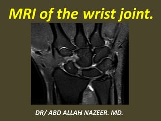
  • 1. MRI of the wrist joint. DR/ ABD ALLAH NAZEER. MD.
  • 2. 1. Acute and chronic wrist instability. 2. Dorsal or ulnar-sided wrist pain. 3. Wrist symptoms in adolescent gymnasts and other athletes. 4. Unexplained chronic wrist pain. 5. Acute wrist trauma. 6. Avascular necrosis. 7. Wrist malalignments. 8. Limited or painful range of motion. 9. Unexplained wrist swelling, mass, or atrophy. 10. Planning for diagnostic or therapeutic arthroscopy. 11. Recurrent, residual, or new symptoms following wrist surgery. Indications for MRI of the wrist joint.
  • 3.
  • 4. MR Sequences images. T1WI (Lower Egypt). T21WI (Upper Egypt).
  • 6.
  • 7.
  • 8.
  • 9.
  • 10.
  • 11.
  • 12.
  • 13.
  • 15.
  • 16.
  • 17.
  • 18. The transverse carpal ligament (short arrows) extends from the hook of the hamate (long arrow) to the tubercle of the trapezium (arrowhead), forming the floor of the carpal tunnel. At the radial aspect of the carpal tunnel, the flexor digitorum tendons are arranged in two rows (separated by dotted line), the profundus tendons deep to the superficialis tendons. The flexor pollicis longus tendon (star) is positioned at the ulnar aspect of the tunnel, separated from the flexor carpi radialis tendon (curved arrow) by a ligamentous reflection of the transverse carpal ligament. The median nerve is indicated (asterisk). - See more at: http://radsource.us/palmar-bursae-and-flexor-tendon-sheaths/#sthash.mSFX1u0c.dpuf
  • 19.
  • 20.
  • 21.
  • 22.
  • 23.
  • 24.
  • 25.
  • 26.
  • 27.
  • 28.
  • 29.
  • 30.
  • 32.
  • 33.
  • 34. CT images with small cyst formation within the fracture line.
  • 35. MR Protocol with pre and post-contrast images for fracture.
  • 36.
  • 37. Scaphoid waist fracture with subtle collapse of the proximal pole, related AVN .
  • 38.
  • 39. Diffuse enhancement of scaphoid proximal pole has good correlation with stage group I, patchy enhancement have correlation with grade II and III and enhancement with complete necrosis.
  • 40.
  • 41. Preiser's disease. Coronal inversion recovery (A) and fast spin echo (B) images: 1. a bone marrow edema pattern in the scaphoid (black arrow) and 2. avascular necrosis of the proximal pole (white arrow). No fracture is visible.
  • 42. Idiopathic avascular necrosis of the scaphoid (Preiser’s disease)
  • 43.
  • 44.
  • 45.
  • 46.
  • 47.
  • 49.
  • 51. Intraosseous ganglion. Cor. T2FFE. Rounded high signal intensity lesion in the radial aspect of the lunate with cortex erosion.
  • 53.
  • 54.
  • 55. A simple approach is to consider the lunate which is usually the easiest carpal bone to visualize on a lateral wrist image. If the lunate is abnormally tilted in a dorsal direction on a standard lateral wrist image, a DISI should be considered. If the lunate is abnormally tilted in a volar direction a VISI should be considered. DISI is due to disruption of the scapho-lunate articulation. VISI is secondary to disruption of the luno- triquetral articulation.
  • 56.
  • 57.
  • 58.
  • 59.
  • 62. DISI.
  • 63.
  • 64.
  • 65.
  • 66.
  • 67. Volar intercalated segmental instability(VISI).
  • 68. VISI with volar tilting of the lunate.
  • 69. VISI with luno-triquetrum ligament tear and volar tilting of the lunate.
  • 70. VISI deformity secondary to Lunotriquetral ligament tear.
  • 71.
  • 72.
  • 73. Scapholunate dissociation. Note the widened scapholunate space (“Terry Thomas” sign) and the foreshortened scaphoid with a “scaphoid ring” sign (arrows). Adjacent is British comedian Terry Thomas and his famous teeth.
  • 75. Scapho - lunate ligament tear
  • 76.
  • 77.
  • 78.
  • 79.
  • 81. 1. Scapholunate ligament tear, dorsal angulation of the lunate and proximal migration of the capitate consistent with a DISI deformity. 2. Tear of the lunotriquetral and triscaphe ligaments.
  • 82.
  • 83.
  • 84.
  • 85.
  • 86.
  • 88.
  • 89. Acute TFCC tear on FS T2-WI (A) and T1-WI (B).
  • 90. Ulnar variance may be : Neutral (both the ulnar and radial articular surfaces at the same level) Positive (ulna projects more distally) Negative (ulna projects more proximally) Causes trauma or mechanical distal radius/ulnar fractures with shortening (e.g. impaction) & angulation DRUJ ligamentous injuries (e.g. Galeazzi & Essex-Lopresti) surgical shortening of ulna or radius growth arrest (e.g. previous Salter-Harris fracture) Congenital Madelung deformity/reverse Madelung deformity Associations positive ulnar variance is associated with ulnar impaction syndrome. negative ulnar variance is associated with Keinbock,s disease and ulnar impingement syndrome
  • 91. Positive ulnar variance secondary to TFC tear.
  • 92. Positive ulnar variance secondary to TFC tear.
  • 93. TFCC 1B tear (arrow) on coronal T1-WI (B) and coronal FS T2-WI (C).
  • 94. Negative ulnar variance secondary to TFC tear.
  • 96.
  • 97.
  • 98.
  • 99.
  • 100.
  • 101.
  • 102. Swelling, deformity, abnormal signal of the median nerve.
  • 104. Carpal tunnel syndrome caused by cysticercosis.
  • 105. Sarcoidosis with carpal tunnel syndrome.
  • 106.
  • 107.
  • 108.
  • 109.
  • 110. Lipoma at the Ulnar tunnel.
  • 111.
  • 112.
  • 113.
  • 114.
  • 115.
  • 116.
  • 117.
  • 118.
  • 119.
  • 121.
  • 122. Tenosynovitis. Tenosynovitis is an inflammation of the tendon sheath , either primary or secondary. Marked inflammatory tenosynovitis.
  • 123. Tenosynovitis of Extensor Carpi Ulnaris.
  • 124. Extensor carpi ulnaris tenosynovitis. Axial FS T2-WI shows fragmentation into multiple tendon fragments of the extensor carpi ulnaris tendon (arrowheads). Note increased fluid and debris within the tendon sheath.
  • 125. Tenosynovitis and partial thickness tearing of the FCR tendon (arrow).
  • 126. Tenosynovitis of the flexor tendons.
  • 127. Tenosynovitis of all flexor tendons FDS, FDP, FPL
  • 128. Tenosynovitis with partial tears of the EPL, ECRB and ECRL
  • 129. Moderate tenosynovitis of the extensor digitorum tendon (yellow arrowhead).
  • 130. Tenosynovitis of the flexor polices tendon.
  • 132. Tenosynovitis in a 32-year-old woman with early rheumatoid arthritis of the wrist (5 months duration) and normal radiographic findings. Axial T1-weighted (a), fat-suppressed T2-weighted (b), and contrast- enhanced fat-suppressed T1-weighted (c) MR images show marked tenosynovitis (arrows) involving both the dorsal extensor and volar flexor compartments. Note that the tenosynovitis and periscaphoid joint synovitis (*) have the same signal intensity in a (intermediate) and b (high) and similar enhancement
  • 133. Flexor Tendon Rice Body Tenosynovitis.
  • 135. Sarcoidosis. (a, b) Axial T2 FS and coronal T1. Increased fluid is present within the flexor tendon sheath with multiple loose bodies (rice bodies) present (blue arrows). (c, d) Axial and coronal T1 FS PG. Thick synovial enhancement is present.
  • 136.
  • 137.
  • 138. De Quervain tenosynovitis of abductor pollicis longus (APL) and extensor pollicis longus (EPL).
  • 140. De Quervain syndrome. (a, b) Axial T2 FS and coronal T2 FS. Increased fluid is present within the APB and EPB tendon sheaths (blue ellipses). Surrounding edema is present, as evidenced by increased T2 weighted signal.
  • 141. SOFT TISSUE HEMANGIOMA OF THE WRIST.
  • 142. Giant cell tumor of the tendon sheath in wrist.
  • 143. Synovial Sarcoma of Wrist Area.