2. INTRODUCTION
Cysticercosis is a disease caused by the presence of
Cysticercus cellulosae and Cysticercus racemose, the
larval forms of Taenina solium
Cysticercus bovis, the larval form of T. saginata
occurs very rarely in man
A new species, T. asiatica was described in 1993
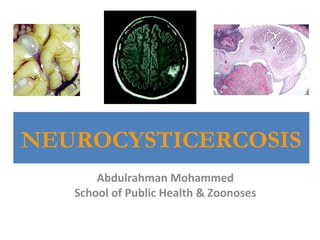
Neurocysticercosis – cysticerci (larval form) form in the
brain
3. NEUROCYSTICERCOSIS
• NCC is due to the development of Taenina solium
cysticerci in the human central nervous system,
where parasites can be found in the parenchyma, the
subarachnoid tissue, and the ventricles
• 26.3% to 53.8% active epilepsy cases in the
developing world including India and Latin America
are due to NCC. It is also becoming more common in
the developed world because of increased migration
of people with the disease or Taenia solium carriers
and frequent travel to the endemic countries.
5. HISTORY
• Cysticercosis was first described in pigs by
Aristophanes and Aristotle in 3rd century BC.
• Later it was noticed in human by Parunoli in 1550.
• Cysticercosis has also been described in ancient Indian
medical book, the Charak Sanhita.
• NCC was first reported in a coolie from Madras, who
died due to seizure and was found to be infected with
cyst on autopsy (Armstrong 1888).
• In 1912, Krishnaswamy (1912) reported cysticerci
related case of muscle pains and
subcuataneousnodules with abundant cysticerci in the
muscles, heart and brain at autopsy.
• In 1934, high rate of new onset epilepsy related to
cysticercosis in the British army deployed in India was
noticed (MacArthur 1934).
6. Two different theories explain the
origin of tapeworms.
1) Domestication of animals 10,000 years ago
7. Two different theories explain the
origin of tapeworms.
2) Scavenging of food 2 million years ago by African
hominids
10. The scolex of T. solium
The scolex of T. solium
has four suckers and an
armed-rostellum. The
scolex of T. saginata
looks similar but lacks
hooks. These two
species can be
differentiated by
counting the number of
uterine branches in the
proglottids; T. solium
has between 7 to 13 per
side, while T. saginata
has 15 to 20.
20. Life cycle of Taenia saginata
Humans are the only definitive hosts for Taenia saginata.
The adult tapeworms (length: usually 5 m or less, but up to
25 m) reside in the small intestine, where they attach by
their scolex. They produce proglottids (each worm has
1,000 to 2,000 proglottids), which mature, become gravid,
detach from the tapeworm, and migrate to the anus or are
passed in the stool (approximately 6 per day). The eggs
contained in the gravid proglottids (80,000 to 100,000 eggs
per proglottid) are released after the proglottid becomes
free and are passed with the feces. The eggs can survive
for months to years in the environment. Cattle and other
herbivores become infected by ingesting vegetation
contaminated with eggs (or proglottids). In the animal's
intestine, the eggs release the oncosphere, which
evaginates, invades the intestinal wall and migrates to the
striated muscles, where its develops into a cysticercus. The
cysticercus can survive for several years in the animal.
Humans become infected by ingesting raw or undercooked
infected meat. In the human intestine, the cysticercus
develops over 2 months into an adult tapeworm, which can
survive for more than 30 years.
22. Life cycle of Taenia solium
The life cycle of Taenia solium is similar to that of
T. saginata. The adults (length 2 to 7 m; less
than 1,000 proglottids, which are less active than
in T. saginata, and each with 50,000 eggs;
longevity up to 25 years) develop not only in
humans but also some other animal species
(monkeys, hamsters). The cysticercus develops
not only in striated muscle, but also in the brain,
liver, and other tissues of pigs and other animals,
including humans. Humans develop taeniasis
when they ingest undercooked pork meat
containing cysticerci. They develop cysticercosis
by ingesting T. solium eggs, either by ingestion of
fecally contaminated food, or by autoinfection. In
the latter case, a human infected with adult T.
solium ingests eggs produced by that tapeworm,
either through fecal contamination or, more
arguably, from proglottids carried into the
stomach by reverse peristalsis.
23.
24.
25. T.saginata T.solium
D.H Human Human Human
I.H Cattle Swine Human
Habitation Small intestine Small intestine Tissue(brain, eye,
skin etc.)
Infective stage Cysticercus bovis
Cysticercus
Cellulosae
Egg
Disease Taeniasis Taeniasis Cysticercosis
26. • Cysticercosis (Intrinsic or extrinsic auto-infection; Cross
infection due to T.solium egg only; Pathogenic factor:
cysticercus cellulosae)
– Symptoms vary with site & intensity of infection
– Clinical aspects: headache, dizziness, epilepsy,
blurred vision, subcutaneous nodule etc
27. Prevalece
• Most common infection of human nervous
system by parasites.
• Most frequent preventable cause of epilepsy in
developing world.
• Affects 50 million people worldwide.
• 50,000 deaths in highly endemic places.
• Interesting that a lot of developed countries also
are endemic for this disease – raw meat, but rate
is going down due to stricter rules for meat
inspection.
28. Epidemic factors
– Egg or gravid proglottid contamination of grass
and soil
– Method of raising domestic animals
– Unhygienic dinning habit of eating raw or
undercooked meat
30. TYPES OF CYSTS
Cysticercus cellulosae
• Less virulent form
• Small (<2cm), round, thin
walled
• Lodges in the
parenchyma or the
subarachnoid space
• Provokes only a minor
inflammation
• Often remain silent
31. TYPES OF CYSTS
Cysticercus racemose
• Large lobulated cysts with predilection
for basal cisterns
• Causes cysticercotic arachnoiditis and
presents as meningitis
• Causes obstruction of 4th ventricle and
resultant raised ICP and
hydrocephalus
• Can cause occlusion of vessels and
vasculits resulting in stroke
• Causes intense inflammatory reaction
and seizures
32. MODE OF INFECTION
• HETEROINOCULATION
– eggs may come from the environment
• INTERNAL AUTOINOCULATION
– regurgitated from proglottids into the stomach
• EXTERNAL AUTOINOCULATION
– from the fingers of an infected person
Humans only acquire cysticercosis when they consume eggs
in food handled by people infected by adult T. solium or
through the faecal-oral route
33. TARGET TISSUES
• Predilection for migration to eyes, CNS and
striated muscles, probably due to high
glycogen and glucose content of these tissues.
• CNS and Eye involvement is termed as
Neurocysticercosis.
34. PRESENTATION
The manifold and diverse clinical presentation of NC
is determined by
• Location of cysts
• Size of cysts
• Cyst load (number of cysts)
• Host’s immune response
36. CLINICAL SIGNS
• The manifestations of NCC are polymorphic;
no symptom or sign is specific.
• Acute symptomatic seizures are the most
common manifestation of human NCC;
• the other clinical conditions include headache,
hydrocephalus, chronic meningitis, focal
neurological defi cits, psychological disorders,
dementia,ocular and spinal cysts
38. CLINICAL PRESENTATION
• Intraventricular NC
– 5- 10% of all cases
– 4th ventricle most common site for obstruction
– Cysts in lateral ventricles less likely to cause
obstruction
– Hydrocephalus and acute, subacute or
intermittent signs of raised ICP without localizing
signs
39. CLINICAL PRESENTATION
• Meningeal NC
– Meningeal irritation resembling TBM
– Raised ICP from oedema, inflammation and
presence of cyst obstructing flow of CSF
45. STAGES OF NC
• Cystic or vesicular stage
• Cyst wall & scolex do not enhance
• Cyst is viable & has a well defined, fluid-filled membrane contains only
one scolex.
• Colloid stage
• Enhancing walls with perilesional oedema
• Earliest stage in the involution of the cyst.
• the fluid contents of the cyst become more turbid and the scolex begins
to degenerate.
• Necrotic, granular stage
• Characterized by parasite necrosis and surrounding inflammation
• Gives an appearance of an eosinophilic structure in which the bladder and
scolex are in various stages of disintegration
• Oedema and/or necrosis of the surrounding neural tissue may be present
in some cases
• Fibro-calcified nodule
• With time, fibrosis develops, progressively occupying the entire lesion
46. DIAGNOSTIC CRITERIA
• Absolute criteria
– Demostration of cysticerci by histologic or microscopic examination of biopsy material
– Visualization of the parasite in the eye by fundoscopy
– Neuroradiologic demostration of cystic lesions containing a characteristic scolex
• Major criteria
– Neuroradiologic lesions suggestive of NC
– Demostration of antibodies to cysticerci in serum by enzyme linked
immunoelectrotransfer blot
– Resolution of intracranial cystic lesions spontaneously or after therapy with albendazole
or praziquantel alone
• Minor criteria
– Lesions compatible with NC detected by neuroimaging studies
– Clinical manifestations suggestive of NC
– Demonstration of antibodies to cysticerci or cysticercal antigen in CSF by ELISA
– Evidence of cysticercosis outside the CNS (eg. Cigar shaped soft tissue calcification)
• Epidemiologic criteria
– Residence in a cysticercosis-endemic area
– Frequent travel to a cysticercosis- endemic area
– Household contact with an individual infected with T. solium
47. DIAGNOSTIC CRITERIA
• Definitive
– 1 absolute
– 2 major
– 1 major + 2 minor + 1 epidemiological
• Probable
– 1 major + 2 minor
– 1 major + 1 minor + 1 epidemiological
– 3 minor + 1 epidemiological
• Possible
– 1 major
– 2 minor
– 1 minor + 1 epidemiological
48. SUGGESTED DIAGNOSTIC CRITERIA
• Absolute criteria
• Histopathological demostration of the parasite in the tissues obtained from the
biopsy of a brain or spinal cord lesion
• Multiple cystic lesions with or without scolex on CT or MRI
• Major Criteria
• Lesion highly suggestive of NC in neuroimaging studies
• Spontaneous resolution or eventual calcification
• Positive serum EITB assay for the detection of antibodies against T. solium
• Minor criteria
• Presence of a characteristic clinical picture
• Positive CSF ELISA
• Cysticercosis outside the CNS
• Aggravation of existing symptoms or appearance of a new symptom following
anticysticercal therapy
• Diagnosis with caution
• Old age
• Patients with pre existing systemic tuberculosis or malignancy
• HIV infection
• Grossly abnormal neurological examination
Garg
49. DDX: Tuberculoma Versus Cysticercus
Granuloma
Cysticercus Granuloma
• Round in shape
• Cystic
• 20mm or less with ring
enhancement or visible
scolex
• Cerebral edema not
enough to produce
midline shift or focal
neurological deficit
Tuberculoma
• Irregular in shape
• Solid
• Greater than 20mm
• Associated with severe
perifocal edema and focal
neurological deficit
Target lesions:
Lesions with central nidus of calcification or a dot enhancement
50. TREATMENT
• Praziquantel three doses of 25-30 mg/kg at 2-
hour intervals on a single day equally effective
to the 50mg/kg 8 hourly dose for 15 days.
• Albendazole 15mg/kg/day in 2 divided doses
for 1 week equally effective to the 15
mg/kg/day 12 hrly for a 1-month period.
51. Surgery restricted to:
• Placement of ventriculo-peritoneal shunts for
hydrocephalus
• Excision of single big cysts causing mass effect
• Endoscopical excision of intraventricular parasites.
• Unfortunately shunts are frequently occluded by the high protein
content and debris in the CSF of patients with extraparenchymal
neurocysticercosis, requiring multiple revisions.
• Deaths due to shunt dysfunction may occur in up to 50% of cases,
mainly in the initial 1–2 years after placement.
52. PREVENTION.
• Changing domestic pig-raising practices.
Keeping/raising pigs that have access to human
faeces
Lack of latrines or latrines accessible by pigs
Eating undercooked or raw pork
• Mass chemotherapy of porcine cysticercosis.
• Community health education.
• S3PVAC Porcine vaccine against cysticercosis is
available (pig farmers cannot afford it though) –
eliminates infection.
53.
54.
55. INDIAN SCENARIO
• Cysticercosis has been designated as a “biological marker”of the
social and economic development of a community
• Recent Indian studies using neuroimaging techniques suggest that
the disease burden in India surpasses many other developing
countries
• All the biological markers for transmission of T. solium taeniasis and
cysticercosis exist in India
• Disease is under reported in India because due attentionhas not
been given to this neglected disease and systematic population-based
studies are lacking
• There are great disparities within the country in geography,
ethnicity, religious rituals, income, food habits, personal hygiene,
level of education and standards of living, which are likely to
influence the disease burden.
• There are wide variations in the frequency of cysticercosis in India
57. • There are only few reports from the State of Kerala,where the
level of education and standards of hygiene are high, and
from Jammu and Kashmir, a Muslim majority State due to
prohibition of pork consumption by religion
• Before the era of CT scan and magnetic resonance imaging
(MRI),National Institute of Mental Health and Neuro Sciences
(NIMHANS), Bangalore reported diagnosis of NCC in 2% of an
unselected series of epileptics (Mani et al 1974)
• At atertiary referral centre in New Delhi, NCC constituted
2.5%of all intracranial space occupying lesions (Wani et al
1981).
• With the availability of CT and MRI, the proportion of NCC in
seizure disorders dramatically increased. Sawhney et al
(1996) reported cerebral cysticercosis in 31% of patientsin
whom CT was done.
• In a community survey of 50,617 individuals from South India,
the prevalence of active epilepsy was 3.83 per 1000 and NCC
was detected in 28.4% of them by CT (Rajshekhar et al 2006).
58. • Cysticercosis appears to be more prevalent in the northern States Bihar,
Uttar Pradesh through Punjab. In a recent study based on 30 cluster
sampling approach suggested by WHO in the rural pig farming community
of Mohanlalganj block, Lucknow district, Uttar Pradesh, the prevalence of
taeniasis was 18.6 (Prasad et al 2007).
• In the same community active epilepsy was identifi ed and clinically confi
rmed in 5.8% of the populations during door to door survey and 48.3% of
them fulfilled either defi nitive or probable diagnostic criteria of NCC.
Epilepsy in the family and no separate place for pig were identifi ed as risk
factors for NCC clustering. (Prasadet al 2008)
• In a study of 156 histologically proven cases of cysticercosis from Patiala,
Punjab, 88% patients presented with solitary lesion and the most frequent
site being the upper arm, chest wall, eye, abdomen wall and neck (Saigal
et al 1984)
• The prevalence of taeniasis ranged from 0.5-2% in hospitalized patients in
northern India, 12–15% in labour colonies where pigs are raised (Mahajan
et al 1982).
• The treatment gap in rural India is above 90% (Prasad et al 2008b) and the
probable reasons for such high gap are socioeconomic, lack of knowledge
and medical facilities, social prejudice to modern medicine and faith in
alternative treatment modalities.
59. Cysticercosis in Swine
• Cysticercosis also appears to be widespread in swine in
India. In and around Chandigarh, 8-10% of the pigs
slaughtered had cysticerci in their muscles and around
0.5% of the pigs reared in Government farms were
found to be infected (Mahajan et al 1982).
• Another survey in slaughter houses of Kolkata (West
Bengal) revealed cysticercosis in muscles of 7% of the
slaughtered pigs (Ratnam et al 1983).
• Prasad et al (2002) reported a high frequency of
cysticercosis (26%) in swine from Mohanlalganj block
of Lucknow district in the State of Uttar Pradesh and
40% ofthem had cysticerci in the brain.
60. Neurocysticercosis-more than a
neglected disease (Nash et al., 2013)
• Neurocysticercosis (NCC) is the most common cause of adult-acquired epilepsy worldwide
and one the most frequent parasitic infections associated with chronic morbidity in the
United States.
• Despite its importance worldwide morbidity due to NCC is underappreciated and research is
underfunded, and therefore researchers are unable to capitalize on recent advances that
hold great promise to prevent millions of cases of epilepsy and to effectively treat viable
brain infections.
• By conservative estimates, greater than 5 million cases of epilepsy worldwide, which are all
preventable, are caused by NCC.
• The study of NCC is inherently difficult. The life cycle is near impossible to maintain in the
laboratory, and for practical purposes purposes most basic research cannot be performed in
developed, non-endemic countries. The high cost of the logistics to establish and maintain
the stages of the life cycle required for basic experiments is compounded by the difficult
challenge of obtaining financial support for a neglected disease.
• It is easy to forget that a few decades ago the diagnosis of NCC was made infrequently,
medical treatments were unavailable, and its role as a primary cause of adult onset epilepsy
unknown
• Controlled human treatment trials and observational studies are difficult to conduct because
of their cost, need for frequent imaging, and requirement for long periods of follow up.
61.
62. Areas of study
1) Determine the extent and burden of disease worldwide.
2) Understand that great strides can be made with relatively
few resources, for example:
a. Development of methods to test for new and effective
drugs.
b. Use of existing, licensed immunomodulators to control
treatment- induced inflammation and damaging
inflammation in parenchymal and subarachnoid disease.
c. Effective use of vaccination in pigs.
3) Boost support for the relatively few programs devoted to
NCC.
4) In the United States, realize that health care and research is
hindered because those with disease have the least access to
health care; they are commonly undocumented Central and
South American immigrants. Often, the most minimal care
and testing is allowed or given.