Gallstone disease rufi
•Download as PPT, PDF•
19 likes•2,838 views

evaluation & management of gallstone diseases in emergency

Recommended
More Related Content
What's hot
What's hot (20)
Viewers also liked
Viewers also liked (20)
Similar to Gallstone disease rufi
Similar to Gallstone disease rufi (20)
More from Dr Abdul sherwani
More from Dr Abdul sherwani (13)
Recently uploaded
Recently uploaded (20)
Gallstone disease rufi
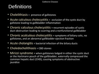
- 1. Gallstone Disease Definitions • Cholelithiasis = presence of gallstones • Acute calculous cholecystitis = occlusion of the cystic duct by gallstone leading to gallbladder inflammation • Chronic calculous cholecystitis = recurrent episodes of cystic duct obstruction leading to scarring and a nonfunctional gallbladder • Chronic acalculous cholecystitis = symptoms of biliary colic, no gallstones, and an abnormal gallbladder ejection fraction • Acute cholangitis = bacterial infection of the biliary ducts • Choledocholithiasis = CBD stones • Mirizzi syndrome = when gallstones lodged in either the cystic duct or the Hartmann pouch of the gallbladder, externally compressed the common hepatic duct (CHD), causing symptoms of obstructive jaundice
- 2. Gallstone Disease Types of Gallstones •Pure cholesterol (70%)-radioluscent •Pigmented (30%)-radiopaque • PURE 20%-Black stones (contain Ca bilirubinate, a/w cirrhosis and hemolysis) • MIXED 10%-Brown stones (a/w biliary tract infection)
- 3. Gallstone Disease Risk Factors Calculous • “Female, Fat, Forty, Fertile” • Oral contraceptives • Obesity • Fatty diet • Hemolytic states • Cirrhosis • Bile duct stasis (biliary stricture, congenital cysts, pancreatitis, sclerosing cholangitis) • IBD • Vagotomy • Hyperlipidemia Acalculous • Rapid weight loss (gastric bypass pts.) • DM • Prolonged fasting • TPN • Ileal resection • Old age • Severe illness • Burns ,trauma
- 4. Gallstone Disease Gallstone Complications • Gallstone ileus, gallstone pancreatitis • Acute cholecystitis: 10-20% of pts. w/ symptomatic gallstones • GB gangrene • GB perforation • GB empyema (pus in the GB) • Emphysematous cholecystitis (a/w GB vascular compromise, stones, impaired immune system, infection w/gas-forming organisms - clostridium, E. coli, Klebsiella) • Cholecystoenteric fistula • Choledocholithiasis: 8-15% of pts. w/ symptomatic gallstones • Cirrhosis • Cholangitis • Pancreatitis
- 5. Gallstone Disease RUQ DDx • Gallbladder: cholecystitis, Choledocholithiasis, cholangitis • Duodenal ulcer • Hepatitis • Appendicitis (atypical presentation) • Pancreatitis
- 6. Gallstone Disease Strasberg S. N Engl J Med 2008;358:2804-2811 Diagnostic Criteria for Acute Cholecystitis, According to Tokyo Guidelines
- 7. Gallstone Disease CLUE • Provocation/Timing: meals (50%), night time • Quality: constant • Radiation: RUQ to the R scapula (Boas’ sign) • Severity: “severe”
- 8. Gallstone DiseaseComplication History Examination Blood tests Biliary Colic - Intermittent RUQ/epigastric pain (minutes/hours) into back or right shoulder - N&V -Tender RUQ -No peritonism -Murphy’s – -Apyrexial, HR and BP (N) -WCC (N) CRP (N) - LFT (N) Acute Cholecystitis -Constant RUQ pain into back or right shoulder -N&V -Feverish -Tender RUQ -Periotnism RUQ (guarding/rebound) -Murphy’s + -Pyrexia, HR (↑) -WCC and CRP (↑) -LFT (N or mildly (↑) Empyema -Constant RUQ pain into back or right shoulder -N&V -Feverish -Tender RUQ -Peritonism RUQ -Murphy’s + -Pyrexia, HR (↑), BP (↔ or ↓) -More septic than acute cholecystitis -WCC and CRP (↑) -LFT (N or mildly (↑) Obstructive Jaundice -Yellow discolouration -Pale stool, dark urine -painless or assocaited with mild RUQ pain -Jaundiced -Non-tender or minimally tender RUQ -No peritonism -Murphy’s – -Apyrexial, HR and BP (N) -WCC and CRP (N) -LFT: obstructive pattern bili (↑), ALP (↑), GGT (↑), ALT/AST (↔) -INR (↔ or ↑) Ascending Cholangitis Charcot”s triad -RUQ pain (constant) -Jaundice -Rigors -Jaundiced -Tender RUQ -Peritonism RUQ -Spiking high pyrexia (38-39) -HR (↑), BP (↔ or ↓) -Can develop septic shock -WCC and CRP (↑) -LFT : obstructive pattern bili (↑), ALP (↑), GGT (↑), ALT/AST (↔) -INR (↔ or ↑) Acute Pancreatitis -Severe upper abdominal pain (constant) into back -Profuse vomiting -Tender upper abdomen -Upper abdominal or generalised peritonism -Usually apyrexial, HR (↑), BP (↔ or ↓) -WCC and CRP (↑) -LFT: (N) if passed stone or obstructive pattern ifstone still in CBD -Amylase (↑) -INR/APTT (N) or (↑) if DIC Gallstone Ileus - 4 cardinal features of SBO -distended tympanic abdomen -hyperactive/tinkling bowel sounds
- 9. Gallstone Disease Labs • Order: BMP, amylase/lipase, LFTs, CBC, coags • Acute cholecystitis: increased WBC, increased alk phos, slight increase in amylase and T bili
- 10. Gallstone Disease Imaging • KUB - only 15% of gallstones are radiopaque • U/S - gallstone identification false(-) rate is 5-15%. It identifies bile duct dilatation w/ 80% accuracy. Look for • thickened GB wall (>3.5mm)-independent predictor of acute cholecystitis .(ppv 95%) • pericholecystic fluid, distended GB, •Murphy’s sign-sensitivity as high as 88%
- 11. Gallstone Disease Strasberg S. N Engl J Med 2008;358:2804-2811 Ultrasonographic Images of Three Gallbladders
- 12. Gallstone Disease • CT scan - used to diagnose complications • MRI - can detect gallstones and common duct stones • ERCP - to look for CBD stones • HIDA scan - radionuclide IV, extracted from blood, excreted into bile
- 13. Gallstone Disease Thomas L et al. N Engl J Med 1999;341:1134-1138 CT Scan of the Abdomen
- 14. Gallstone Disease Strasberg S. N Engl J Med 2008;358:2804-2811 Hepatobiliary Scintigraphy
- 15. Gallstone Disease Management • biliary colic: oral or parenteral opiate analgesics and NSAIDS( ketorolac). • complicated: analgesia, antiemetics , cessation of oral intake, volume and electrolyte replacement, antibiotics(cefotaxime or ceftriaxone, 1 gram IV) and metronidazole; or a fluoroquinolone and metronidazole) • ERCP is undertaken for patients with common bile duct stones or dilated common bile ducts
- 16. Gallstone Disease Case 1 • HPI: 46y F p/w 4hr h/o nausea and RUQ pain radiating to the R scapula. Symptoms began 1 hr. after a fatty meal. Pt. currently has no pain. No prior episodes. • PMHx/PSHx None • PE: RUQ minimally TTP, (-)Murphy’s • Labs: WBC 8, LFT normal
- 17. Gallstone Disease Case 1 What is the diagnosis? → → ►
- 18. Gallstone Disease Case 1: Continued • Dx: symptomatic cholethiasis • Plan: NPO, IVF, cholecystectomy
- 19. Gallstone Disease Case 2 • 46yF p/w h/o >24hr of RUQ pain radiating to the R scapula, started after fatty meal, a/w nausea, vomiting, fever • Exam: Febrile, RUQ +, (+)Murphy’s sign • Labs: WBC 13, Mild ↑LFT
- 20. Gallstone Disease Case 2: Continued ◄
- 21. Gallstone Disease Case 2: Continued • → denotes the GB wall thickening • ► denotes the fluid around the GB • GB also appears distended • ?DIAGNOSIS → ►
- 22. Gallstone Disease Case 2: Continued • Dx: acute calculous cholecystitis • Persistent cystic duct obstruction leads to GB distension, wall inflammation & edema • Risk of: empyema, gangrene, rupture • Treatment: • NPO • IVF • ABX: • Common organisms: E coli, Bacteroides fragilis, Klebsiella, Enterococcus, and Pseudomonas Sanford guide:2006/11 • Piperacillin/tazobactam (Zosyn), ampicillin/sulbactam (Unasyn), or meropenem • Cholecystectomy
- 23. Gallstone Disease Case 3 • 87y M critically ill, on long-term TPN c/o RUQ pain • PE: febrile, RUQ + • U/S: GB wall thickening, pericholecystic fluid, no gallstones • What is the diagnosis?
- 24. Gallstone Disease Case 3: Continued • Dx: acute acalculous cholecystitis • Caused by gallbladder stasis from lack of enteral stimulation by cholecystokinin • Risk of: gangrene, empyema, perforation due to ischemia • TX: rapid progression of acute acalculous cholecystitis to gangrene & perforation, early recognition and intervention are required.
- 25. Gallstone Disease Case 4 • 46y F p/w RUQ pain, jaundice, acholic stools, dark tea-colored urine, w/o fever • PMHx: Cholelithiasis • Exam: unremarkable • WBC 8, T.Bili 8, AST/ALT NL, Hep B/C neg • U/S: gallstones, CBD stone, dilated CBD > 1cm • What is the diagnosis?
- 26. Gallstone Disease Case 4: Continued • DX: Choledocholithiasis • Similar presentation as Cholelithiasis, except with the addition of jaundice • DDx: Cholelithiasis, hepatitis, cholangitis, CA, choledochal cyst, bile duct stricture, UC, pancreatitis • Plan: • Endoscopic retrograde cholangiopancreatography (ERCP) w/ stone extraction and sphincterotomy • Interval cholecystectomy after recovery from ERCP
- 27. Gallstone Disease Case 5 • 46y F p/w 4hr h/o nausea and RUQ pain radiating to the R scapula. Symptoms began 1 hr. after a fatty meal. Pt. currently has no pain. Has had multiple similar episodes. • PMHx/PSHx None • PE: RUQ minimally TTP, (-)Murphy’s • Labs: WBC 6, LFT normal • Studies: RUQ U/S w/Cholelithiasis without GB wall thickening or pericholecystic fluid • Diagnosis: ?
- 28. Gallstone Disease Case 5: Continued • Dx: chronic calculous cholecystitis • Recurrent inflammatory process due to recurrent cystic duct obstruction leading to scarring/wall thickening • Treatment: cholecystectomy
- 29. Gallstone Disease Case 6 • 46y F p/w persistent epigastric & back pain • PMHx: symptomatic gallstones • SHx: no ETOH • PE: Tender epigastrum • Labs: Amylase 2000, ALT 150 • U/S: gallstones • What is the diagnosis? • What is the plan?
- 30. Gallstone Disease Case 6: Continued • Dx: gallstone pancreatitis • 35% of acute pancreatitis secondary to stones • Pathophysiology: reflux of bile into pancreatic duct and/or obstruction of ampulla by stone • ALT >150 (3-fold elevation) has 95% PPV for diagnosing gallstone pancreatitis • Treatment: • ABC, resuscitate, NPO/IVF, pain medication • ERCP once pancreatitis resolves • Cholecystectomy before d/c
- 31. Gallstone Disease Take Home Points • Start with ABCs • Cholelithiasis = “Female, Fat, Forty, Fertile” • Stone formation based on the relative concentration of cholesterol, bile salts, and phospholipid • Cholecystitis PE = Murphy’s sign • RUQ evaluation: U/S, HIDA, CT, MRI, ERCP • Acalculous cholecystitis a/w TPN, ICU setting • Cholangitis = Charcot’s triad, Reynold’s pentad
Editor's Notes
- Pure cholestrol_-cholestrol monohydrate crystals-bile super saturation/motility decreases formation of sludge Composition of bile:61% biles salts(tarocolic acis& chenodeoxycholic acid),12 % fatty acids 9% cholestrol phospholipids,bilirubin 3% protein 7% others 5%
- CRP : 1-3mg/l
- SBO: PAIN ,VOMITTING,CONTIPATION,DISTENTION& BOWEL SOUND CHANGES
- Figure 1. Ultrasonographic Images of Three Gallbladders. A normal, sonolucent gallbladder (Panel A) is characterized by a thin wall and an absence of acoustic shadows. In a patient with symptomatic gallstones (Panel B), the gallbladder contains small echogenic objects with posterior acoustic shadows that are typical of gallstones (arrow), with a normal wall thickness. In a patient with acute calculous cholecystitis (Panel C), thickening is visible in the gallbladder wall (arrow), along with a large gallstone (arrowhead).
- Uptake by liver, GB, CBD, duodenum w/in 1hr = normal Slow uptake = hepatic parenchymal disease Filling of GB/CBD w/delayed or absent filling of intestine = obstruction of ampulla Non-visualization of GB w/ filling of the CBD and duodenum = cystic duct obstruction and acute cholecystitis (95% sensitivity & specificity)
- Figure 1. CT Scan of the Abdomen. There is an abnormal air density within the gallbladder fossa and the gallbladder wall, with associated pneumobilia. The findings are consistent with the presence of emphysematous cholecystitis and cholangitis.
- Figure 2. Hepatobiliary Scintigraphy. In Panel A, a normal liver is visible 10 minutes after the intravenous injection of a technetium-labelled analogue of iminodiacetic acid. In Panel B, at 55 minutes after tracer injection, filling of the bile duct (arrow) and gallbladder (arrowhead) can be seen. In Panel C, at 1 hour after tracer injection in a patient with acute cholecystitis and obstruction of the cystic duct, there is filling of the bile duct (arrow) but no filling of the gallbladder.
- Untreated acute cholecystitis may lead to severe complications such as ascending cholangitis, emphysematous cholecystitis, gangrenous cholecystitis, or pancreatitis.
- Studies: RUQ U/S w/Cholelithiasis without GB wall thickening or pericholecystic fluid → denotes gallstones ► denotes the acoustic shadow due to absence of reflected sound waves behind the gallstone
- Curved arrow Two small stones at GB neck Straight arrow Thickened GB wall ◄ pericholecystic fluid = dark lining outside the wall
- STANDFORD GUIDELINE
- If pt. is too sick, percutaneous cholecystectomy tube followed by cholecystectomy
- CBD ON ULTRASOUND 6-7 MM
- MAIN AIM TO TO RELIEVE THE OBSTRUCTION OR ATLEAST DRAINAGE OF THE BILE
- Reynolds pentad: shock & altered mental status