Viral infections of oral cavity - Dr. Abhishek Solanki
•Download as PPTX, PDF•
180 likes•65,443 views


Recommended
More Related Content
What's hot
What's hot (20)
Viewers also liked
Similar to Viral infections of oral cavity - Dr. Abhishek Solanki
Similar to Viral infections of oral cavity - Dr. Abhishek Solanki (20)
Recently uploaded
Recently uploaded (20)
Viral infections of oral cavity - Dr. Abhishek Solanki
- 2. VIRAL INFECTION HERPES SIMPLEX VIRUS ENTEROVIRUS VARICELLA RUBEOLA HERPES ZOSTER RUBELLA INFECTIOUS MUMPS MONONUCLEOSIS HIV CYTOMEGAVIRUS
- 3. HERPES SIMPLEX VIRUS Members of the Herpes Virus Family which are some of the most common human viruses The Type 1 virus causes cold sores. Most people get Type 1 infections during infancy or childhood. The Type 2 virus causes genital sores. Most people get Type 2 infections following sexual contact with an infected person.
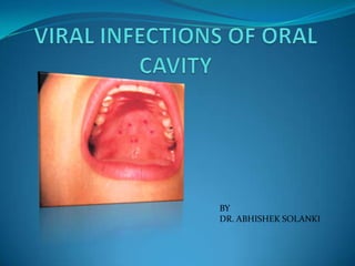
- 4. Acute Herpetic Gingivostomatitis 6 mon – 5 yrs (peak 2-3 yrs) before 6 months rare because of protection by maternal anti- HSV antibodies. Onset is abrupt & accompanied by anterior cervical lymphadenopathy, chills, fever ( 103 to 105 F).
- 5. Pharyngotonsillitis Sore throat, Fever, Malaise & Headache. Numerous vesicles develops on the tonsils & posterior pharynx. Vesicles ruptures to form ulcers which coalsce.
- 6. Herpes Labialis "labia" = "lip” Age: Adults Sex: No predilection Most common recurrent site for HSV-1 is vermilion border & adjacent skin of lip.
- 7. In some pt UV light & trauma trigger recurrence. Pain, Burning, Itching, tingling, Localized warmth, erythema of involved epithelium. Multiple small, erythematous papules develop & form clusters of fluid filled vesicles. Persistent herpes labialis is indicative of immunocompromised status, including HIV infection.
- 8. DIFFERENTIAL DIAGNOSIS Impetigo Contact dermatitis
- 9. HERPETIC WHITLOW A/k/a herpetic paronychia Medical & Dental personnel infect their digits by contact with infected patients. Can cause permanent scarring
- 10. Herpes gladiaotorum a/k/a scrumpox Ocular involvement may occur d/t self inoculation Pt with diffuse chronic skin disease, such as eczema, pemphigus and Darier’s disease may develop life threatening HSV infection ka ECZEMA HERPETICUM (KAPOSI’s VARICELLIFORM ERUPTION).
- 11. COMPLICATIONS Meningitis Encephalitis Eczema herpetiform-- widespread herpes across the skin)
- 12. Keratoconjunctivitis-- Infection of the eye Pneumonia Infection of the trachea Keratitis-- Corneal infection, irritations, and inflammations
- 13. H/P Infected epithelial cells exhibit acantholysis, nuclear clearing, nuclear enlargement which has been termed ballooning degeneration. Tzanck cells (multinucleated giant cells) Multinucleated, infected epithelial cells, infected cells are formed when fusion occurs between adjacent cells.
- 15. Diagnosis Clinical presentation Viral isolation from tissue culture HSV antigens. Serologic test for HSV antibodies (4-8 wks after infection)
- 16. TREATMENT & PROGNOSIS Acyclovir suspensions Viscous lidocaine (not in pediatrics) NSAIDS Anti-viral vaccines (studies)
- 17. VARICELLA VZV or HHV – 3 DNA virus Two clinically distinct syndromes Chickenpox Shingles. Acquired by inhalation or contact, with primary infection of conjunctiva or upper airway mucosa
- 19. Primary Varicella (Chicken Pox) Age: Children Sex: No predilection Dermal vesicular exanthem incubation period lasts 2 to 3 weeks
- 20. Early onset of vesicles that rapidly rupture & leave erosions with a surface pseudomembrane lesions located on the trunk and face, are vesicular with an erythematous boundary, and are extremely pruritic. Fever, malaise mild generalized lymphadenopathy lesions resolve within 5 to 8 days.
- 21. H/P Superficial intraepithelial vesicle formation. vesicular contents contain eosinophilic exudate, inflammatory cells and epithelial cells. Nuclear ballooning Superficial submucosal inflammatory cell infiltrate DIFFERENTIAL DIAGNOSIS Herpes Coxsackieviruses Aphthae.
- 22. Herpes zoster Age: Adults and elderly people Sex: Slight male predilection VZV spreads from skin/mucosa into sensory nerve endings
- 23. Virus travels to dorsal root ganglion and becomes latent Reactivation occurs with decreased cell-mediated immunity Initial replication occurs in affected ganglion after reactivation
- 24. Clinical Features Prodrome: Headache photophobia malaise fever abnormal skin sensations pain
- 25. Rash: Vesicular eruption follows the distribution of sensory nerves, being segmental and unilateral. Thoracic , cervical, ophthalmic involvement most common Initially erythematous, maculopapular Vesicles form over several days, then crust over Full resolution in 2-4 weeks
- 26. Complications of Herpes Zoster Postherpetic Neuralgia (PHN) Herpes Zoster Ophthalmicus VZV viremia Dermatologic complications
- 27. Histopathologic Features Same as HSV Treatment and prevention Vaccination Acyclovir VZIG as post-exposure prophylaxis in individuals at high risk Exclude kids from school until sixth day of rash
- 28. Infectious Mononucleosis Aka Glandular Fever & Kissing Disease because adult contract the virus through direct salivary transfer like straws or kissing 7-10 days incubation period. Acute self-limiting infection Epstein-Barr Virus
- 29. Clinical Features Age : Young Adults Sex : no prediliction Petechiae on hard palate Lymphadenopathy, Pharyngitis, Tonsillitis. Sore throat, fever, rash
- 30. NUG is common. Malaise, lethargy, extreme tiredness Liver and spleen involvement and enlargement Hematology: High WBC, over 20% atypical reactive lymphocytes also known as Downey cells.
- 31. Histopathologic feature Downey cells – atypical lymphocytes diagnostic feature DIFFERENTIAL DIAGNOSIS Platelet disorders Hereditary hemorrhagic telangiectasia.
- 32. T/t Supportive Bed rest and high liquid intake. Mild analgesic and antipyretic
- 33. Cytomegalovirus HHV-5 Transmission occurs from person to person. Close intimate contact Sexual contact During delivery Breast milk Organ transplant Blood transfusion
- 34. Clinical features Symptoms resemble IM In babies may cause life threatening illness Patients with deficient immune systems AIDS patients Transplant patients Common in AIDS pt.
- 35. 90 % of CMV are infections are assymptomatic Typical Features Hepatosplenomegaly Thrombocytopenia • Fever • Malaise • Myalgia
- 36. H/P Scattered infected cells are extremely swollen, showing both intracytoplasmic and intranuclear inclusions and prominent nuclioli - Owl Eye
- 37. Diagnosis Clinical Features Viral Antigen Treatment CMV infection resolve spontaneously Gancyclovir in immunocompromised patient
- 38. Enteroviruses Genus of the picornavirus family which replicate mainly in the gut. Single stranded RNA virus
- 39. Divided into 5 groups Polioviruses Coxsackie A viruses & Coxsackie B viruses Echoviruses Enteroviruses Herpanginia, Hand-foot-and-mouth disease, Acute lymphonodular pharyngiitis
- 40. Herpangina Caused by Coxakievirus A 1 to 6, 8, 10, 22 Coxakievirus A7, 9 or 16; Coxakievirus B 2 to 6; Echovirus 9,16,17; enetrovirus 71. Age: Children Sex: No predilection
- 41. Most cases arise in summer with crowding & poor oral hygiene. Fecal-oral route : major path of transmission
- 42. Clinical Features Sore throat Dysphagia Fever, cough Rhinorrhea Anorexia.
- 43. Vomiting, diarrhea and headache. Mostly soft palate or tonsillar pillars involved affected areas begin as red macules which form fragile vesicles that rapidly ulcerate.
- 44. H/P Intraepithelial vesicles contain eosinophilic exudate. Nuclear ballooning degeneration of epithelial cells. DIFFERENTIAL DIAGNOSIS Infection by Herpes virus & Varicella zoster
- 45. Hand-Foot-and-Mouth Disease Coxsakievirus A 5,9,10,16 Age: Children and young adults Sex: No predilection
- 46. Like herpangina skin rash & oral lesions with flu like symptoms like fever, dysphagia, sore throat associated with cough, anorexia, vomiting, diarrhea, headache. Without prodomal symptoms
- 47. Buccal mucosa, labial mucosa and tongue most affected. after a short incubation period, vesicles with an erythematous halo appear in the oral cavity, on the hands, and on the feet
- 48. H/P Intraepithelial vesicles – early stages with intra- cytoplasmic eosinophilic inclusion bodies. Later stages - shallow ulcerations and erosions with regeneration of the marginal epithelium. Superficial inflammatory cell infiltrate in submucosa.
- 49. DIFFERENTIAL DIAGNOSIS Herpetic gingivostomatitis, Herpangina, Varicella, and Aphthous stomatitis
- 50. Acute Lymphonodular pharyngitis Clinical Features Coxsakievirus A 10 Sore throat, fever, mild headache Yellow to dark pink nodules on soft palate and tonsillar pillars
- 51. H/P Affected epithelium exhibit intracellular & intercellular edema leads to intraepithelial vesicle. Vesicle enlarges and ruptures through the epithelial basal cell layer which leads to subepithelial vesicle. Diagnosis Virus Isolation Serology
- 52. Treatment Most cases self limited Symptomatic relief Topical anaesthetics Nonaspirin antipyretics
- 53. Rubeola (Measles) Paramyxo RNA virus Highly contagious Primarily respiratory infection Incubation approximately 10 days, ranges from 8-13. Rash appears at about day 14.
- 54. Prodromal Symptoms irritability, runny nose, eyes that are red and sensitive to light, cough, and high fever Koplik’s spot- small, red, irregular with blue white centres on mouth and conjunctiva Rash on forhead, face, neck, limbs
- 55. Complications bronchitis bronchiolitis pneumonia conjunctivitis myocarditis Hepatitis encephalitis
- 56. T/t Self limiting vaccines
- 57. Rubella (Germen Measles) RNA virus – Toga virus Incubation 2- 3 weeks Highly contagious, spread through respiratory tract. Rubella vaccine has resulted in 99% decline in infections.
- 58. Mumps(Endemic Parotitis) Age: Children Sex: No predilection Single stranded RNA virus. Mumps is transmitted by direct contact with saliva and discharges from the nose and throat incubation 16-18 days.
- 59. Virus can infect many parts of body, especially parotid salivary glands & Submandibular also common. Glands usually become increasingly swollen & painful over a period of 1 to 3 days Pain is moderate to severe Both left & right parotid glands may affected
- 60. DIFFERENTIAL DIAGNOSIS Bacterial or occlusive salivary inflammatory disease Sjögren’s syndrome Complications Inflammation and swelling of the brain Orchitis Oophoritis Infection in pregnant women may result in increased risk for fetal death
- 61. Laboratory Testing Complement fixation Hemagglutination inhibition ELISA
- 62. T/t MMR vaccine No specific therapy exists for mumps. Warm or cold packs for the parotid gland tenderness and swelling is helpful. Pain relievers acetaminophen , ibuprofen are also helpful.
- 64. Introduction Human Immuno Deficiency Virus Etiologic agent of Acquired Immunodeficiency Syndrome (AIDS). Characterized by severe depletion of CD4 cells.
- 65. MODES OF TRANSMISSION SEXUAL TRANSMISSION BLOOD OR BLOOD PRODUCTS MATERNAL-FETAL TRANSMISSION INFECTED NEEDLES
- 66. CDC CLASSIFICATION FOR HIV INFECTED PATIENTS CD4 Cell Clinical Categories Categories A B C Asymptomatic, Acute HIV, or Symptomatic Conditions, AIDS-Indicator PGL not A or C Conditions ≥500 cells/µL A1 B1 C1 200-499 A2 B2 C2 cells/µL <200 cells/µL A3 B3 C3
- 67. CLASSIFICATION OF CLINICAL MANIFESTATIONS GROUP I : ACUTE INFECTION GROUP II : CHRONIC ASYMPTOMATIC INFECTIONS GROUP III : PERSISTENT GENERALIZED LYMPHADENOPATHY GROUP IV : AIDS RELATED COMPLEX
- 68. Acute Infections IM HEPATITIS MENINGITIS MENINGOCEPHALITIS
- 69. CHRONIC ASYMPTOMATIC INFECTIONS MOST DANGEROUS GROUP SEROPOSITIVE PT WHO IS APARENTLY HEALTHY CAPABLE OF INFECTION ENLARGED AXILLARY GLANDS HEMATOLOGICAL & IMMUNOLOGICAL ABNORMILITIES
- 70. PERSISTENT GENERALISED LYMPHADENOPATHY LYMPHADENOPATHY in 2 or more extrainguinal sites persisting for more than 3 months in the absence of disease
- 71. AIDS RELATED COMPLEX OPPORTUNISTIC INFECTIONS - Pneumonia, Cryptococcosis, Viral Infections, Toxoplasmosis, TB etc. NEOPLASMS - KS, Lymphoma, SCC
- 72. NEUROLOGIC DISEASES - Meningocephalitis OTHERS - Encephalopathy, Purpura, Thrombocytopenia
- 73. Oral Manifestations of AIDS INFECTION ORAL DISEASE FUNGAL CANDIDIASIS HISTOPLASMOSIS CRYPTOCOCCOSIS VIRAL HERPES SIMPLEX HERPES ZOSTER CMV EBV(HAIRY LEUKOPLAKIA) HHV-8 (KS) ORAL WARTS(HUMAN PAPILOMA VIRUS) BACTERIAL LINEAR GINGIVAL ERYTHMA NUP TUBERCULOSIS
- 74. Oral Manifestations of AIDS Contd.. TYPE OF LESION DISEASE NEOPLASTIC KAPOSI SARCOMA LYMPHOMA SCC LYMHADENOPATHY CERVICAL OTHERS HIV- Necrotizing Ulceration HIV-Salivary Gland Disease / Xerostomia Thrombocytopenic Purpura Abnormal Mucosal Pigmentation APHTHOUS ULCERS
- 75. CANDIDIASIS
- 76. PSEUDOMEMBRANOUS ERYTHEMATOUS ANGULAR CHEILITIS
- 77. HISTOPLASMOSIS Histoplasma capsulatum Nodules over the mucosa which undergoes ulceration Gingiva, tongue, palate, buccal mucosa
- 78. LINEAR GINGIVAL ERYTHMA Very fine red band along gingival margin and attached gingiva with profuse bleeding
- 79. NECROTIZING ULCERATIVE PERIODONTITIS Advanced destruction of peridontium, rapid bone loss, loss of PDL
- 80. HAIRY LEUKOPLAKIA Soft painless plaque on the tongue
- 81. WART (HPV) Painless papule or nodule with papillary projections or rough surface Pedunculated or Sessile
- 82. APHTHOUS ULCER (MINOR) Single or multiple recurrent ulcers with whitish pseudomembrane & surrounded by Erythamatous halo mostly seen on cheek, tongue, soft palate, tonsils.
- 84. KAPOSI’S SARCOMA Predominantly in homosexuals. lesions are vascular, angiomatous neoplasms that begin as red macule & progress to large tumefactive red & purple lesions. Oral lesions: multifocal & typically seen on palate & gingiva
- 85. LYMPHOMA Most are of B cell origin and Epstein-Barr virus occurs in cells from several cases. Lymphoma can occur anywhere in the oral cavity & there may be soft tissue involvement with or without involvement of underlying bone.
- 86. DIAGNOSIS CLINICAL FEATURES WESTERN BLOT ANALYSIS ELISA PCR IMMUNOLOGICAL TEST VIRAL CULTURE
- 87. TREATMENT HAART - ZIDOVUDINE, STAVUDINE, LAMIVUDINE, DIDAN OSINE SYMPTOMATIC TREATMENT PRECAUTIONS