9. CAUSES OF INFECTIVE ENDOCARDITIS Causes by gram- ve, gram+ve bact & fungi. Most often caused by Staph and strep cocci an recently recognise bact S lugdensis affect previously normal valve Patient wd native valve endocarditis: 70% a-hemolytic strep 25% S aures and S epidermidis 5% othe bact and fungi Endocaditis in drug addicts : S aureus (55%),S viridans (15%),gram neg-ve bact 1 Prosthetic valvular endocarditis (PVE):
10.
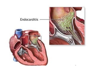
11. The aortic valve with a large, irregular, reddish tan vegetation Here, infective endocarditis on the mitral valve has spread into the septum all the way to the tricuspid valve, producing a fistula.
12. Microscopically, the valve in infective endocarditis demonstrates friable vegetations of fibrin and platelets (pink) mixed with inflammatory cells and bacterial colonies (blue). The friability explains how portions of the vegetation can break off and embolize.
13. Here is a valve with infective endocarditis. The blue bacterial colonies on the lower left are extending into the pink connective tissue of the valve. Valves are relatively avascular, so high dose antibiotic therapy is needed to eradicate the infection.
14. Acute bacterial endocarditis caused by Staphylococcus aureus with perforation of the aortic valve and aortic valve vegetations.
15. Acute bacterial endocarditis caused by Staphylococcus aureus with aortic valve ring abscess extending into myocardium.
16.
17. The small pink vegetation on the rightmost cusp margin represents the typical finding with non-bacterial thrombotic endocarditis (or so-called "marantic endocarditis"). This is non-infective. It tends to occur in persons with a hypercoagulable state (Trousseau's syndrome, a paraneoplastic syndrome associated with malignancies) and in very ill persons
18. Bartonella henselae bacilli in cardiac valve of a patient with blood culture-negative endocarditis The bacilli appear as black granulations.
19.
20. Petechial rash. He was diagnosed with right-sided staphylococcal endocarditis. Osler nodes
21. Osler's nodes on a finger and foot. Janeway lesions are Flat, painless, erythematous lesions seen on the palm of this patient's hand. Frequently associated with bacterial endocarditis.
22. Roth spots: red center with pale periphery; top and left (The one on right is the optic disc, as it has pale center and red periphery)
23.
24. Seen here in the finger at the right are small splinter hemorrhages in a patient with infective endocarditis. These hemorrhages are subungual, linear, dark red streaks. Similar hemorrhages can also appear with trauma.
25.
26.
27.
28.
29.
30.
31. Here is another marantic vegetation on the leftmost cusp. These vegetations are rarely over 0.5 cm in size. However, they are very prone to embolize.
32. The valve is seen on the left, and a bland vegetation is seen on the right. It appears pink because it is composed of fibrin and platelets. It displays about as much morphologic variation as a brown paper bag. Such bland vegetations are typical of the non-infective forms of endocarditis.
33.
34. Libman-sacks endocarditis. Here are flat, pale tan, spreading vegetations over the mitral valve surface and even on the chordae tendineae.
![[object Object],[object Object]](data:image/gif;base64,R0lGODlhAQABAIAAAAAAAP///yH5BAEAAAAALAAAAAABAAEAAAIBRAA7)