SlideShare a Scribd company logo
1 of 33
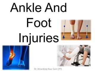
Ankle And
Foot
Injuries
Dr. Amardeep Kaur Saini (PT)
Anatomy
• The ankle joint (or
talocrural joint) is a
synovial joint located in
the lower limb. It is
formed by the bones of
the leg (tibia and fibula)
and the foot (talus).
• Functionally, it is a
hinge type joint,
permitting dorsiflexion
and plantarflexion of
the foot.
Dr. Amardeep Kaur Saini (PT)
Ligaments
Medial Ligament
• The medial ligament (or
deltoid ligament) is attached
to the medial malleolus (a
bony prominence projecting
from the medial aspect of the
distal tibia).
• It consists of four ligaments,
which fan out from the
malleolus, attaching to the
talus, calcaneus and navicular
bones. The primary action of
the medial ligament is to resist
over-eversion of the foot.
Dr. Amardeep Kaur Saini (PT)
Lateral Ligament
1. The lateral ligament
originates from the lateral
malleolus (a bony
prominence projecting from
the lateral aspect of the
distal fibula).
2. It resists over-inversion of
the foot, and is comprised of
three distinct and separate
ligaments:
• Anterior talofibular – spans
between the lateral malleolus
and lateral aspect of the talus.
• Posterior talofibular – spans
between the lateral malleolus
and the posterior aspect of
the talus.
• Calcaneofibular – spans
between the lateral malleolus
and the calcaneus.
Dr. Amardeep Kaur Saini (PT)
Forces At Ankle
o Inversion(adduction) : inward twisting of ankle.
o Eversion(abduction): outward twisting of ankle.
o Supination : inversion plus adduction of the foot so
that the sole faces medially.
o Pronation : eversion plus abduction of the foot so
that the sole faces laterally.
o Rotation(external or internal): a rotatory movement
of the foot so that the talus is subjected to a rotatory
force along its vertical axis.
o Vertical compression: a force along the long axis of
the tibia.
Vertical compression
injury
Clinical features
• History of a twisting injury to ankle
followed by PAIN and SWELLING.
• Swelling and Tenderness may be
localized to the area of injury (bone or
ligament).
• Crepitus may be noticed if there is a
fracture. The ankle may be lying
deformed (adducted or abducted ,
with or without rotation).
Radiological Examination
 Antero-posterior and
lateral X-rays of ankle are
sufficient in most cases.
 Fracture line of the
medial and malleoli
should be studied in
order to elevate the type
of the ankle injury (lauge
- hansen classification ).
Small avulsion # from
malleoli are sometimes
missed. These have often
attached to whole
ligament.
Tibio - fibular syndesmosis: All ankle injuries
where the fibular # is above the mortise, the
syndesmosis is bound to have been disrupted.
In injuries where fibular # is at level of the
syndesmosis, one must carefully look for any
lateral subluxation of talus; if it is so, width of
the joint space b/w medial malleolus and the
talus will be more than the space b/w the
weight bearing surfaces of tibia and talus.
Posterior subluxation of the talus should be
looked for, on the lateral x-ray.
Soft tissue swelling
Treatment
Principle of treatment :
The complexity of the
forces involved produce a
variety of combinations of
#s and #- dislocation
around the ankle.
Fractures without
displacements: it is usually
sufficient to protect the
ankle in below – knee
plaster for 3-6 weeks.
Good, readymade braces
can be used in place of
rather uncomfortable
plaster cast.
# With Displacement
Operative method : ORIF( open
reduction internal fixation).
MEDIAL MALLEOLUS #
• Transverse # - compression screw,
tension band wiring.
• Oblique #- compression screws
• Avulsion #- tension band wiring
LATERAL MALLEOLUS #
• Transverse # -tension band wiring
• Spiral #- compression screws
• Comminuted #- BUTTRESS PLATING
• # of lower third of fibula -4-hole plate
Tibio fibular syndesmosis disruption – needs to
be stabilized by inserting a long screw from
fibula into tibia.
Conservative methods: below knee plaster cast
is applied. If the check X-ray shows satisfactory
position, the plaster cast is continued for 8-10
weeks.
The patient is not allowed to bear weight on the
leg during this period .
After removing plaster cast, patient taught
physiotherapy to regain ankle movement.
• External fixation: this is required in open #
with bad crushing of the muscles and tendons,
with skin loss around the ankle.
Complications
Stiffness of ankle
Osteoarthritis
Sprained Ankle
• Ligament injury of the ankle.
• Commonly, It is inversion injury, and lateral
collateral ligament is sprained.
• Sometimes , eversion force may result in
sprain of medial collateral ligament.
• Clinical features:
 swelling and tenderness
 If a torn ligament is subjected to stress by
following maneuvers, patient experiences
severe pain:
 Inversion of plantar flexed foot for anterior
talo-fibular ligament sprain.
 Inversion in neutral position for complete
lateral collateral ligament sprain .
 Eversion in neutral position for medial
collateral ligament sprain.
Radiological Examination
• X-rays of ankle (AP and lateral) are usually
normal.
• In some case, stress x-rays may be done to
judge the severity of the sprain.
• A tilt of the talus greater than 20° on forced
inversion or eversion indicates a complete tear
of the lateral or medial collateral ligament
respectively.
Dr. Amardeep Kaur Saini (PT)
Treatment
It depends upon the grade of sprain:
• Grade I: Below-knee plaster cast for 2 weeks followed by
mobilization.
• Grade II: Below-knee cast for 4 weeks followed by mobilization.
• Grade III: Below-knee cast for 6 weeks followed by
mobilization.
Current trend is to treat ligament injuries, in general, by
‘functional’ method i.e., without immobilization. Treatment
consists of rest, ice packs, compression, and elevation (RICE) for
the first 2-3 days. The patient begins early protected range of
motion exercises. Methods are devised by which during
mobilisation, stress is avoided on ‘healing’ ligaments, and the
muscles around the joint are built up. For this approach, a well
developed physiotherapy unit is required. For grade III ligament
injury to the ankle, especially in young athletic individuals,
operative repair is preferred by some surgeons.
Dr. Amardeep Kaur Saini (PT)
CHRONIC ANKLE SPRAIN
• Chronic recurrent sprain ankle is a disabling
condition. If a course of physiotherapy and
modification in shoe has not helped, a detailed
evaluation with MRI and arthroscopy may be
necessary.
• Pain in a number of these so-called chronic ankle
sprains is in fact due to impingement of the
scarred capsule or chondromalacia of the talus.
Arthroscopy is a good technique for diagnosis and
treatment of such cases.
Dr. Amardeep Kaur Saini (PT)
Fractures Of The Calcaneum
Anatomy
The calcaneum forms the bone of the heel.
Its upper surface articulates with the talus,
and the front surface with the cuboid.
Its inferior surface is prolonged backwards
as the tuber calcanei. Normally, the angle
between the superior articular surface
(between talus and calcaneum) and the
upper surface of the tuberosity is 35°
(tuber-joint angle).
It is reduced in most fractures of the
calcaneum.
Dr. Amardeep Kaur Saini (PT)
Pathoanatomy
Fractures of the calcaneum are caused by fall from height
onto the heels, thus both heels may be injured at the same
time. The fracture may be:
(i) an isolated crack fracture, usually in the region of the
tuberosity; or
(ii) more often a compression injury where the bone is
shattered like an egg shell.
The degree of displacement varies according to the severity
of trauma.
• Undisplaced fracture resulting from a minimal trauma
• Extra-articular fracture, where the articular surfaces
remain intact, and the force splits the calcaneal tuberosity
vertically.
• Intra-articular fracture, where the articular surface of the
calcaneum fails to withstand the stress. It is shattered and is
driven downwards into the body of the bone, crushing the
delicate trabeculae of the cancellous bone into powder. This
is the commonest type of fracture.
Dr. Amardeep Kaur Saini (PT)
Clinical features:
 The patient often gives a history of a fall from
height, landing on their heels (e.g. a thief jumping
from the first floor of a house). There is pain and
swelling in the region of the heel. The patient is
not able to bear weight on the affected foot.
 On examination, there is marked swelling and
broadening of the heel. If first seen after a day or
two, there will be ecchymosis around the heel
and on the sole. Movement at the ankle is not
appreciably impaired.
 Many cases of compression fractures of the
calcaneum are associated with a compression
fracture of a vertebral body (usually in the
dorsolumbar region), fractures of the pubic rami,
or an atlanto-axial injury. One must look for these
injuries in a case of a fracture of the calcaneum.
Dr. Amardeep Kaur Saini (PT)
Radiological examination
It is possible to diagnose most
calcaneum fractures on a lateral X-
ray of the heel. In some cases, an
additional axial view of the
calcaneum may be required. Very
often, rather than a clear fracture
extending through the calcaneum,
there occurs crushing of the bone.
This can be diagnosed on a lateral
X-ray of the heel by reduction in the
tuber-joint angle.
Dr. Amardeep Kaur Saini (PT)
TREATMENT
Undisplaced fracture: Below-knee plaster cast
for 4 weeks followed by mobilisation exercises.
Compression fracture: This is a serious injury
which inevitably leads to permanent impairment of
functions. Many different methods of treatment
have been advocated with no appreciable
difference in results. The following method is one
used most widely.
The foot is elevated in a well padded below-knee
plaster slab for 2-3 weeks. Once pain and swelling
subside, the slab is removed and ankle and foot
mobilisation begun.
Leg elevation is continued, and a compression
bandage (crepe bandage) applied for a period of 4-
6 weeks in order to avoid gravitational oedema.
Weight bearing is not permitted for a period of
12 weeks.
Dr. Amardeep Kaur Saini (PT)
Complications
1. Stiffness of the subtalar and mid-tarsal joints: Some
amount of stiffness of the subtalar joint, resulting in limitation to
the inversion-eversion motion of the foot is inevitable in most
compression fractures of the calcaneum. Stiffness can be kept to
minimum by early physiotherapy.
2. Osteoarthritis. Because of the irreparable distortion of the
subtalar joint surface, osteoarthritis is an expected complication.
It results in pain and stiffness, most noticeable while walking on
an uneven surface. A patient with a severe disability may
require fusion of the subtalar joint (arthrodesis).
Dr. Amardeep Kaur Saini (PT)
Fractures Of The Talus
Anatomy
Blood supply to the talus: This is
the only bone of the foot without
any muscle attachment. The main
blood supply to the talus is from the
anastomotic ring of blood vessels,
the osseous vessels entering its neck
and running postero-laterally within
the bone to supply its body.
Therefore, blood supply to the body
of the talus is often cut off
following fractures occurring
through the neck. Dr. Amardeep Kaur Saini (PT)
Mechanism
Fracture of the neck of the talus results from forced
dorsiflexion of the ankle. Typically, this injury is
sustained in an aircraft crash where the rudder bar is
driven forcibly against the middle of the sole of the
foot (Aviator's fracture), resulting in forced
dorsiflexion of the ankle; the neck, being a weak area,
gives way. This may be associated with dislocation of
the body of the talus backwards, out of the ankle-
mortise. Vascularity of the body of the talus may be
compromised.
Diagnosis
Unless carefully examined on a lateral X-ray of the
ankle, this fracture is frequently missed because of
the overlapping of the tarsal bones.
Dr. Amardeep Kaur Saini (PT)
Treatment
It depends upon the displacement. If Undisplaced, a below knee plaster cast
for 8-10 weeks is sufficient. In a displaced fracture, open reduction and
internal fixation of the fracture with a screw is required.
Complications
1. Avascular necrosis and non-union: Because of the poor blood supply,
after a fracture through the neck, the body of the talus becomes avascular. The
avascular fragment fails to unite with rest of the bone and gradually collapses,
leading to deformation of the bone, and eventually osteoarthritis of the ankle.
2. Osteoarthritis: Besides avascular necrosis of the talus, an associated injury
to its articular cartilage may lead to osteoarthritis of the ankle. The patient
complains of pain and stiffness. Treatment is mostly by physiotherapy and
fomentation. In severe cases, an ankle arthrodesis may be needed.
Dr. Amardeep Kaur Saini (PT)
INJURIES OF THE TARSAL BONES
Fractures and dislocations of other
tarsal bones are uncommon. Most of
the fractures can be treated by a
below-knee plaster cast. Most
dislocations at any of the tarsal joints
(subtalar, talo-navicular or inter-
tarsal) can be treated by manipulation
and immobilization in a plaster cast.
Sometimes, an open reduction and
internal fixation with K-wires may be
required.
Dr. Amardeep Kaur Saini (PT)
FRACTURES OF THE
METATARSAL BONES
Most metatarsal fractures are caused by direct
violence from a heavy object falling onto the foot.
A metatarsal fracture may be caused by repeated
stress without any specific injury (march fracture).
FRACTURE OF THE BASE OF
5TH METATARSAL
(Jones' fracture)
This is a fracture at the base of the 5th metatarsal,
caused by the pull exerted by the tendon of the
peroneus brevis muscle inserted on it. Clinically,
there is pain, swelling and tenderness at the outer
border of the foot, most marked at the base of the
5th metatarsal. Diagnosis is easily confirmed on X-
ray Treatment is by a below-knee walking plaster
cast for 3 weeks.
Dr. Amardeep Kaur Saini (PT)
FRACTURE OF THE
METATARSAL SHAFTS
One or more metatarsal shafts may be
fractured, mostly following a crush injury.
Treatment is by below-knee plaster cast for 3-4
weeks.
March Fracture
It is a ‘fatigue’ fracture of third metatarsal,
resulting from long continued or often repeated
stress, particularly from prolonged walking or
running in those not accustomed to it. Thus, it
may occur in army recruits freshly committed
to marching – hence the term ‘March fracture’.
The fracture heals spontaneously, so treatment
is purely symptomatic.
FRACTURES OF PHALANGES
OF THE TOES
These are common injuries, most
often resulting from fall of a
heavy object, or twisting of the
toes. The great toe is injured most
commonly. Satisfactory general
alignment is maintained in most
cases and little or no treatment is
required. The injured toe is
covered with a soft woolly
dressing and strapped to the toe
adjacent to it.
Dr. Amardeep Kaur Saini (PT)

More Related Content

What's hot

Complications of fractures
Complications of fracturesComplications of fractures
Complications of fracturesSubhanjan Das
 
Recurrent Dislocation of patella -PAWAN
Recurrent Dislocation of patella -PAWANRecurrent Dislocation of patella -PAWAN
Recurrent Dislocation of patella -PAWANPawan Yadav
 
Proximal tibia fracture
Proximal tibia fractureProximal tibia fracture
Proximal tibia fracturevisheshrohatgi
 
Septic arthritis
Septic arthritisSeptic arthritis
Septic arthritisgroup7usmkk
 
Volksmann contracture
Volksmann contracture Volksmann contracture
Volksmann contracture Kimberly Walsh
 
Colles' fracture
Colles' fracture Colles' fracture
Colles' fracture akond1986
 
Osteoarthritis knee
Osteoarthritis  kneeOsteoarthritis  knee
Osteoarthritis kneeNarula Gandu
 
Lower limb fractures
Lower limb fracturesLower limb fractures
Lower limb fracturesairwave12
 
Devlopmental dysplasia of hip(DDH) by DR.NAVEEN RATHOR
Devlopmental dysplasia of hip(DDH) by DR.NAVEEN RATHORDevlopmental dysplasia of hip(DDH) by DR.NAVEEN RATHOR
Devlopmental dysplasia of hip(DDH) by DR.NAVEEN RATHORDR.Naveen Rathor
 
Spinal fractures (injury)
Spinal fractures (injury)Spinal fractures (injury)
Spinal fractures (injury)kajalgoel8
 
Neuropathic (Charcots) joints
Neuropathic (Charcots) joints Neuropathic (Charcots) joints
Neuropathic (Charcots) joints Subodh Pathak
 
Meniscal injury
Meniscal injuryMeniscal injury
Meniscal injurymanoj das
 
Proximal Femoral Nail
Proximal Femoral NailProximal Femoral Nail
Proximal Femoral NailAlex Bertino
 
MCL,LCL & ALL injuries of the knee
MCL,LCL & ALL injuries of the knee MCL,LCL & ALL injuries of the knee
MCL,LCL & ALL injuries of the knee Mohamed Abulsoud
 

What's hot (20)

Complications of fractures
Complications of fracturesComplications of fractures
Complications of fractures
 
Recurrent Dislocation of patella -PAWAN
Recurrent Dislocation of patella -PAWANRecurrent Dislocation of patella -PAWAN
Recurrent Dislocation of patella -PAWAN
 
Proximal tibia fracture
Proximal tibia fractureProximal tibia fracture
Proximal tibia fracture
 
Septic arthritis
Septic arthritisSeptic arthritis
Septic arthritis
 
Volksmann contracture
Volksmann contracture Volksmann contracture
Volksmann contracture
 
SPRAINED ANKLE
SPRAINED ANKLESPRAINED ANKLE
SPRAINED ANKLE
 
Bursitis
BursitisBursitis
Bursitis
 
Colles' fracture
Colles' fracture Colles' fracture
Colles' fracture
 
Osteoarthritis knee
Osteoarthritis  kneeOsteoarthritis  knee
Osteoarthritis knee
 
Lower limb fractures
Lower limb fracturesLower limb fractures
Lower limb fractures
 
Devlopmental dysplasia of hip(DDH) by DR.NAVEEN RATHOR
Devlopmental dysplasia of hip(DDH) by DR.NAVEEN RATHORDevlopmental dysplasia of hip(DDH) by DR.NAVEEN RATHOR
Devlopmental dysplasia of hip(DDH) by DR.NAVEEN RATHOR
 
Foot injuries
Foot injuriesFoot injuries
Foot injuries
 
Spinal fractures (injury)
Spinal fractures (injury)Spinal fractures (injury)
Spinal fractures (injury)
 
Neuropathic (Charcots) joints
Neuropathic (Charcots) joints Neuropathic (Charcots) joints
Neuropathic (Charcots) joints
 
Meniscal injury
Meniscal injuryMeniscal injury
Meniscal injury
 
Supracondylar Fractures
Supracondylar FracturesSupracondylar Fractures
Supracondylar Fractures
 
Proximal humerus fractures
Proximal humerus fracturesProximal humerus fractures
Proximal humerus fractures
 
Proximal Femoral Nail
Proximal Femoral NailProximal Femoral Nail
Proximal Femoral Nail
 
Ankle fractures
Ankle fracturesAnkle fractures
Ankle fractures
 
MCL,LCL & ALL injuries of the knee
MCL,LCL & ALL injuries of the knee MCL,LCL & ALL injuries of the knee
MCL,LCL & ALL injuries of the knee
 

Similar to Ankle and foot injuries

Ligamentous Injuries Around Knee by Dr Arun C Raj, Ortho Resident, KIMS Hubli
Ligamentous Injuries Around Knee by Dr Arun C Raj, Ortho Resident, KIMS HubliLigamentous Injuries Around Knee by Dr Arun C Raj, Ortho Resident, KIMS Hubli
Ligamentous Injuries Around Knee by Dr Arun C Raj, Ortho Resident, KIMS HubliArunCRaj1
 
Soft Tissue Injuries of Ankle & Foot.pptx
Soft Tissue Injuries of Ankle & Foot.pptxSoft Tissue Injuries of Ankle & Foot.pptx
Soft Tissue Injuries of Ankle & Foot.pptxNaolShibiru
 
PPT trauma muskuloskeletal dr. Satria, Sp.OT.pptx
PPT trauma muskuloskeletal dr. Satria, Sp.OT.pptxPPT trauma muskuloskeletal dr. Satria, Sp.OT.pptx
PPT trauma muskuloskeletal dr. Satria, Sp.OT.pptxSebastianMihardja1
 
ankle sprain presentation.pdf
ankle sprain presentation.pdfankle sprain presentation.pdf
ankle sprain presentation.pdfAshrafHussein36
 
PT Management of Fractures of Condyles of Femur
PT Management of Fractures of Condyles of FemurPT Management of Fractures of Condyles of Femur
PT Management of Fractures of Condyles of FemurNavKalsi1
 
Ankle Instability and Pain
Ankle Instability and PainAnkle Instability and Pain
Ankle Instability and PainSummit Health
 
elbow and wrist and hand fracture with management
elbow and wrist and hand fracture with managementelbow and wrist and hand fracture with management
elbow and wrist and hand fracture with managementkajalgoel8
 
ankle fracture F2.pptx
ankle fracture F2.pptxankle fracture F2.pptx
ankle fracture F2.pptxFuadNaji1
 
Tibia and fibula diaphysis, ankle and foot injuries
Tibia and fibula diaphysis, ankle and foot injuriesTibia and fibula diaphysis, ankle and foot injuries
Tibia and fibula diaphysis, ankle and foot injuriesJoyce Mwatonoka
 
Orthopedics 5th year, 7th/part two & 8th lectures (Dr. Ali A.Nabi)
Orthopedics 5th year, 7th/part two & 8th lectures (Dr. Ali A.Nabi)Orthopedics 5th year, 7th/part two & 8th lectures (Dr. Ali A.Nabi)
Orthopedics 5th year, 7th/part two & 8th lectures (Dr. Ali A.Nabi)College of Medicine, Sulaymaniyah
 
Osteoarthritis of Knee Joint
Osteoarthritis of Knee JointOsteoarthritis of Knee Joint
Osteoarthritis of Knee JointDr.Anshu Sharma
 
Acute knee ligament injuries
Acute knee ligament injuriesAcute knee ligament injuries
Acute knee ligament injuriesMilind Merchant
 
SPORTS INJURIES OF ANKLE AND FOOT original.pptx
SPORTS INJURIES OF ANKLE AND FOOT original.pptxSPORTS INJURIES OF ANKLE AND FOOT original.pptx
SPORTS INJURIES OF ANKLE AND FOOT original.pptxMeghaPrakash9
 

Similar to Ankle and foot injuries (20)

Ligamentous Injuries Around Knee by Dr Arun C Raj, Ortho Resident, KIMS Hubli
Ligamentous Injuries Around Knee by Dr Arun C Raj, Ortho Resident, KIMS HubliLigamentous Injuries Around Knee by Dr Arun C Raj, Ortho Resident, KIMS Hubli
Ligamentous Injuries Around Knee by Dr Arun C Raj, Ortho Resident, KIMS Hubli
 
Soft Tissue Injuries of Ankle & Foot.pptx
Soft Tissue Injuries of Ankle & Foot.pptxSoft Tissue Injuries of Ankle & Foot.pptx
Soft Tissue Injuries of Ankle & Foot.pptx
 
Ankle fracture
Ankle fractureAnkle fracture
Ankle fracture
 
Sports Injury.pptx
Sports  Injury.pptxSports  Injury.pptx
Sports Injury.pptx
 
L12 ankle fxs
L12 ankle fxsL12 ankle fxs
L12 ankle fxs
 
PPT trauma muskuloskeletal dr. Satria, Sp.OT.pptx
PPT trauma muskuloskeletal dr. Satria, Sp.OT.pptxPPT trauma muskuloskeletal dr. Satria, Sp.OT.pptx
PPT trauma muskuloskeletal dr. Satria, Sp.OT.pptx
 
Ankle seminar
Ankle seminarAnkle seminar
Ankle seminar
 
orthopaedics surgery by dr shubham patel
orthopaedics surgery by dr shubham patelorthopaedics surgery by dr shubham patel
orthopaedics surgery by dr shubham patel
 
Ankle Sprain
Ankle SprainAnkle Sprain
Ankle Sprain
 
ankle sprain presentation.pdf
ankle sprain presentation.pdfankle sprain presentation.pdf
ankle sprain presentation.pdf
 
PT Management of Fractures of Condyles of Femur
PT Management of Fractures of Condyles of FemurPT Management of Fractures of Condyles of Femur
PT Management of Fractures of Condyles of Femur
 
Ankle Instability and Pain
Ankle Instability and PainAnkle Instability and Pain
Ankle Instability and Pain
 
elbow and wrist and hand fracture with management
elbow and wrist and hand fracture with managementelbow and wrist and hand fracture with management
elbow and wrist and hand fracture with management
 
ankle fracture F2.pptx
ankle fracture F2.pptxankle fracture F2.pptx
ankle fracture F2.pptx
 
Ankle injuries
Ankle injuriesAnkle injuries
Ankle injuries
 
Tibia and fibula diaphysis, ankle and foot injuries
Tibia and fibula diaphysis, ankle and foot injuriesTibia and fibula diaphysis, ankle and foot injuries
Tibia and fibula diaphysis, ankle and foot injuries
 
Orthopedics 5th year, 7th/part two & 8th lectures (Dr. Ali A.Nabi)
Orthopedics 5th year, 7th/part two & 8th lectures (Dr. Ali A.Nabi)Orthopedics 5th year, 7th/part two & 8th lectures (Dr. Ali A.Nabi)
Orthopedics 5th year, 7th/part two & 8th lectures (Dr. Ali A.Nabi)
 
Osteoarthritis of Knee Joint
Osteoarthritis of Knee JointOsteoarthritis of Knee Joint
Osteoarthritis of Knee Joint
 
Acute knee ligament injuries
Acute knee ligament injuriesAcute knee ligament injuries
Acute knee ligament injuries
 
SPORTS INJURIES OF ANKLE AND FOOT original.pptx
SPORTS INJURIES OF ANKLE AND FOOT original.pptxSPORTS INJURIES OF ANKLE AND FOOT original.pptx
SPORTS INJURIES OF ANKLE AND FOOT original.pptx
 

Recently uploaded

Call Girls Whitefield Just Call 7001305949 Top Class Call Girl Service Available
Call Girls Whitefield Just Call 7001305949 Top Class Call Girl Service AvailableCall Girls Whitefield Just Call 7001305949 Top Class Call Girl Service Available
Call Girls Whitefield Just Call 7001305949 Top Class Call Girl Service Availablenarwatsonia7
 
Call Girls Jayanagar Just Call 7001305949 Top Class Call Girl Service Available
Call Girls Jayanagar Just Call 7001305949 Top Class Call Girl Service AvailableCall Girls Jayanagar Just Call 7001305949 Top Class Call Girl Service Available
Call Girls Jayanagar Just Call 7001305949 Top Class Call Girl Service Availablenarwatsonia7
 
Mumbai Call Girls Service 9910780858 Real Russian Girls Looking Models
Mumbai Call Girls Service 9910780858 Real Russian Girls Looking ModelsMumbai Call Girls Service 9910780858 Real Russian Girls Looking Models
Mumbai Call Girls Service 9910780858 Real Russian Girls Looking Modelssonalikaur4
 
High Profile Call Girls Jaipur Vani 8445551418 Independent Escort Service Jaipur
High Profile Call Girls Jaipur Vani 8445551418 Independent Escort Service JaipurHigh Profile Call Girls Jaipur Vani 8445551418 Independent Escort Service Jaipur
High Profile Call Girls Jaipur Vani 8445551418 Independent Escort Service Jaipurparulsinha
 
97111 47426 Call Girls In Delhi MUNIRKAA
97111 47426 Call Girls In Delhi MUNIRKAA97111 47426 Call Girls In Delhi MUNIRKAA
97111 47426 Call Girls In Delhi MUNIRKAAjennyeacort
 
Noida Sector 135 Call Girls ( 9873940964 ) Book Hot And Sexy Girls In A Few C...
Noida Sector 135 Call Girls ( 9873940964 ) Book Hot And Sexy Girls In A Few C...Noida Sector 135 Call Girls ( 9873940964 ) Book Hot And Sexy Girls In A Few C...
Noida Sector 135 Call Girls ( 9873940964 ) Book Hot And Sexy Girls In A Few C...rajnisinghkjn
 
Hematology and Immunology - Leukocytes Functions
Hematology and Immunology - Leukocytes FunctionsHematology and Immunology - Leukocytes Functions
Hematology and Immunology - Leukocytes FunctionsMedicoseAcademics
 
Call Girls Electronic City Just Call 7001305949 Top Class Call Girl Service A...
Call Girls Electronic City Just Call 7001305949 Top Class Call Girl Service A...Call Girls Electronic City Just Call 7001305949 Top Class Call Girl Service A...
Call Girls Electronic City Just Call 7001305949 Top Class Call Girl Service A...narwatsonia7
 
Book Call Girls in Kasavanahalli - 7001305949 with real photos and phone numbers
Book Call Girls in Kasavanahalli - 7001305949 with real photos and phone numbersBook Call Girls in Kasavanahalli - 7001305949 with real photos and phone numbers
Book Call Girls in Kasavanahalli - 7001305949 with real photos and phone numbersnarwatsonia7
 
Call Girls Thane Just Call 9910780858 Get High Class Call Girls Service
Call Girls Thane Just Call 9910780858 Get High Class Call Girls ServiceCall Girls Thane Just Call 9910780858 Get High Class Call Girls Service
Call Girls Thane Just Call 9910780858 Get High Class Call Girls Servicesonalikaur4
 
call girls in green park DELHI 🔝 >༒9540349809 🔝 genuine Escort Service 🔝✔️✔️
call girls in green park  DELHI 🔝 >༒9540349809 🔝 genuine Escort Service 🔝✔️✔️call girls in green park  DELHI 🔝 >༒9540349809 🔝 genuine Escort Service 🔝✔️✔️
call girls in green park DELHI 🔝 >༒9540349809 🔝 genuine Escort Service 🔝✔️✔️saminamagar
 
Book Call Girls in Yelahanka - For 7001305949 Cheap & Best with original Photos
Book Call Girls in Yelahanka - For 7001305949 Cheap & Best with original PhotosBook Call Girls in Yelahanka - For 7001305949 Cheap & Best with original Photos
Book Call Girls in Yelahanka - For 7001305949 Cheap & Best with original Photosnarwatsonia7
 
Call Girls Jp Nagar Just Call 7001305949 Top Class Call Girl Service Available
Call Girls Jp Nagar Just Call 7001305949 Top Class Call Girl Service AvailableCall Girls Jp Nagar Just Call 7001305949 Top Class Call Girl Service Available
Call Girls Jp Nagar Just Call 7001305949 Top Class Call Girl Service Availablenarwatsonia7
 
Call Girls Hsr Layout Just Call 7001305949 Top Class Call Girl Service Available
Call Girls Hsr Layout Just Call 7001305949 Top Class Call Girl Service AvailableCall Girls Hsr Layout Just Call 7001305949 Top Class Call Girl Service Available
Call Girls Hsr Layout Just Call 7001305949 Top Class Call Girl Service Availablenarwatsonia7
 
Glomerular Filtration and determinants of glomerular filtration .pptx
Glomerular Filtration and  determinants of glomerular filtration .pptxGlomerular Filtration and  determinants of glomerular filtration .pptx
Glomerular Filtration and determinants of glomerular filtration .pptxDr.Nusrat Tariq
 
Call Girls Service in Bommanahalli - 7001305949 with real photos and phone nu...
Call Girls Service in Bommanahalli - 7001305949 with real photos and phone nu...Call Girls Service in Bommanahalli - 7001305949 with real photos and phone nu...
Call Girls Service in Bommanahalli - 7001305949 with real photos and phone nu...narwatsonia7
 
Call Girls Service Nandiambakkam | 7001305949 At Low Cost Cash Payment Booking
Call Girls Service Nandiambakkam | 7001305949 At Low Cost Cash Payment BookingCall Girls Service Nandiambakkam | 7001305949 At Low Cost Cash Payment Booking
Call Girls Service Nandiambakkam | 7001305949 At Low Cost Cash Payment BookingNehru place Escorts
 
Low Rate Call Girls Mumbai Suman 9910780858 Independent Escort Service Mumbai
Low Rate Call Girls Mumbai Suman 9910780858 Independent Escort Service MumbaiLow Rate Call Girls Mumbai Suman 9910780858 Independent Escort Service Mumbai
Low Rate Call Girls Mumbai Suman 9910780858 Independent Escort Service Mumbaisonalikaur4
 
Housewife Call Girls Bangalore - Call 7001305949 Rs-3500 with A/C Room Cash o...
Housewife Call Girls Bangalore - Call 7001305949 Rs-3500 with A/C Room Cash o...Housewife Call Girls Bangalore - Call 7001305949 Rs-3500 with A/C Room Cash o...
Housewife Call Girls Bangalore - Call 7001305949 Rs-3500 with A/C Room Cash o...narwatsonia7
 
Call Girl Surat Madhuri 7001305949 Independent Escort Service Surat
Call Girl Surat Madhuri 7001305949 Independent Escort Service SuratCall Girl Surat Madhuri 7001305949 Independent Escort Service Surat
Call Girl Surat Madhuri 7001305949 Independent Escort Service Suratnarwatsonia7
 

Recently uploaded (20)

Call Girls Whitefield Just Call 7001305949 Top Class Call Girl Service Available
Call Girls Whitefield Just Call 7001305949 Top Class Call Girl Service AvailableCall Girls Whitefield Just Call 7001305949 Top Class Call Girl Service Available
Call Girls Whitefield Just Call 7001305949 Top Class Call Girl Service Available
 
Call Girls Jayanagar Just Call 7001305949 Top Class Call Girl Service Available
Call Girls Jayanagar Just Call 7001305949 Top Class Call Girl Service AvailableCall Girls Jayanagar Just Call 7001305949 Top Class Call Girl Service Available
Call Girls Jayanagar Just Call 7001305949 Top Class Call Girl Service Available
 
Mumbai Call Girls Service 9910780858 Real Russian Girls Looking Models
Mumbai Call Girls Service 9910780858 Real Russian Girls Looking ModelsMumbai Call Girls Service 9910780858 Real Russian Girls Looking Models
Mumbai Call Girls Service 9910780858 Real Russian Girls Looking Models
 
High Profile Call Girls Jaipur Vani 8445551418 Independent Escort Service Jaipur
High Profile Call Girls Jaipur Vani 8445551418 Independent Escort Service JaipurHigh Profile Call Girls Jaipur Vani 8445551418 Independent Escort Service Jaipur
High Profile Call Girls Jaipur Vani 8445551418 Independent Escort Service Jaipur
 
97111 47426 Call Girls In Delhi MUNIRKAA
97111 47426 Call Girls In Delhi MUNIRKAA97111 47426 Call Girls In Delhi MUNIRKAA
97111 47426 Call Girls In Delhi MUNIRKAA
 
Noida Sector 135 Call Girls ( 9873940964 ) Book Hot And Sexy Girls In A Few C...
Noida Sector 135 Call Girls ( 9873940964 ) Book Hot And Sexy Girls In A Few C...Noida Sector 135 Call Girls ( 9873940964 ) Book Hot And Sexy Girls In A Few C...
Noida Sector 135 Call Girls ( 9873940964 ) Book Hot And Sexy Girls In A Few C...
 
Hematology and Immunology - Leukocytes Functions
Hematology and Immunology - Leukocytes FunctionsHematology and Immunology - Leukocytes Functions
Hematology and Immunology - Leukocytes Functions
 
Call Girls Electronic City Just Call 7001305949 Top Class Call Girl Service A...
Call Girls Electronic City Just Call 7001305949 Top Class Call Girl Service A...Call Girls Electronic City Just Call 7001305949 Top Class Call Girl Service A...
Call Girls Electronic City Just Call 7001305949 Top Class Call Girl Service A...
 
Book Call Girls in Kasavanahalli - 7001305949 with real photos and phone numbers
Book Call Girls in Kasavanahalli - 7001305949 with real photos and phone numbersBook Call Girls in Kasavanahalli - 7001305949 with real photos and phone numbers
Book Call Girls in Kasavanahalli - 7001305949 with real photos and phone numbers
 
Call Girls Thane Just Call 9910780858 Get High Class Call Girls Service
Call Girls Thane Just Call 9910780858 Get High Class Call Girls ServiceCall Girls Thane Just Call 9910780858 Get High Class Call Girls Service
Call Girls Thane Just Call 9910780858 Get High Class Call Girls Service
 
call girls in green park DELHI 🔝 >༒9540349809 🔝 genuine Escort Service 🔝✔️✔️
call girls in green park  DELHI 🔝 >༒9540349809 🔝 genuine Escort Service 🔝✔️✔️call girls in green park  DELHI 🔝 >༒9540349809 🔝 genuine Escort Service 🔝✔️✔️
call girls in green park DELHI 🔝 >༒9540349809 🔝 genuine Escort Service 🔝✔️✔️
 
Book Call Girls in Yelahanka - For 7001305949 Cheap & Best with original Photos
Book Call Girls in Yelahanka - For 7001305949 Cheap & Best with original PhotosBook Call Girls in Yelahanka - For 7001305949 Cheap & Best with original Photos
Book Call Girls in Yelahanka - For 7001305949 Cheap & Best with original Photos
 
Call Girls Jp Nagar Just Call 7001305949 Top Class Call Girl Service Available
Call Girls Jp Nagar Just Call 7001305949 Top Class Call Girl Service AvailableCall Girls Jp Nagar Just Call 7001305949 Top Class Call Girl Service Available
Call Girls Jp Nagar Just Call 7001305949 Top Class Call Girl Service Available
 
Call Girls Hsr Layout Just Call 7001305949 Top Class Call Girl Service Available
Call Girls Hsr Layout Just Call 7001305949 Top Class Call Girl Service AvailableCall Girls Hsr Layout Just Call 7001305949 Top Class Call Girl Service Available
Call Girls Hsr Layout Just Call 7001305949 Top Class Call Girl Service Available
 
Glomerular Filtration and determinants of glomerular filtration .pptx
Glomerular Filtration and  determinants of glomerular filtration .pptxGlomerular Filtration and  determinants of glomerular filtration .pptx
Glomerular Filtration and determinants of glomerular filtration .pptx
 
Call Girls Service in Bommanahalli - 7001305949 with real photos and phone nu...
Call Girls Service in Bommanahalli - 7001305949 with real photos and phone nu...Call Girls Service in Bommanahalli - 7001305949 with real photos and phone nu...
Call Girls Service in Bommanahalli - 7001305949 with real photos and phone nu...
 
Call Girls Service Nandiambakkam | 7001305949 At Low Cost Cash Payment Booking
Call Girls Service Nandiambakkam | 7001305949 At Low Cost Cash Payment BookingCall Girls Service Nandiambakkam | 7001305949 At Low Cost Cash Payment Booking
Call Girls Service Nandiambakkam | 7001305949 At Low Cost Cash Payment Booking
 
Low Rate Call Girls Mumbai Suman 9910780858 Independent Escort Service Mumbai
Low Rate Call Girls Mumbai Suman 9910780858 Independent Escort Service MumbaiLow Rate Call Girls Mumbai Suman 9910780858 Independent Escort Service Mumbai
Low Rate Call Girls Mumbai Suman 9910780858 Independent Escort Service Mumbai
 
Housewife Call Girls Bangalore - Call 7001305949 Rs-3500 with A/C Room Cash o...
Housewife Call Girls Bangalore - Call 7001305949 Rs-3500 with A/C Room Cash o...Housewife Call Girls Bangalore - Call 7001305949 Rs-3500 with A/C Room Cash o...
Housewife Call Girls Bangalore - Call 7001305949 Rs-3500 with A/C Room Cash o...
 
Call Girl Surat Madhuri 7001305949 Independent Escort Service Surat
Call Girl Surat Madhuri 7001305949 Independent Escort Service SuratCall Girl Surat Madhuri 7001305949 Independent Escort Service Surat
Call Girl Surat Madhuri 7001305949 Independent Escort Service Surat
 

Ankle and foot injuries

  • 2. Anatomy • The ankle joint (or talocrural joint) is a synovial joint located in the lower limb. It is formed by the bones of the leg (tibia and fibula) and the foot (talus). • Functionally, it is a hinge type joint, permitting dorsiflexion and plantarflexion of the foot. Dr. Amardeep Kaur Saini (PT)
  • 3. Ligaments Medial Ligament • The medial ligament (or deltoid ligament) is attached to the medial malleolus (a bony prominence projecting from the medial aspect of the distal tibia). • It consists of four ligaments, which fan out from the malleolus, attaching to the talus, calcaneus and navicular bones. The primary action of the medial ligament is to resist over-eversion of the foot. Dr. Amardeep Kaur Saini (PT)
  • 4. Lateral Ligament 1. The lateral ligament originates from the lateral malleolus (a bony prominence projecting from the lateral aspect of the distal fibula). 2. It resists over-inversion of the foot, and is comprised of three distinct and separate ligaments: • Anterior talofibular – spans between the lateral malleolus and lateral aspect of the talus. • Posterior talofibular – spans between the lateral malleolus and the posterior aspect of the talus. • Calcaneofibular – spans between the lateral malleolus and the calcaneus. Dr. Amardeep Kaur Saini (PT)
  • 5.
  • 6. Forces At Ankle o Inversion(adduction) : inward twisting of ankle. o Eversion(abduction): outward twisting of ankle. o Supination : inversion plus adduction of the foot so that the sole faces medially. o Pronation : eversion plus abduction of the foot so that the sole faces laterally. o Rotation(external or internal): a rotatory movement of the foot so that the talus is subjected to a rotatory force along its vertical axis. o Vertical compression: a force along the long axis of the tibia.
  • 7.
  • 9. Clinical features • History of a twisting injury to ankle followed by PAIN and SWELLING. • Swelling and Tenderness may be localized to the area of injury (bone or ligament). • Crepitus may be noticed if there is a fracture. The ankle may be lying deformed (adducted or abducted , with or without rotation).
  • 10. Radiological Examination  Antero-posterior and lateral X-rays of ankle are sufficient in most cases.  Fracture line of the medial and malleoli should be studied in order to elevate the type of the ankle injury (lauge - hansen classification ). Small avulsion # from malleoli are sometimes missed. These have often attached to whole ligament.
  • 11. Tibio - fibular syndesmosis: All ankle injuries where the fibular # is above the mortise, the syndesmosis is bound to have been disrupted. In injuries where fibular # is at level of the syndesmosis, one must carefully look for any lateral subluxation of talus; if it is so, width of the joint space b/w medial malleolus and the talus will be more than the space b/w the weight bearing surfaces of tibia and talus. Posterior subluxation of the talus should be looked for, on the lateral x-ray. Soft tissue swelling
  • 12. Treatment Principle of treatment : The complexity of the forces involved produce a variety of combinations of #s and #- dislocation around the ankle. Fractures without displacements: it is usually sufficient to protect the ankle in below – knee plaster for 3-6 weeks. Good, readymade braces can be used in place of rather uncomfortable plaster cast.
  • 13. # With Displacement Operative method : ORIF( open reduction internal fixation). MEDIAL MALLEOLUS # • Transverse # - compression screw, tension band wiring. • Oblique #- compression screws • Avulsion #- tension band wiring LATERAL MALLEOLUS # • Transverse # -tension band wiring • Spiral #- compression screws • Comminuted #- BUTTRESS PLATING • # of lower third of fibula -4-hole plate
  • 14. Tibio fibular syndesmosis disruption – needs to be stabilized by inserting a long screw from fibula into tibia. Conservative methods: below knee plaster cast is applied. If the check X-ray shows satisfactory position, the plaster cast is continued for 8-10 weeks. The patient is not allowed to bear weight on the leg during this period . After removing plaster cast, patient taught physiotherapy to regain ankle movement.
  • 15. • External fixation: this is required in open # with bad crushing of the muscles and tendons, with skin loss around the ankle. Complications Stiffness of ankle Osteoarthritis
  • 16. Sprained Ankle • Ligament injury of the ankle. • Commonly, It is inversion injury, and lateral collateral ligament is sprained. • Sometimes , eversion force may result in sprain of medial collateral ligament. • Clinical features:  swelling and tenderness  If a torn ligament is subjected to stress by following maneuvers, patient experiences severe pain:  Inversion of plantar flexed foot for anterior talo-fibular ligament sprain.  Inversion in neutral position for complete lateral collateral ligament sprain .  Eversion in neutral position for medial collateral ligament sprain.
  • 17. Radiological Examination • X-rays of ankle (AP and lateral) are usually normal. • In some case, stress x-rays may be done to judge the severity of the sprain. • A tilt of the talus greater than 20° on forced inversion or eversion indicates a complete tear of the lateral or medial collateral ligament respectively. Dr. Amardeep Kaur Saini (PT)
  • 18. Treatment It depends upon the grade of sprain: • Grade I: Below-knee plaster cast for 2 weeks followed by mobilization. • Grade II: Below-knee cast for 4 weeks followed by mobilization. • Grade III: Below-knee cast for 6 weeks followed by mobilization. Current trend is to treat ligament injuries, in general, by ‘functional’ method i.e., without immobilization. Treatment consists of rest, ice packs, compression, and elevation (RICE) for the first 2-3 days. The patient begins early protected range of motion exercises. Methods are devised by which during mobilisation, stress is avoided on ‘healing’ ligaments, and the muscles around the joint are built up. For this approach, a well developed physiotherapy unit is required. For grade III ligament injury to the ankle, especially in young athletic individuals, operative repair is preferred by some surgeons. Dr. Amardeep Kaur Saini (PT)
  • 19. CHRONIC ANKLE SPRAIN • Chronic recurrent sprain ankle is a disabling condition. If a course of physiotherapy and modification in shoe has not helped, a detailed evaluation with MRI and arthroscopy may be necessary. • Pain in a number of these so-called chronic ankle sprains is in fact due to impingement of the scarred capsule or chondromalacia of the talus. Arthroscopy is a good technique for diagnosis and treatment of such cases. Dr. Amardeep Kaur Saini (PT)
  • 20. Fractures Of The Calcaneum Anatomy The calcaneum forms the bone of the heel. Its upper surface articulates with the talus, and the front surface with the cuboid. Its inferior surface is prolonged backwards as the tuber calcanei. Normally, the angle between the superior articular surface (between talus and calcaneum) and the upper surface of the tuberosity is 35° (tuber-joint angle). It is reduced in most fractures of the calcaneum. Dr. Amardeep Kaur Saini (PT)
  • 21. Pathoanatomy Fractures of the calcaneum are caused by fall from height onto the heels, thus both heels may be injured at the same time. The fracture may be: (i) an isolated crack fracture, usually in the region of the tuberosity; or (ii) more often a compression injury where the bone is shattered like an egg shell. The degree of displacement varies according to the severity of trauma. • Undisplaced fracture resulting from a minimal trauma • Extra-articular fracture, where the articular surfaces remain intact, and the force splits the calcaneal tuberosity vertically. • Intra-articular fracture, where the articular surface of the calcaneum fails to withstand the stress. It is shattered and is driven downwards into the body of the bone, crushing the delicate trabeculae of the cancellous bone into powder. This is the commonest type of fracture. Dr. Amardeep Kaur Saini (PT)
  • 22. Clinical features:  The patient often gives a history of a fall from height, landing on their heels (e.g. a thief jumping from the first floor of a house). There is pain and swelling in the region of the heel. The patient is not able to bear weight on the affected foot.  On examination, there is marked swelling and broadening of the heel. If first seen after a day or two, there will be ecchymosis around the heel and on the sole. Movement at the ankle is not appreciably impaired.  Many cases of compression fractures of the calcaneum are associated with a compression fracture of a vertebral body (usually in the dorsolumbar region), fractures of the pubic rami, or an atlanto-axial injury. One must look for these injuries in a case of a fracture of the calcaneum. Dr. Amardeep Kaur Saini (PT)
  • 23. Radiological examination It is possible to diagnose most calcaneum fractures on a lateral X- ray of the heel. In some cases, an additional axial view of the calcaneum may be required. Very often, rather than a clear fracture extending through the calcaneum, there occurs crushing of the bone. This can be diagnosed on a lateral X-ray of the heel by reduction in the tuber-joint angle. Dr. Amardeep Kaur Saini (PT)
  • 24. TREATMENT Undisplaced fracture: Below-knee plaster cast for 4 weeks followed by mobilisation exercises. Compression fracture: This is a serious injury which inevitably leads to permanent impairment of functions. Many different methods of treatment have been advocated with no appreciable difference in results. The following method is one used most widely. The foot is elevated in a well padded below-knee plaster slab for 2-3 weeks. Once pain and swelling subside, the slab is removed and ankle and foot mobilisation begun. Leg elevation is continued, and a compression bandage (crepe bandage) applied for a period of 4- 6 weeks in order to avoid gravitational oedema. Weight bearing is not permitted for a period of 12 weeks. Dr. Amardeep Kaur Saini (PT)
  • 25. Complications 1. Stiffness of the subtalar and mid-tarsal joints: Some amount of stiffness of the subtalar joint, resulting in limitation to the inversion-eversion motion of the foot is inevitable in most compression fractures of the calcaneum. Stiffness can be kept to minimum by early physiotherapy. 2. Osteoarthritis. Because of the irreparable distortion of the subtalar joint surface, osteoarthritis is an expected complication. It results in pain and stiffness, most noticeable while walking on an uneven surface. A patient with a severe disability may require fusion of the subtalar joint (arthrodesis). Dr. Amardeep Kaur Saini (PT)
  • 26. Fractures Of The Talus Anatomy Blood supply to the talus: This is the only bone of the foot without any muscle attachment. The main blood supply to the talus is from the anastomotic ring of blood vessels, the osseous vessels entering its neck and running postero-laterally within the bone to supply its body. Therefore, blood supply to the body of the talus is often cut off following fractures occurring through the neck. Dr. Amardeep Kaur Saini (PT)
  • 27. Mechanism Fracture of the neck of the talus results from forced dorsiflexion of the ankle. Typically, this injury is sustained in an aircraft crash where the rudder bar is driven forcibly against the middle of the sole of the foot (Aviator's fracture), resulting in forced dorsiflexion of the ankle; the neck, being a weak area, gives way. This may be associated with dislocation of the body of the talus backwards, out of the ankle- mortise. Vascularity of the body of the talus may be compromised. Diagnosis Unless carefully examined on a lateral X-ray of the ankle, this fracture is frequently missed because of the overlapping of the tarsal bones. Dr. Amardeep Kaur Saini (PT)
  • 28. Treatment It depends upon the displacement. If Undisplaced, a below knee plaster cast for 8-10 weeks is sufficient. In a displaced fracture, open reduction and internal fixation of the fracture with a screw is required. Complications 1. Avascular necrosis and non-union: Because of the poor blood supply, after a fracture through the neck, the body of the talus becomes avascular. The avascular fragment fails to unite with rest of the bone and gradually collapses, leading to deformation of the bone, and eventually osteoarthritis of the ankle. 2. Osteoarthritis: Besides avascular necrosis of the talus, an associated injury to its articular cartilage may lead to osteoarthritis of the ankle. The patient complains of pain and stiffness. Treatment is mostly by physiotherapy and fomentation. In severe cases, an ankle arthrodesis may be needed. Dr. Amardeep Kaur Saini (PT)
  • 29. INJURIES OF THE TARSAL BONES Fractures and dislocations of other tarsal bones are uncommon. Most of the fractures can be treated by a below-knee plaster cast. Most dislocations at any of the tarsal joints (subtalar, talo-navicular or inter- tarsal) can be treated by manipulation and immobilization in a plaster cast. Sometimes, an open reduction and internal fixation with K-wires may be required. Dr. Amardeep Kaur Saini (PT)
  • 30. FRACTURES OF THE METATARSAL BONES Most metatarsal fractures are caused by direct violence from a heavy object falling onto the foot. A metatarsal fracture may be caused by repeated stress without any specific injury (march fracture). FRACTURE OF THE BASE OF 5TH METATARSAL (Jones' fracture) This is a fracture at the base of the 5th metatarsal, caused by the pull exerted by the tendon of the peroneus brevis muscle inserted on it. Clinically, there is pain, swelling and tenderness at the outer border of the foot, most marked at the base of the 5th metatarsal. Diagnosis is easily confirmed on X- ray Treatment is by a below-knee walking plaster cast for 3 weeks. Dr. Amardeep Kaur Saini (PT)
  • 31. FRACTURE OF THE METATARSAL SHAFTS One or more metatarsal shafts may be fractured, mostly following a crush injury. Treatment is by below-knee plaster cast for 3-4 weeks. March Fracture It is a ‘fatigue’ fracture of third metatarsal, resulting from long continued or often repeated stress, particularly from prolonged walking or running in those not accustomed to it. Thus, it may occur in army recruits freshly committed to marching – hence the term ‘March fracture’. The fracture heals spontaneously, so treatment is purely symptomatic.
  • 32. FRACTURES OF PHALANGES OF THE TOES These are common injuries, most often resulting from fall of a heavy object, or twisting of the toes. The great toe is injured most commonly. Satisfactory general alignment is maintained in most cases and little or no treatment is required. The injured toe is covered with a soft woolly dressing and strapped to the toe adjacent to it. Dr. Amardeep Kaur Saini (PT)
  • 33. Dr. Amardeep Kaur Saini (PT)