
Recommended
More Related Content
What's hot
What's hot (20)
Similar to Digestive System Anatomy and Functions
Similar to Digestive System Anatomy and Functions (20)
More from ambika bagora
More from ambika bagora (6)
Recently uploaded
Recently uploaded (20)
Digestive System Anatomy and Functions
- 1. DIGESTIVE SYSTEM SUBMITTED BY- Ambika Bagora (RN)
- 2. The digestive system consist of gastrointestinal tract (alimentary canal) and its gland. The functions of G.I.T. are - a. Ingestion b. Digestion c. Absorption e. Excretion of waste products.
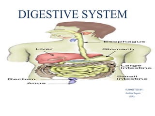
- 3. • Parts of digestive system- Digestive system consist of following parts- 1. Mouth 2. Pharynx 3. Oesophagus 4. Stomach 5. Small intestine 6. Large intestine 7. Rectum 8. Anus
- 4. MOUTH – It is upper expanded portion which forms the beginning of alimentary canal. The important structure of mouth are: a. Tongue b. Teeth c. Salivary gland
- 5. 1. Tongue- Tongue lie in the floor of the mouth and it is attached to hyoid bone. Tongue contains: * A root at which blood vessel and nerves pass. * There are four types of taste bud present on the upper surface- a. Circumvallate papillae b. Fungiform papillae c. Filiform papillae d. Folate papillae
- 6. ssssss
- 7. • TEETH- Teeth are concerned with mastication. Depending on the age at which they are arises, teeth can be classified as- a. Permanent teeth- 32 b. Temporary teeth- 20 Each half of the upper and lower jaw contains 8 Teeth. They are: 2 incisors, 1 canine, 2 premolars, 3 molars
- 9. Salivary gland- There are three pair of salivary glands in mouth. They are: 1. Parotid- One on each side is present below and anteriorly front of each ear. These are largest of the salivary gland 2. Submandibular- It is a major pair pair of salivary gland located beneath the lower jaws. 3. Sublingual- It is situated inferior to the tongue, and anterior to the submandibular glands.
- 11. Processes of the mouth- • Mastication (chewing) of food. • Mixing masticated food with saliva to produce easy digested food called bolus. • Saliva contain enzyme amylase which convert starch into maltose. • Initiation of swallowing by tongue. • Allowing for the sense of taste.
- 12. PHARYNX • Nasopharynx- It is not the part of digestive system • Oropharynx- It is situated posterior to oral cavity. • Laryngopharynx- It is situated below the oropharynx and connected to the oesophagus.
- 13. OESOPHAGUS It runs from pharynx to stomach. It is about 25 cm. It is a mucus muscular membrane lined tube. They perform peristalsis movement (involuntary rhythmic muscle contraction).
- 15. STOMACH It is located on the left side of the abdominal cavity. Region of stomach- Cardiac region Fundus region Body Pyloric region Food empties into the small intestine at the pyloric sphincter.
- 17. CELLS IN STOMACH: 1. Mucus cells- It secrete the alkaline mucous for protecting the epithelium from hydrochloric acid. 2. Parietal cells- It secrete hydrochloric acid; the acid activates release of pepsin for protein digestion. The acid also kills micro-organisms swallowed with the food. 3. Chief cells- It secrete pepsin. These cells are located in the fundic region. 4. G-Cells- It secrete gastrin which stimulates the secretion of hydrochloric acid.
- 18. PANCREAS The pancreas is closely associated with duodenum of the small intestine. The head of pancreas is located in C-shaped curve of the duodenum and its tail is against the spleen.
- 19. • PANCREATIC JUICE- It contains enzymes that digest carbohydrate, fats, and protiens. 1. Amylase- It breaks starch into glucose. 1. Lipase- It breaks fat into fatty acids and glycerol. 1. Tripsin & chymotrypsin- It is proteolytic enzymes that digest protien.
- 20. LIVER • It is reddish brown structure, located in the upper right quadrant of the abdominal cavity. • Liver is highly vascuarized, enclosed in a fibrous capsule and divided into 4 lobe.
- 21. Liver Functions- a. It store glycogen, iron, and vitamins A, B12 and D. It can also store 200-400ml of blood. b. Liver’s role in digestion is formation of bile. BILE- It is yellowish green liquid that hepatic cells continuously secrete. Bile emulsify fats and aid in absorption of fatty acids.
- 22. GALL BLADDER * It is pear shaped sac like structure locater in a depression on the inferior surface of liver. It store approx. 30-50 ml bile.
- 23. SMALL INTESTINE • Small intestine is the part of alimentary canal which extended from the pyloric end of stomach to caecum (first part of large intestine). • Following are the parts of small intestine- a. Duodenum b. Jejunum c. Ileum
- 25. 1. Duodenum- It is C-shaped fixed structure which is attached to posterior abdominal wall by peritoneum. The bile duct and pancreatic duct open together at duodenum. 2. Jejunum- It is the continuation of duodenum and it is the middle portion of small intestine. 3. Ileum- It forms the last part of small intestine.
- 26. Digestion in small intestine- The acidic chyme from the stomach enters into the duodenum. There it mixes with – • The alkaline intestinal juice called succus entericus (the clear to pale yellow watery secretions from the glands lining the small intestine walls.) • The alkaline secretions from liver (bile) and pancreas (amylase, lipase etc.)
- 27. • Absorption in small intestine- The absorption of digested food occurs in small intestine through intestinal villi. Villi- They are minute finger like projections which are present in the inner mucous coat of the intestine .
- 28. LARGE INTESTINE It extends from the end of ileum to rectum. Large intestine consist of following parts- a. Caecum b. Appendix c. Ascending colon d. Transverse colon f. Descending colon e. Sigmoid colon
- 30. Functions of large intestine- 1. Digestion- This is carried out by microorganism of colon. They are act on the undigested and unabsorbed residue from small intestine. 2. Absorption- All carbohydrate, protiens and fat are already absorbed in small intestine. Only water and glucose are absorbed in the colon. 3. Secretion- Mucin is the only secretion. It lubricates the colon and facilitates the passage of fecal matter. 4. Excretion- Iron and some purgatives are excreted in large intestine.
- 31. RECTUM It occupies the lower posterior part of pelvis. It extends between sigmoid colon and anus. The lower part of rectum is dilated and it is called RECTAL AMPULLA. ANUS It is a small canal measuring about one inch in length. The opening of anus is guarded by a sphincter called anal sphincter. This sphincter is under voluntary control.
- 32. DEFECATION It is defined as evacuation of the fecal matter of the rectum. Defecation is a reflex mechanism. But this reflex is under voluntary control. The reflex for defecation occurs when sufficient quantity of feces accumulates in the rectum. • This produces stretching of rectal walls and also increases pressure in the rectum. • When the pressure exceeds 40mm Hg the nerve endings of rectum are stimulated. • The impulses for defecation are carried to the rectum through motor nerves.
- 33. PERITONEUM It is a serous membrane which lines the abdomen and covers the abdominal organs. It consists of the following two layers- a. Parietal peritoneum b. Visceral peritoneum