Lung sonography vs light pletismography
•
0 likes•36 views

In questo caso clinico di un paziente Sla ricoverato per un adattamento alla ventilazione non invasiva e alla macchina della tosse abbiamo confrontato due dispositivi di valutazione respiratoria non invasivi come l'ecografia del torace e la pletismografia corporea Pneumacare per evidenziarne pregi e difetti.
Recommended

Recommended
More Related Content
What's hot
What's hot (20)
Similar to Lung sonography vs light pletismography
Similar to Lung sonography vs light pletismography (20)
More from Angelo Longoni
More from Angelo Longoni (12)
Recently uploaded
Recently uploaded (20)
Lung sonography vs light pletismography
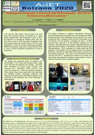
- 1. “Diaphragmatic+Sonography+vs+Structured+Light+Plethysmography+++after+NIV+an+ Mechanical+I=Exsufflator+treatments“ A. Longoni*/** , P. Pozzi **, A. Paddeu** * S.R.R.F. - ** Cardio-Respiratory Rehabilitation Center “Paola Giancola Foundation”, S. Anthony Abate Hospital Cantù, ASST Lariana, Italy angelo.longoni@asst-lariana.it ! !+ A 56 year-old male patient, former smoker and truck driver that practised activity as bicyclist for 6 years started suffering from right emiparesis in 2013; in 2014 the diagnosis of motoneuron disease (ALS) was set; ALS went on and on 2016 the patient devoleped chest wall muscles deficit and ipovalid cough. In 2017 he was hospitalized to start non invasive mechanical ventilation (NIMV) and assisted cough with a Cough Machine (CM) in order to prevent respiratory insufficiency as well as pneumonia. !! Case history The rehabilitative treatments Conclusions The difference in US diaphragmatic excursion was 0,4 cm to 1,5 cm with forced breath and 1,5 cm to 4 cm under Caugh Machine. The Pneumacare values in forced breath were: upper left 61%, upper right 78%, lower left 90%, lower right 70% to upper left 22%, upper right 23%, lower left 24%, lower right 30%. Our study suggests that the Ultrasonography and the Structured Light Plethysmography are a safe, reliable, useful and complementary modality that provides clinicians with a non-invasive way, when the patient is no longer able to perform pulmonary function testings, of observing active, real-time regional and selective respiratory function like the movement of the chest wall and the diaphragm escursion.They can be used to set the NIMV and the Cough-assist machine at the bedside, in conscious or unconscious patient and they can help clinicians and respiratory therapists in offering patients a tailored therapy. The patient has performed cycles of nighttime and diurnal NIMV in S/T mode with oronasal mask, single circuit with leak and esternal hot humidifier and three daily treatments of diurnal cough machine with Pressure I/E=+- 35cmH2O, (Time ins=2,5s, Time exp=1,5s, Pause=1s) =1 cycle, for three/four repetitions of five cycles. Once a day it performed motor exercises with assisted minibike. Respiratory evaluation with Ultrasound and Structured Light Plethysmography were performed at the admission and at the discharge in sitting position. The patient underwent a nighttime pulsoximetry. Pulmonary function testings were not possible and we evaluated the patient through the study of the diaphragm muscle excursion and chest movements with a ultrasound (US) and the Pneumacare Thora 3-D System. The US machine, a portable one with a 1-5 MHz Convex probe, was set in B and M-Mode modality and the valutation was performed in an anterior subcostal approach on semi-recumbent position. PneumaCare (called Structured Light Plethysmography - SLP) consisted of a visible white light that projected a grid pattern onto the patient’s chest. Two cameras filmed the movement at high speed (30 frames per second) and the software generated a 3D view of the chest wall movement over the time and it calculated the volume of air moved. The examination was performed in sitting or lying position in order to measure the tidal volume in different settings. Investigations 25!Gennaio!2020! SLP: 7+4 = 11 VS US: 5+7 = 12 SLP VS US