Call Girls Jayanagar Just Call 7001305949 Top Class Call Girl Service Available
Pediatric Supracondylar Fractures
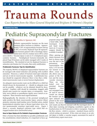
1. P A R T N E R S O R T H O P A E D I C
Trauma Rounds
Case Reports from the Mass General Hospital and Brigham & Women’s Hospital
A Quarterly Case Study Volume 3, Fall 2011
Pediatric Supracondylar Fractures
Samantha A. Spencer, MD propriate setup with
a hand table, sterile
Pediatric supracondylar fractures are the most tourniquet, C-arm
common elbow fractures in children. Approxi- and hand instru-
mately 7-10% of supracondylar fractures and up ment set. A vascular
to 50% of severely displaced Type III supracon- surgeon should be
dylar fractures present with a neurologic injury: available if needed.
radial nerve (41.2%); median nerve (36%); ulnar
nerve (22.8%). Vascular injury is seen in 1% of displaced supra- When opening pe-
condylar fractures. Nondisplaced fractures/minimally dis- diatric fractures, it is
placed Type II fractures can be safely managed with 3 weeks of best to always open
immobilization. The standard of care for displaced fractures is over the tear in the
reduction/pin fixation for 3-4 weeks, then early mobilization. periosteum. For
supracondylar frac-
Problematic Fractures: Tips for Identification tures, a 3-5 cm ante-
The majority (90-95%) of displaced supracondylar fractures can rior incision in the
be managed with closed reduction and pinning with excellent elbow crease usu-
outcomes. However, a subset of fractures need open reduction ally allows easy ex-
and are at risk for neurovascular sequelae. A problematic frac- posure of the frac-
ture should be suspected whenever there is less than a fully ture and the neuro-
intact neurovascular exam or severe fracture displacement. vascular structures.
These are often
An adequate neurovascular exam can be difficult in a child but
tented over the
should always be documented, or – should an adequate exam
proximal fracture
not be possible - whatever can be obtained should be docu-
fragment. Once
mented. Capillary refill should be immediate; sluggish refill
any entrapped mus-
should raise concern for vascular injury or entrapment. Simi-
cle and/or nerves/
larly, nerve deficits or paresthesias signify nerve stretch or en-
vessels are cleared,
trapment. These fractures need urgent treatment.
the fracture can be
Radiographically, the direction of the proximal metaphyseal open reduced and
spike predicts the likely neurovascular injury: anterior (direct Figure 1: Elbow x-ray demonstrating severely
pinned in the usual displaced supracondylar fracture.
posterior extension type)-median nerve/brachial artery, medial fashion. The nerves
(posterolateral extension type or flexion type)-ulnar nerve, lat- and vessels can then be assessed with the tourniquet down. It
eral (posteromedial extension type)-radial nerve. Figure 1 often takes warming and dripping vasodilative agents on the
shows a severely displaced extension type which had en- brachial artery for 10-15 minutes to relieve vasospasm. If pulsa-
trapped median nerve and brachial artery. tile flow returns - which is common - standard closure and bi-
How to Open Reduce & Fix Pediatric Supracondylar Fractures valved casting can proceed. If flow does not return or an arte-
Once a fracture has been identified as possibly problematic and rial injury is visible, a vascular surgery assessment for need of
has unsatisfactory closed reduction, it is important to have ap- brachial artery repair must occur.
Trauma Rounds, Volume 3, Fall 2011
1
2. P A R T N E R S O R T H O P A E D I C T R A U M A R O U N D S
Figure 2: Postoperative AP and
Lateral x-rays of pin
configurations.
After either closed or open
reduction and pinning of a
supracondylar fracture (Fig-
ure 2), children should be
comfortable with little nar-
cotic requirement and no
negative change to their pre-
operative neurologic exam.
Significant pain and increas-
ing pain medicine require-
ments are the best indicators
in children of evolving com-
partment syndrome or missed
arterial injury or entrapped
nerve. Entrapment should
particularly be suspected if
pain increases and nerve function is decreased after closed reduc- Bibliography
1. White L, Mehlman CT, Crawford AH. Perfused, pulseless, and puzzling: a sys-
tion and pinning. These issues require emergent surgical explora- tematic review of vascular injuries in pediatric supracondylar humerus fractures
tion. and results of a POSNA questionnaire: J Pediatr Orthop 2010; 30(4):328-35.
Conclusions 2. Campbell CC, et al, Neurovascular injury and displacement in type III supracon-
dylar humerus fractures: J Pediatr Orthop 1995; 15(1):47-52.
The majority of displaced supracondylar fractures can be man- 3. Kasser JR and Beaty JH, Supracondylar Fractures of the Distal Humerus: Chap
aged with closed reduction and pin fixation in a regularly 14 In Rockwood and Wilkins, Fractures in Children, 6th ed. Lippincott Wil-
scheduled OR time. However, displaced fractures with preop- liams & Wilkins; Philadelphia, PA. 2006: 543-589.
erative neurovascular deficits should raise concern for neuro-
vascular entrapment and injury. Indications for open reduction New England Regional Fracture Summit, Stowe, VT
of closed pediatric supracondylar fractures include inadequate
The popular AO Fracture Summit will be held January 13 – 16, 2012 in
hand perfusion after pinning, inability to obtain an adequate
Stowe, VT. The course is chaired by Drs Mark Vrahas, Jesse Jupiter and
reduction, and evidence of iatrogenic neurovascular injury Raymond White, and features several BWH and MGH Orthopaedic
postoperatively. When open reduction is performed, an ante- Faculty. This year’s special guest is Dr Joseph Schatzker.
rior antecubital crease incision affords access to the torn perios- The course uses an informal, discussion-based, highly interactive format.
teum as well as the neurovascular structures. The chief aim is to educate community orthopaedic surgeons who are
Dr. Samantha Spencer is a pediatric orthopaedist at Children's Hospital, Boston actively involved in the treatment of patients with fractures. Partici-
specializing in trauma, lower extremity, vascular anomalies, osteogenesis imper- pants are invited to bring their own cases for discussion.
fecta and skeletal dysplasias. Samantha.Spencer@childrens.havard.edu Registration is still open!
For more information: www.aona.org
AchesAndJoints.org/Trauma
Please share your comments online, or by email:
Trauma Faculty Michael Weaver, MD — 617-525-8088
Mark Vrahas, MD / mvrahas@partners.org
BWH Orthopedic Trauma
Mark Vrahas, MD — 617-726-2943 Yawkey Center for Outpatient Care, Suite 3C
mjweaver@partners.org
Partners Chief of Orthopaedic Trauma 55 Fruit Street, Boston, MA 02114
mvrahas@partners.org Jesse Jupiter, MD — 617-726-5100
MGH Hand & Upper Extremity Service Editor in Chief
Mitchel B Harris, MD — 617-732-5385 jjupiter@partners.org Mark Vrahas, MD
Chief, BWH Orthopedic Trauma
mbharris@partners.org David Ring, MD — 617-724-3953
MGH Hand & Upper Extremity Service
Program Director
R Malcolm Smith, MD, FRCS — 617-726-2794 dring@partners.org Suzanne Morrison, MPH
Chief, MGH Orthopaedic Trauma (617) 525-8876
Brandon E Earp, MD — 617-732-8064 smmorrison@partners.org
rmsmith1@partners.org
BWH Hand & Upper Extremity Service
David Lhowe, MD — 617-724-2800 bearp@partners.org Editor, Publisher
MGH Orthopaedic Trauma George Dyer, MD — 617-732-6607 Arun Shanbhag, PhD, MBA
dlhowe@partners.org BWH Hand & Upper Extremity Service www.MassGeneral.org/ortho
gdyer@partners.org www.BrighamAndWomens.org/orthopedics
2
Trauma Rounds, Volume 3, Fall 2011