
Recommended
More Related Content
What's hot
What's hot (20)
Similar to Oral cancer screening
Similar to Oral cancer screening (20)
Recently uploaded
Recently uploaded (20)
Oral cancer screening
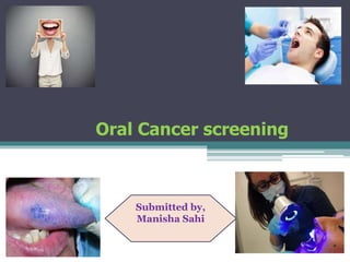
- 1. Oral Cancer screening Submitted by, Manisha Sahi
- 2. Definition of cancer: “Abnormal mass of tissue the growth of which exceeds and is uncoordinated with neighbouring tissue and growth persist in same exceesive manner even after the ceassation of the stimuli which evoked the change.” –ROBBINS Cancer occuring an any part of oral cavity is termed as oral cancer.
- 3. Oral cancer is the sixth deadliest cancer in the world. While most cancer survival rates have increased over the past 40 years., the five year survival rate of oral cancer has remained below 50%.a
- 4. Pathogenesis: The pathogenesis of oral cancer reflects an accumulation of genetic changes that occur over a period of years. (Genetic susceptibilty) The major genes involved in oral cancer are proto onco genes and tumor suppressor genes. Other factors that play role in the progression of the disease may include allelic loss at other chromosome region,mutations to proto oncogenes and TSGs, or epigenetic changes such as deoxyribonucleic acid(DNA) methylation or histone deacetylation.
- 5. Protooncogenes code for growth factors,growth factor receptors,protein kinase, signal transducers, nuclear phosphoproteins and transcription factors. Overall effect is they increase cell growth and differentiation. TSGs negatively regulate cell growth and differentiation. Both copies of TSG must be inactivated or lost (LOH) for loss of function ( “two-hit” hypothesis).LOH commonly seen in chromosome arms 3p,4q,8p,9p,11q,13q,and 17p.
- 7. Risk Factors: • Chemical irritants(tobacco,alcohol,mouthwashes with high alcohol content) • Physical irritants(Denture use, prolonged denture irritaion, irregular teeth or restoraion, chronic cheek biting habits) • Nutritional factors(defficiency of vitamin A and caretenoid supplementation)
- 8. • Prolonged sun exposure • Hormonal effects • Increased cellular aging • Decreeased immunologic surveillance with aging • Viruses(HSV-1,HSV-2.HPV)
- 9. Increased risk: Patient age 40 and older(95% of cases) 18-39 years of age combined with the following: • Tobacco use • Chronic alcohol consumption • Oral HPV infection Highest risk: Patient age 65 and older with lifestyle risk factors Patients with history of other cancer
- 11. Signs and symptoms or oral cancer • A sore in the mouth that doesn’t heal • Persistent mouth pain. • A lump or thickening in the cheek. • A white or red patch on the gums, tongue, tonsil or lining of the mouth. • A sore throst or feeling that soomething is caught in the throat that doesn’t go away. • Difficulty in swallowing or chewing. • Difficulty in moving the jaw or tongue.
- 12. • Numbness of the tongue or elsewhere in the mouth. • Jaw swelling that makes denture hurt or fit pooryly. • Loosening of the teeth. • Pain in the teeth or jaw. • Voice change. • A lump in the neck. • Significant weight loss. • Persistent bad breath.
- 13. Oral cancer screening: It is an examination performed by the dentist of the doctor to look for the signs of cancer or precancerous conditions in your mouth. The goal of oral cancer screening is to identify mouth cancer early, when there is a greater chance for a cure. It can be done by examination of mouth during a routine dental check up or by use of additional tests to aid in identifying areas of abnormal cells in the mouth.
- 14. Screening preparation Overview • Head and neck exam should be routine. • Review history of alcohol and tobacco use • Follow up on suspicious signs. Armamentarium • Proper lighting,Probe mouth mirror tweezer • Gauze sponges • Disposable gloves • 3 to 5 minutes of time
- 15. Points to remember when screening for oral cancer • Tell your patient whar are you doing with each procedure and why?? • Special attention should be focused on the lesions of lateral border of tongue and floor of mouth . • Always note any changes in colour texture of all soft tissue or any swelling, if you detect an abnormality determine the historu of the lesion if the abnormality has been of more than 2 weeks duration,take appropriate action to obtain a biopsy.
- 16. • Follow up to ensure a definitive diagnosis of an abnormality. • Teach your patient about signs and symptoms of oral cancer. • If patient uses tobacco products provide appropriate counselling or refer patient for counselling. • Remove all removable prosthesis before starting the examination.
- 17. Dianostic aids Standard screening aids (conventional intra and extra oral examination) Established screening adjuncts (oral cytology,oral brush cytology) Vital staining (toluedine blue,methylene blue,lugol’s iodine)
- 18. Extra oral examination: General appraisal Skull Facial form Skin Eyes Neurological deficit Lymph nodes of head and neck TMJ Masticatory musscles
- 22. Drawbacks of conventional examination: It cannot discrimate between the lesion that are progressive or malignant and those non progressive counter parts. It doesn’t identify all potentially premalignant lesions.
- 23. Oral Cytology It is the study of cells which exfoliate or abrade from body surface.When epithelium becomes seat of any pathology cells lose their cohesiveness and cells in deeper layers may shed along with superficial cells. Drawback: the sensitivity and specificity differs due to its subjectivity or due to poor technique in obtaining cells and smear preparation.
- 25. Brush Biopsy(Oral CDx) It utilizes a stiff brush to colled the sample cells from the basal layer cells non invasively and assess the dysplasia by computer asssisted neural network.Its accurancy can be increased by using DNA cytometry, silver nucleolar organisation regions analysis,immunocytochemistry and fluorescent insitu hybridization(FISH).
- 27. Vital Stains: 1. Toluedene blue: It is a topical dye,which gets concentrate in cells with abundant nucleic acid.It has high sensitivity for detecting carcinoma. Interpretation Dark blue colour :positive for lesions suspicious malignancy Light blue: positive for premalignant lesions proved otherwise by biopsy.
- 28. Advantages: Specify area for biopsy. Cheap and non invasive,disposable Has high sensitivity for carcinoma Disadvantages: It is distasteful. Its sensitivity for identifying dysplasia is poor. The colour of the dye remains in the mouth for 4 to 6 hours. It may give false positive results(inflammatory and ulcerative conditions).
- 29. 2. Methylene Blue • It is a heterotropic aromatic chemical compound.At room temperature it is solid,odourless dark green powder which yields a blue colour when dissolve in water. Advantages: It is cheaper and less cyto toxic.
- 30. Disadvantage: the result is not confirmatory. Application: Early detection of suspected oral cancer Treat Alzhiemer disease Examine(RNA) or (DNA) under microscope.
- 32. 3.Lugol’s iodine It consist of 10 parts of potassium iodide to 5 parts of iodine.Application of iodine results in brown or black colour staining in areas containing glycogen. In areas lacking glycogen iodine isn’t absorped and such areas remain colourless or turn yellow. Advantages: Used for non keratinized stratified squamous epithelium. It is simple,low costs. High sensitivity with low false negative Result are fast
- 33. Disadvantage: It is an irritant cause abdominal pain,heart burn and nausea. Allergic reaction to iodine. Less accurate when used in post menopausal women. Application: Used routinely for the patient with head and neck cancer. Heavy smokers and drinkers.
- 34. Velscope The velscope handpiece emits a safe blue light into the oral cavity causing tissue fluorescence from the surface of epithelium through to the basal membrane-where pre malignant changes typically start.
- 35. By utilizing special optical filters in the velscope hand piece the clinician is able to immediately view the different fluorescence signatures in the oral tissues to help differentiate between normal and abnormal cellular activity. Abnormal tissue typically appears as an irregular, dark area that stands out against the green fluorescence pattern of surrounding healthy tissue.
- 37. Advantages: It takes only 1-2 mins and is painless and non invasive,with no stains and rinses required. Improves the distinction between normal and abnormal tissues. Thorough visual and digital soft tissue examination. Useful benefit in the determination of surgical border and post surgical evaluation. It covers large surface area so small lesions can also be identified.
- 38. Disadvantage Heat from prolonged and close tissue examination may cause patient discomfort. Lack of methodologically sound clinical trails. Insufficient use of histologic and molecular mapping.
- 40. Orascoptic DK: This instrument work in conjunction with a mild acedic acid rinse to improve the visualization of oral lesions. This examination enhances the ability to identify potentially cancerous at its earliest stages. Early detection of precancerous tissue can minimize or eliminate the disfiguring effects of oral cancer and possibly save a life
- 41. It includes a transilllumination instrument and lightened mirror. It uses a battery powered handled LED light source and three interchangeable diagnostic instruments.
- 42. Microlux diagnostic Light With refractive light technology,this aid help save lives in the detection of pre cancerous abnoramalities and makes this advance in patient care simple and inexpensive.
- 43. After noting any acetowhite soft tissue lesions during a routine exam,simply have the patient rinse with Microlux DL 1% acetic acid solution for 1 minute. Then repeat the oral exam using Microlux DL. When used in conjunction with conventional oral mucosal exams,it improves identification evaluation and monitoring of soft tissue abnormalities and changes in all patients
- 44. The irregular cells take on a whitish hue which contrasts with surrounding tissue, helping to identify abnormalities which require further testing. Since the acetic acid dehydrates the cytoplasm of aceto white lesions the lesions refractive properties are changed.Under diffused light from the special Microlux DL fiber optic light guide, acetowhite or leukoplakic lesions become more visible.
- 45. ViziLite Plus with TBlue630 • ViziLite Plus with TBlue630 is an oral lesion identification and marking system that is used as an adjunct to the conventional head and neck examination. • It is comprised of a chemiluminescent light source (ViziLite) to improve the identification of lesions and a blue phenothiazine dye to mark those lesions identified by ViziLite.
- 46. • Similarly in the oral cavity, after rinsing with a dilute acetic acid solution, abnormal squamous epithelium tissue will appear acetowhite when viewed under ViziLite's diffuse low-energy wavelength light. • Normal epithelium will absorb the light and appear dark. ViziLite can assist a dentist or hygienist in identifying an abnormality in the oral cavity.
- 47. How ViziLite Plus Works