SlideShare a Scribd company logo
1 of 113
Dr. Ashok Ayer
Assistant Professor
Department of Conservative Dentistry & Endodontics
B.P.Koirala Institute of Health Sciences, Dharan, Nepal
Contents:
 Introduction
 Coronal and radicular pulp
 Apical foramen
 Accessory canal
 Functions of dental pulp
 Components of dental pulp
 Functions of pulpal extracellular matrix
 Organization of cells in the pulp
 The principle cells of the pulp
 The pathways of collagen synthesis
 Matrix and ground substances
 Vasculature and lymphatic supply
 Innervation of Dentin- pulp complex
 Disorders of the dental pulp
 Advances in pulp vitality testing
 Conclusion
Dental Pulp
 Occupies the center of each tooth.
 Soft connective tissue that supports the dentin.
 Total 52 pulp organs; 32: Permanent, 20: Primary
 Total Volume of all permanent teeth pulp organs is
0.38 cc.
 Mean volume of a single adult human pulp is 0.02 cc.
Maxillary
(Cubic Centimeter)
Mandibular
(Cubic Centimeter)
Central Incisor 0.012 0.006
Lateral Incisor 0.011 0.007
Canine 0.015 0.014
First Premolar 0.018 0.015
Second Premolar 0.017 0.015
First Molar 0.068 0.053
Second Molar 0.044 0.032
Third Molar 0.023 0.031
Orban’s Oral histology & embryology: Pulp; Department of Oral Surgery,
Newcastle - Tyne, England
Coronal Pulp:
 Six surfaces
 Pulp horns, depends
on the cuspal
number.
 Radicular Pulp:
 The radicular portion of the pulp organs are
continuous with the periapical connective tissue
through the apical foramen or foramina.
 As growth proceeds, more dentin is formed, so that
when the root of teeth are matured the radicular pulp
is narrower.
 The apical pulp canal becomes smaller also because of
apical cementum deposition.
 Apical foramen:
 Average size of apical foramen of the maxillary teeth
in the adult is 0.4 mm
 Mandibular teeth 0.3 mm
 Sometimes it is found on the lateral side of the apex
although the root itself is not curved.
 Frequently there are two or more foramina separated
by a portion of dentin and cementum or by cementum
only.
 Accessory canal:
 Leading from the radicular pulp laterally through the
root dentin to the periodontal tissue.
 May be seen anywhere along the root but are most
numerous in the apical third of the root.
 Clinically significant in spread of infection, either
from the pulp to the periodontal ligament or vice
versa.
 Occur in areas where there is premature loss of root
sheath cells; these cells induce the formation of
odontoblasts which form dentin.
 May also occur where the developing root encounters
a blood vessel.
Functions of dental pulp
 Inductive:
 Interact with the oral epithelial cells
 Differentiation of the dental lamina and enamel
organ formation.
 Cells of pulp + blood vessels & nerves
provides the tooth vitality
 Formative:
 Produces dentin that surrounds and protects the pulp.
 Pulpal odontoblasts develop the organic matrix and
function in its calcification.
 Nutritive:
 Blood vascular system of the pulp; nourishes dentin
through the odontoblasts and their processes.
 Protective:
 Sensory nerve respond to pain
 Nerves initiate reflexes that control circulation in the
pulp.
 Defensive or reparative:
 First line of defense to injuries and infection of
dentine
 Tertiary dentine
 Immuno-competent
 Clearance of toxic substances
Components of dental pulp
Cells + (extracellular) Matrix
Fiber Ground substance
• Collagen
• Elastin
Structural Adhesive
• Fibronectin
• Laminin
• HS
• DS
• CS
GAG Proteoglycan
• Decorin
• Versican
Components of dental pulp
 CELLS (odontoblast, fibroblast,
undifferentiated cell, macrophage,
dendritic cell)
 FIBERS AND GLYCOPROTEIN (collagen
type I, III, no elastic fiber, fibronectin)
 GROUND SUBSTANCES
(glycosaminoglycans, chondroitin sulfate
proteoglycan)
 BLOOD VESSELS, NERVES, LYMPH
VESSELS
 Maintain tissue’s physical properties and integrity
 Control of growth and development and repairs
 Control of cell migration
 Control of diffusion of macromolecules
Functions of pulpal extracellular matrix
Collagen in dental pulp
 Concentration varies from species to species, 32% in human
pulp.
 Higher content in the middle and apical pulp.
 Total collagen decreases with age.
 Interestingly high level of collagen type III. (43%) : vascular
content, tissue extensibility (cf. Elastin)
 Absence of elastin (except in b.v.).
Adhesive glycoproteins in dental pulp
 Fibronectin found in
predentine NOT mature
dentine.
 Fibronectin present in
pulp and dental papilla.
 Fibroblasts synthesize
pulpal fibronectin.
 Fibronectin is expressed
during reparative
dentinogenesis.
Immunoreactive fibronectin molecules detected
along the border of predentine and between
odontoblast (Yoshiba et al., 1994)
Glycosaminoglycans in dental pulp
 Chondroitin sulfate, dermatan sulfate, hyaluronic
acid present
 Amount of uronic acid decreases with age
 Total GAG decreases with reduced dentinogenic
activity
 Decorin may involve in mineral nucleation at the
mineralization front
Organization of cells in the pulp
tight junction
nerve terminals
Four distinct zones:
1. The odontoblastic zone at the pulp periphery
2. A cell free zone of Weil beneath the odontoblast;
prominent in the coronal pulp
3. A cell rich zone; high cell density
4. The pulp core; major vessels and nerves
 The principle cells of the pulp:
 Odontoblasts
 Fibroblast
 Undifferentiated mesenchymal cells
 Macrophages
 Immunocompetent cells
 Odontoblasts:
 The most distinctive cells of the dental pulp
 Form a layer lining the periphery of the pulp and have
a process extending into the dentin
 Arranged in palisade pattern of three to five cells deep
 59,000 to 76,000 per square milimeter in coronal
dentin, with a lesser number in root dentin.
 Active cells:
 Elongated, basal nucleus, much basophilic cytoplasm,
promonent golgi zone.
 Resting cell:
 Stubby, little cytoplasm, more hematoxophilic nucleus.
 Odontoblast process begins at the neck of the cells
just above the apical junctional complex where the cell
gradually begins to narrow as it enters predentin.
 The process is devoid of major organelles but does
display an abundance of microtubules and filaments
arranged in a linear pattern along its length.
 The pathways of collagen synthesis:
 The spherical distensions contain free polypeptides
that assemble as a triple helix in the cylindrical
distensions to form the procollagen molecule.
 The cylindrical distension bud off as secretory
granules.
 Secretory granules that are transported toward the
odontoblast process, where their content is released.
Synthesis of collagen and its assembly into fibrils and fiber
Some types (of 15) of known collagen
Type Molecular Tissue distribution
Fibril-forming I [a1(I)]2 a2(I) bone, skin, tendon, ligaments
(90%) of body collagen
II [a1(II)]3
cartilage, intervertebral disc,
notochord, vitreous humor of eye
III [a1(III)]3 skin, blood vessels, internal organs
V [a1(V]2 a2(V) as type I
XI [a1(XI] a2(XI) a3(XI) as type II
Fibril-associated IX [a1(IX] a2(IX) a3(IX) cartilage (with type II)
XII [a1(XII)]3 tendon, ligaments (with some type I)
Network-forming IV [a1(IV)]2 a2(IV) basal laminae
VII [a1(VII)]3 anchoring fibrils beneath stratified
squmous epithelia
RGD = cell-binding domain
The structure of a fibronectin dimer.
Structure of a GAG
Structure of proteoglycans
Aggrecan mechanical support
(cartilage)
Betaglycan binds TGF-beta
(cell surface*, matrix)
Decorin binds type I and (CNT)
TGF-beta
Perlecan basal laminae
(basal laminae)
Syndecan-1 binds FGF
(cell surface*)
* = Integral membrane proteoglycan
Some known proteoglycans:
 Junctions occur between adjacent odontoblasts
involving
 Gap junctions
 Occluding zones (Tight junctions)
 Desmosomes
 The actin filaments inserting into the adherent
junction are prominent and form a terminal cell web.
 This junctional complex does not form a zonula,
completely encircling the cell, as occurs in epithelia;
(it is focal, and there is some debate whether it can
restrict the passage of molecules and ions from the
pulp into the dentin layer)
 Serum proteins seem to pass freely between
odontoblasts and are found in dentin
Fibroblasts:
 Greatest number in the pulp
 Numerous in coronal pulp where they form the cell-
rich zone.
 The function is to form and maintain pulp matrix.
Undifferentiated Ectomesenchymal Cells:
 Represents the pool from which the connective tissues
of the pulp are derived.
 Depending upon the stimulus these cells may give rise
to odontoblasts and fibroblasts.
 In older pulp they diminish, thereby reducing the
regenerative potential of the pulp.
Macrophages
 Located throughout the pulp center.
 Involved in the elimination of dead cells, the presence
of which indicates that turnover of dental pulp
fibroblast occurs.
Lymphocytes
 In normal pulp T lymphocytes are found, but B
lymphocytes are scare.
Dendritic Cells
 Bone marrow derived, antigen presenting dendritic
cells.
 Beneath the odontoblast layer.
 They capture and present foreign antigen to the T
cells.
 Cells participate in immunosurvillance and increase
in number in carious teeth.
 Infiltrate odontoblast and project their processes into
the tubules.
 8% of total cell population.
Matrix and Ground Substance
 Principally Type I and Type III collagen.
 Composed of glycosaminoglycans, glycoproteins, and
water.
 Overall collagen content increases with age.
 The greatest concentration of collagen generally
occurs in the most apical portion of the pulp.
Significance:
 During pulpectomy; Engaging the pulp with a barbed
broach in the region of apex affords a better
opportunity to remove the tissue intact.
Vasculature and Lymphatic Supply
 Circulation establishes the tissue fluid pressure.
 One or sometimes two vessels of arteriolar size
(about 150µm) enter the apical foramen with the
sensory and sympathetic nerve bundles.
 Smaller vessels, without any accompanying nerve
bundle, enter the pulp through the minor foramina.
Pulp vasculature
The arterioles occupy a central position within the
pulp and, as they pass through the radicular portion of
pulp, give off smaller lateral branches.
 Occasionally U- looping of pulpal arterioles is seen,
and this anatomic configuration is thought to be
related to the regulation of blood flow.
Pulp tissue is highlyvascularized.
40-50 ml/min/100g
(Kim, 1985)
 Some terminal capillary loops extend upward between
the odontoblasts to abut the predentin if
dentinogenesis is occurring.
 Located on the periphery of the capillaries at random
intervals are pericytes.
 Pericytes are contractile cells capable of reducing
the size of the vessel lumen.
 Anastomosis are point of direct communication
between the arterial and venous sides of the
circulation.
 Lymphatic vessels also occur in the pulp tissue, they
exit via one or two large vessels through the apical
foramen.
 Sympathetic adrenergic nerves terminate in relation
to the smooth muscle cells of the arteriolar walls.
 Afferent free nerve endings terminate in relation to
arterioles, capillaries and veins and serve as effectors
by releasing various neuropeptides that exert an
effect on the vascular system.
Dental pulp interstitial fluid (ISF) and exchange of substances between plasma
and ISF. (* values from Tonder and Kvinnsland, 1983; Ciucchi et al., 1995)
(5.5-10.3 mm Hg*)
(43 mm Hg)
(20 mm Hg)
(35 mm Hg)
Hydrostatic pressure
in dental pulp
Innervation of Dentin- Pulp Complex
 Nerve enter the pulp through
apical foramen, along the
afferent blood veessels, and
together from the
neurovascular bundle.
 Each nerve fiber has been
estimated to provide at least
eight terminal branches.
These branches ultimately contribute to an
extensive plexus of nerves in the cell free
zone just below the cell bodies of the
odontoblasts in the crown portion of the
tooth.
Approx. 1800 non
myelinated +
400 myelinated
Intradentinal nerves are mostly found in pulpal
horns.
 This plexus of nerves, which is called the
subodontoblastic plexus of Raschkow, occupies the
cell- free zone of Weil and can be demonstrated in
silver nitrate stained sections under the light
microscope or by immunocytochemical techniques.
 The nerve bundles that enter the tooth pulp consist
principally of :
Sensory afferent nerves of the trigeminal
nerve
and
Sympathetic branches from the superior
cervical ganglion.
As the nerve bundle ascends coronally;
The myelinated axons gradually loose their
mylein coating,
So that a proportional increase in the number
of unmyelinated axons occurs in the more
coronal aspect of the tooth.
A-delta fibers
 Conduction velocity 2-30 m/s
 Lower threshold
 Involved in fast, sharp pain
 Stimulated by hydrodynamic
stimuli
 Sensitive to ischemia
 Sharp pain
C fibers
 Conduction velocity 0-2 m/s
 Higher threshold
 Involved in slow, dull pain
 Stimulated by direct pulp damage
 Sensitive to anesthetics
 Dull pain
Types and properties of pulpal sensory nerve fibers
A-beta fibers
 Conduction velocity 30-70 m/s
 Very low threshold, non-noxious
sensation
 50% of myelinated fibers in pulp
 Functions not fully known
Non-myelinated sympathetic
fibers
 Conduction velocity 0-2 m/s
 Post-ganglionic fibers of superior
cervical ganglion
 Vasoconstriction
 A small number of axons pass between the
odontoblast cell bodies to enter the dentinal tubules
in proximity to the odontoblast process.
Possible mechanisms of dentine sensitivity
Hydrodynamic mechanism
(Gysi, 1900; Brannstrom, 1963)
Pulp venules
STIMULATION
Increased pulp
interstitial fluid
Increased pulp
pressure
Increased tubular
fluid flow
Release of
inflammatory
agents?
Increased blood
viscosity and rbc
congestion in capillary
bed
Increased A-V shunt
blood flow
Outward dentinal
fluid flow and
aspiration of
odontoblasts
CNS, Pain, Reflexes
Vasodilation, Increased permeability
Pulpal axonal reflex due to dentine stimulation
Without infection,
Vascular changes could
be resolved.
Axon
reflex
SP, CGRP
Dentine
Disorders of the Dental Pulp
Pulp Stones
 Pulp stones, or denticles, frequently are found in pulp
tissue.
 Discrete calcified masses that have calcium phosphorus
ratios comparable to that of dentin.
 More frequently at the orifice of the pulp chamber or
within the root canal.
 Concentric layers of mineralized tissue formed by
surface accretion around blood thrombi, dying or
dead cells, or collagen fibers.
 Occasionally a pulp stone may contain tubules and
be surrounded by cells resembling odontoblasts.
 Such stones are rare and, if seen, occur close to the
apex of the tooth. Such stones are referred to as ‘true’
pulp stones as opposed to ‘false’ stones having no cells
associated with them.
 If during the formation of a pulp stone, union occurs
between it and the dentin wall, or if secondary dentin
deposition surrounds the stone, the pulp stone is
called an attached stone.
 The presence of pulp stones is significant in that
They reduce the overall number of cells within
the pulp
and
Act as an impediment to debridement and
enlargement of the root canal system during
endodontic treatment.
Age Changes
 Decrease in the volume of pulp chamber and root
canal brought about by continued dentin deposition.
 On occasion can appear to be obliterated almost
completely.
 From about the age of 20 years, cells gradually
decrease in number until age 70, when the cell density
has decreased by about half.
 Fibrosis is due to aging & Injury.
 Increase in collagen fibers’
bundles which becomes more
evident with the decrease in pulp
size
 Lose and a degeneration of myelinated and
unmyelinated axons that correlate with an age-
related reduction in sensitivity.
 Irregular areas of dystrophic calcification,
especially in central pulp.
 Gradual reduction of tubule diameter.
 The continued deposition often leads to complete
closure of the tubule;
as can be seen readily in a ground section of
dentin, because the dentin becomes translucent
(or sclerotic).
 Sclerotic dentin is found frequently near the root
apex in teeth from middle aged individuals.
Pulpitis
 Acute or chronic.
 Partial or total.
 Open or closed.
 Exudative or suppurative.
 Reversible or irreversible.
 Pulpitis is a dynamic process and presents a
continuous spectrum of changes reflecting
interplay between cause and host
defenses.
 Poor correlation between microscopic
changes & clinical symptoms.
Pulpitis: Clinical Features
 Presents as pain which patient may have difficulty
in localizing to a particular tooth.
 Pain may radiate to adjacent jaw, face, ear, or neck.
 May be continuous for several days or may occur
intermittently over a longer period.
 Pulpitis is often described as acute or chronic
based on duration and severity of symptoms.
Acute pulpitis
 Severe throbbing, lancinating pain on thermal
stimulation or lying down, keeps patient awake.
 Generally lasts 10-15 minutes but may be more or
less continuous (reversible pulpitis).
 With progression, may become spontaneous &
continuous (irreversible pulpitis).
Chronic pulpitis
 Bouts of dull aching which can last for an hour or
more.
 Pain on thermal stimulation or spontaneously.
 Pulpitis may be asymptomatic.
 Most important decision clinically is whether
pulpitis is reversible or irreversible.
 Decision is made based on many factors
including:
1. Severity of symptoms.
2. Duration of symptoms.
3. Size of carious lesion.
4. Pulp tests.
5. Direct observation during operative procedure.
6. Age of patient.
Pulpitis: Etiology
 Microbial:
 Dental caries.
 Traumatic exposure.
 Marginal leakage.
 Cracked tooth
 Coronal fracture.
 Attrition.
 Abrasion.
 Traumatic restorative procedure.
 Invaginated odontome.
 Advanced periodontitis (periodontal-endodontic
lesion).
 Pulpitis starts before leading organisms in
carious dentin reach pulp.
 Pulpitis is not usually seen histologically
until organisms are within 1 mm of the
pulp in permanent teeth, or 2 mm in
deciduous teeth.
Chemical and thermal injury
 During restorative procedures: frictional heat,
irritant substances.
 May respond by reactionary dentin formation.
Barotrauma (aerodontalgia)
 Flying at high altitude in unpressurized aircraft,
or rapid decompression in divers.
 Attributed to formation of nitrogen bubbles in
pulp tissue or vessels.
 Thought not to be a direct cause, but rather an
exacerbating cause in presence of caries.
Pulpitis: Histopathology
 Poor correlation between microscopic changes &
clinical symptoms.
 Inflammatory process may be modified by
several factors:
 Nature, severity and duration of insult.
 Efficiency of host defenses.
 Efficiency of pulpo-dentinal complex defenses.
 Special anatomy of pulp: surrounded by hard
tissue and cannot tolerate edema.
85
 Reactionary dentin may continue to form after
onset of pulpitis if odontoblasts and pulp have not
been irreversibly damaged, and may protect pulp.
 Pulpitis caused by caries starts as a localized area,
but extends throughout pulp if caries is not
treated.
86
If inflammation is severe, local
microcirculation may be compromised,
leading to local necrosis and suppuration
of pulp (pulp abscess), or diffuse
suppuration and necrosis.
Pulpitis: Chronic Hyperplastic Pulpitis
(Pulp Polyp)
Open pulpitis or chronic hyperplastic pulpitis (pulp
polyp):
 Large carious cavities.
 Young molar teeth with wide apices and good
blood supply.
88
 Usually devoid of sensation on gentle probing.
 Polyp consists of chronically inflamed
hyperplastic granulation tissue protruding
from pulp cavity.
 May become epithelialized by spontaneous
grafting of desquamated oral epithelial cells from
saliva.
Pulp Necrosis
 May follow pulpitis or trauma to apical blood
vessels.
 Coagulative necrosis after ischemia.
90
 Liquefactive necrosis after pulpitis;
may become gangrenous with foul odor upon
infection by putrefactive bacteria from caries.
 Pulp necrosis in sickling crisis of sickle cell
anemia.
Restorative factors contributing to pulpal injury
Effects of cavity Preparation:
 Frictional heat
 Desiccation
 Exposure of dentinal tubules
 Direct damage to odontoblast processes
 Chemical treatment to exposed dentinal surface
 Cavity preparation: speed, heat, pressure &
coolant may all cause pulp irritation.
 Aspiration or displacement of odontoblasts into
dentinal tubules, with reduction of numbers.
94
Factors associated with the restorative material & its placement
 Material toxicity
 Insertion pressure
 Thermal effects
 Induced stresses
Effects subsequent to restoration
 Marginal leakage
 Cuspal fracture
Effects of cavity preparation & restorative materials may
further complicate pulpitis caused by caries or other
causes.
Thickness & nature of remaining dentine may affect pulp
response to dental material.
Advances in Pulp Vitality
testing
 Pulse Oximetry
 Dental sensor (a modified finger probe) that can be
successfully applied and adapted to the tooth and well
suited to detect pulsatile absorbance.
 The principle: relates the absorption of light, by a
solute to its concentration and optical properties at a
given light wavelength.
 It also depends on the absorbance
characteristics of haemoglobin in the red
and infra-red range
 In the red region, oxyhaemoglobin
absorbs less light than deoxyhaemoglobin
and vice versa in the infrared region.
Hence one wavelength was sensitive to
changes in oxygenation and the second was
insensitive to compensate for changes in
tissue thickness, haemoglobin content and
light intensity.
 The system consists of a probe containing a diode that
emits light in two wavelengths:
I. Red light of approximately 660 nm
II. Infra-red light of approximately 850 nm
 It is also useful in cases of impact injury where
the blood supply remains intact but the nerve
supply is damaged
Dual Wavelength Spectrophotometry
Dual wavelength spectrophotometry (DWLS) is a
method independent of a pulsatile circulation.
The presence of arterioles rather than arteries in the
pulp and its rigid encapsulation by surrouding dentine
and enamel make it difficult to detect a pulse in
the pulp space.
This method measures oxygenation changes in
the capillary bed rather than in the supply vessels
and hence does not depend on a pulsatile
blood flow.
A major advantage is that it uses visible light
that is filtered and guided to the tooth
by fibreoptics
The test is noninvasive and yields objective results.
Laser doppler flowmetry
Laser Doppler Flowmetry (LDF) is a noninvasive,
electro optical technique,
Which allows the semi-quantitative recording
of pulpal blood flow.
 The Laser Doppler technique measures blood
flow in the very small blood vessels of the
microvasculature.
The technique depends on the Doppler principle;
whereby light from a laser diode incident on the
tissue is scattered by moving RBC's
and
As a consequence, the frequency broadened.
The primary issues in pulp-vitality testing as
follows:
A non-vital post-traumatized incisor has a better
long-term prognosis;
If root canal therapy is completed before the
necrotic pulp gets infected.
The best outcome for the post
traumatized immature incisor is for it;
To revascularize and,
Continue normal root development, including
increased root wall thickness.
 Which is not possible to assess with conventional
electrical and thermal testing
Conclusion
Thus the Preservation of Healthy Pulp during
operative procedures and successful management
in cases of disease are two of the most important
challenges.
References:
1. Seltzer and Bender's Dental Pulp; 2002 by Quintessence Publishing Co, Inc; Rev.
ed. of: The dental pulp / Samuel Seltzer, I.B. Bender. 3rd ed. c1984.
2. Oral histology; Development, Structure and Function: A.R. Ten Cate: 7th Edition
3. Orban’s Oral Histology and Embryology
4. Shafer’s Textbook of Oral Pathology; 5th Edition
5. Yamada, Y., Ito, K., Nakamura, S., Ueda, M. & Nagasaka, T. (2010). Promising cell-
based therapy for bone regeneration using stem cells from deciduous teeth, dental
pulp, and bone marrow. Cell Transplantation. [Epub ahead of print], (October
2010)
6. Gronthos, S., Mangani, M., Brahim, J., Robey, PG. & Shi, S. (2000). Postnatal
human dental pulp stem cells (DPSCs) in vitro and in vivo. Proceedings of the
National Academy of Sciences of the United States of America, Vol.97, No.25,
(December 2000), pp. 13625- 13630, ISSN 0027-8424
7. Miura, M., Gronthos, S., Zhao, M., Lu, B., Fisher, LW., Robey, PG. & Shi, S. (2003).
SHED: stem cells from human exfoliated deciduous teeth. Proceedings of the
National Academy of Sciences of the United Stases of America, Vol.100, No.10,
(May 2003), pp.5807-5812, ISSN 0027-8424
8. Seo, BM., Miura, M., Gronthos, S., Bartold, PM., Batouli, S., Brahim, J.,
Young, M., Robey,PG., Wang, CY. & Shi, S. (2004). Investigation of
multipotent postnatal stem cells from human periodontal ligament.
Lancet, Vol.364, No.9429, (July 2004), pp.149-155, ISSN 0140-6736
9. Sonoyama, W., Liu, Y., Fang, D., Yamaza, T., Seo, BM., Zhang, C., Liu,
H., Gronthos, S.,Wang, CY., Shi, S. & Wang, S. (2006). Mesenchymal
stem cell-mediated functional tooth regeneration in swine. PLoS
One.Vol.1, (December 2006), pp.e79
10. Morsczeck, C., Gotz, W., Schierholz, J., Zeilhofer, F., Kuhn, U., Mohl,
C., Sippel, C. & Hoffmann, KH. (2005). Isolation of precursor cells
(PCs) from human dental follicle of wisdom teeth. Matrix Biology,
Vol.24, No.2, (April 2005), pp.155-165, ISSN 0945- 053X
11. Huang, GT., (2009). Pulp and dentin tissue engineering and
regeneration: current progress. Regenerative Medicine, Vol.4, No.5,
(September 2009), pp.697-707 ISSN 1746-076X
12. D'Aquino, R., De Rosa, A., Laino, G., Caruso, F., Guida, L., Rullo, R.,
Checchi, V., Laino, L., Tirino, V. & Papaccio, G. (2009). Human dental
pulp stem cells: from biology to clinical applications. Journal of
Experimental Zoology Part B: Molecular and Developmental Evolution,
Vol. 312, No.5, (July 2009), pp. 408-15, ISSN 1552-5007
13. Batouli, S., Miura, M., Brahim, J., Tsutsui, TW., Fisher, LW., Gronthos,
S., Robey, PG. & Shi, S. (2003). Comparison of stem cell- mediated
osteogenesis and dentinogenesis. Journal of Dental Research, Vol.82,
No.12, (December 2003), pp. 976–981, ISSN 0022- 0345
14. Laino, G., D'Aquino, R., Graziano, A., Lanza, V., Carinci, F., Naro, F.,
Pirozzi, G., & Papaccio, G. (2005). A new population of human adult
dental pulp stem cells: a useful source of living autologous fibrous bone
tissue (LAB). Journal of Bone and Mineral Research, Vol.20, No.8,
(August 2005), pp.1394-1402, ISSN 0884-0431
15. Nakashima, M. (2005). Bone morphogenetic proteins in dentin
regeneration for potential use in endodontic therapy. Cytokine &
Growth Factor Reviews, Vol.16, No.3, (June 2005), pp.369-376 ISSN 1359-
6101
16. Sun, HH., Jin, T., Yu, Q. & Chen, FM. (2011). Biological approaches
toward dental pulp regeneration by tissue engineering. Journal of
Tissue Engineering and Regenerative Medicine. Vol.5, No.4, (April 2011),
pp. e1-e16
17. Zavan Barbara et al. Dental pulp stem cells and tissue engineering
strategies for clinical application of odontoiatric field. Journal of
Biomaterial science and Engineering.
Thank You

More Related Content

What's hot (20)

Cementum
CementumCementum
Cementum
 
Theories of tooth eruption
Theories of tooth eruptionTheories of tooth eruption
Theories of tooth eruption
 
Cementum
CementumCementum
Cementum
 
Dental Pulp
Dental PulpDental Pulp
Dental Pulp
 
Dentinogenesis
DentinogenesisDentinogenesis
Dentinogenesis
 
Differences between primary and permanent dentition
Differences between primary and permanent dentitionDifferences between primary and permanent dentition
Differences between primary and permanent dentition
 
Dentin
DentinDentin
Dentin
 
Amelogenisis
AmelogenisisAmelogenisis
Amelogenisis
 
Tooth development
Tooth developmentTooth development
Tooth development
 
Amelogenesis
Amelogenesis Amelogenesis
Amelogenesis
 
Dental Pulp
Dental PulpDental Pulp
Dental Pulp
 
Development of tooth
Development of toothDevelopment of tooth
Development of tooth
 
Enamel
EnamelEnamel
Enamel
 
Cementum ppt
Cementum pptCementum ppt
Cementum ppt
 
Tooth Dentin and dentinogenesis ppt
Tooth Dentin and dentinogenesis pptTooth Dentin and dentinogenesis ppt
Tooth Dentin and dentinogenesis ppt
 
Dentin pulp complex
Dentin pulp complexDentin pulp complex
Dentin pulp complex
 
Histology of dentin
Histology of dentinHistology of dentin
Histology of dentin
 
Pulp
Pulp Pulp
Pulp
 
Shedding of deciduous teeth
Shedding of deciduous teeth Shedding of deciduous teeth
Shedding of deciduous teeth
 
Enamel
EnamelEnamel
Enamel
 

Similar to Dental pulp

Dental pulp / rotary endodontic courses by indian dental academy
Dental pulp / rotary endodontic courses by indian dental academyDental pulp / rotary endodontic courses by indian dental academy
Dental pulp / rotary endodontic courses by indian dental academyIndian dental academy
 
9.periodontal ligament ppt
9.periodontal ligament ppt9.periodontal ligament ppt
9.periodontal ligament pptpunitnaidu07
 
Dental pulp /certified fixed orthodontic courses by Indian dental academy
Dental pulp  /certified fixed orthodontic courses by Indian dental academy Dental pulp  /certified fixed orthodontic courses by Indian dental academy
Dental pulp /certified fixed orthodontic courses by Indian dental academy Indian dental academy
 
Connective Tissue in health and disease
Connective Tissue in health and diseaseConnective Tissue in health and disease
Connective Tissue in health and diseaseDr.R.Dhivya.,MDS
 
dental pulp detailed power point presentation
dental pulp detailed power point presentationdental pulp detailed power point presentation
dental pulp detailed power point presentationritukhichar4
 
Periodontal Considerations
Periodontal ConsiderationsPeriodontal Considerations
Periodontal ConsiderationsDina Zohaia
 
digestivse system histology of embriology and cytology
  digestivse system histology of embriology and cytology  digestivse system histology of embriology and cytology
digestivse system histology of embriology and cytologyMuhammadMazharHussai2
 
Periodontal ligament
Periodontal ligamentPeriodontal ligament
Periodontal ligamentRitam Kundu
 
Applied Histology of Pulp and Periradicular Tissues
Applied Histology of Pulp and Periradicular TissuesApplied Histology of Pulp and Periradicular Tissues
Applied Histology of Pulp and Periradicular TissuesHermie Culeen Flores
 

Similar to Dental pulp (20)

Pulp
PulpPulp
Pulp
 
Dental pulp / rotary endodontic courses by indian dental academy
Dental pulp / rotary endodontic courses by indian dental academyDental pulp / rotary endodontic courses by indian dental academy
Dental pulp / rotary endodontic courses by indian dental academy
 
PULP-Theory class.ppt
PULP-Theory class.pptPULP-Theory class.ppt
PULP-Theory class.ppt
 
9.periodontal ligament ppt
9.periodontal ligament ppt9.periodontal ligament ppt
9.periodontal ligament ppt
 
8 pulp
8  pulp8  pulp
8 pulp
 
Dental pulp
Dental pulpDental pulp
Dental pulp
 
Pulp mk.pptxe
Pulp mk.pptxePulp mk.pptxe
Pulp mk.pptxe
 
Dental pulp /certified fixed orthodontic courses by Indian dental academy
Dental pulp  /certified fixed orthodontic courses by Indian dental academy Dental pulp  /certified fixed orthodontic courses by Indian dental academy
Dental pulp /certified fixed orthodontic courses by Indian dental academy
 
Connective Tissue in health and disease
Connective Tissue in health and diseaseConnective Tissue in health and disease
Connective Tissue in health and disease
 
Pulp
PulpPulp
Pulp
 
dental pulp detailed power point presentation
dental pulp detailed power point presentationdental pulp detailed power point presentation
dental pulp detailed power point presentation
 
Periodontal ligament
Periodontal ligamentPeriodontal ligament
Periodontal ligament
 
Periodontal Considerations
Periodontal ConsiderationsPeriodontal Considerations
Periodontal Considerations
 
Dental Pulp.ppt
Dental Pulp.pptDental Pulp.ppt
Dental Pulp.ppt
 
digestivse system histology of embriology and cytology
  digestivse system histology of embriology and cytology  digestivse system histology of embriology and cytology
digestivse system histology of embriology and cytology
 
Pulp
PulpPulp
Pulp
 
Normal periodontium
Normal periodontiumNormal periodontium
Normal periodontium
 
Periodontal ligament
Periodontal ligamentPeriodontal ligament
Periodontal ligament
 
Pulp
PulpPulp
Pulp
 
Applied Histology of Pulp and Periradicular Tissues
Applied Histology of Pulp and Periradicular TissuesApplied Histology of Pulp and Periradicular Tissues
Applied Histology of Pulp and Periradicular Tissues
 

More from Ashok Ayer

MSX1 Polymorphism in an Eastern Nepalese Non Syndromic cleft lip/palate patie...
MSX1 Polymorphism in an Eastern Nepalese Non Syndromic cleft lip/palate patie...MSX1 Polymorphism in an Eastern Nepalese Non Syndromic cleft lip/palate patie...
MSX1 Polymorphism in an Eastern Nepalese Non Syndromic cleft lip/palate patie...Ashok Ayer
 
Salvation of severely fractured anterior tooth: An orthodontic approach
Salvation of severely fractured anterior tooth: An orthodontic approachSalvation of severely fractured anterior tooth: An orthodontic approach
Salvation of severely fractured anterior tooth: An orthodontic approachAshok Ayer
 
Dens evaginatus- a problem based approach
Dens evaginatus- a problem based approachDens evaginatus- a problem based approach
Dens evaginatus- a problem based approachAshok Ayer
 
Microbiology of endodontic disease
Microbiology of endodontic diseaseMicrobiology of endodontic disease
Microbiology of endodontic diseaseAshok Ayer
 
Operative instruments in Conservative Dentistry & Endodontics
Operative instruments in Conservative Dentistry & EndodonticsOperative instruments in Conservative Dentistry & Endodontics
Operative instruments in Conservative Dentistry & EndodonticsAshok Ayer
 
Nickel Titanium Instruments in Endodontics: Part 2
Nickel Titanium Instruments in Endodontics: Part 2Nickel Titanium Instruments in Endodontics: Part 2
Nickel Titanium Instruments in Endodontics: Part 2Ashok Ayer
 
Magnification assisted dentistry
Magnification assisted dentistryMagnification assisted dentistry
Magnification assisted dentistryAshok Ayer
 
Nickel Titanium Instruments in Endodontics: Part-1
Nickel Titanium Instruments in Endodontics: Part-1Nickel Titanium Instruments in Endodontics: Part-1
Nickel Titanium Instruments in Endodontics: Part-1Ashok Ayer
 
Dens evaginatus
Dens evaginatusDens evaginatus
Dens evaginatusAshok Ayer
 
Expanding therapeutic boundaries: Regenerative Endodontics
Expanding therapeutic boundaries: Regenerative EndodonticsExpanding therapeutic boundaries: Regenerative Endodontics
Expanding therapeutic boundaries: Regenerative EndodonticsAshok Ayer
 

More from Ashok Ayer (10)

MSX1 Polymorphism in an Eastern Nepalese Non Syndromic cleft lip/palate patie...
MSX1 Polymorphism in an Eastern Nepalese Non Syndromic cleft lip/palate patie...MSX1 Polymorphism in an Eastern Nepalese Non Syndromic cleft lip/palate patie...
MSX1 Polymorphism in an Eastern Nepalese Non Syndromic cleft lip/palate patie...
 
Salvation of severely fractured anterior tooth: An orthodontic approach
Salvation of severely fractured anterior tooth: An orthodontic approachSalvation of severely fractured anterior tooth: An orthodontic approach
Salvation of severely fractured anterior tooth: An orthodontic approach
 
Dens evaginatus- a problem based approach
Dens evaginatus- a problem based approachDens evaginatus- a problem based approach
Dens evaginatus- a problem based approach
 
Microbiology of endodontic disease
Microbiology of endodontic diseaseMicrobiology of endodontic disease
Microbiology of endodontic disease
 
Operative instruments in Conservative Dentistry & Endodontics
Operative instruments in Conservative Dentistry & EndodonticsOperative instruments in Conservative Dentistry & Endodontics
Operative instruments in Conservative Dentistry & Endodontics
 
Nickel Titanium Instruments in Endodontics: Part 2
Nickel Titanium Instruments in Endodontics: Part 2Nickel Titanium Instruments in Endodontics: Part 2
Nickel Titanium Instruments in Endodontics: Part 2
 
Magnification assisted dentistry
Magnification assisted dentistryMagnification assisted dentistry
Magnification assisted dentistry
 
Nickel Titanium Instruments in Endodontics: Part-1
Nickel Titanium Instruments in Endodontics: Part-1Nickel Titanium Instruments in Endodontics: Part-1
Nickel Titanium Instruments in Endodontics: Part-1
 
Dens evaginatus
Dens evaginatusDens evaginatus
Dens evaginatus
 
Expanding therapeutic boundaries: Regenerative Endodontics
Expanding therapeutic boundaries: Regenerative EndodonticsExpanding therapeutic boundaries: Regenerative Endodontics
Expanding therapeutic boundaries: Regenerative Endodontics
 

Recently uploaded

97111 47426 Call Girls In Delhi MUNIRKAA
97111 47426 Call Girls In Delhi MUNIRKAA97111 47426 Call Girls In Delhi MUNIRKAA
97111 47426 Call Girls In Delhi MUNIRKAAjennyeacort
 
PNEUMOTHORAX AND ITS MANAGEMENTS.pdf
PNEUMOTHORAX   AND  ITS  MANAGEMENTS.pdfPNEUMOTHORAX   AND  ITS  MANAGEMENTS.pdf
PNEUMOTHORAX AND ITS MANAGEMENTS.pdfDolisha Warbi
 
Hematology and Immunology - Leukocytes Functions
Hematology and Immunology - Leukocytes FunctionsHematology and Immunology - Leukocytes Functions
Hematology and Immunology - Leukocytes FunctionsMedicoseAcademics
 
epilepsy and status epilepticus for undergraduate.pptx
epilepsy and status epilepticus  for undergraduate.pptxepilepsy and status epilepticus  for undergraduate.pptx
epilepsy and status epilepticus for undergraduate.pptxMohamed Rizk Khodair
 
Biomechanics- Shoulder Joint!!!!!!!!!!!!
Biomechanics- Shoulder Joint!!!!!!!!!!!!Biomechanics- Shoulder Joint!!!!!!!!!!!!
Biomechanics- Shoulder Joint!!!!!!!!!!!!ibtesaam huma
 
maternal mortality and its causes and how to reduce maternal mortality
maternal mortality and its causes and how to reduce maternal mortalitymaternal mortality and its causes and how to reduce maternal mortality
maternal mortality and its causes and how to reduce maternal mortalityhardikdabas3
 
Wessex Health Partners Wessex Integrated Care, Population Health, Research & ...
Wessex Health Partners Wessex Integrated Care, Population Health, Research & ...Wessex Health Partners Wessex Integrated Care, Population Health, Research & ...
Wessex Health Partners Wessex Integrated Care, Population Health, Research & ...Wessex Health Partners
 
MedDRA-A-Comprehensive-Guide-to-Standardized-Medical-Terminology.pdf
MedDRA-A-Comprehensive-Guide-to-Standardized-Medical-Terminology.pdfMedDRA-A-Comprehensive-Guide-to-Standardized-Medical-Terminology.pdf
MedDRA-A-Comprehensive-Guide-to-Standardized-Medical-Terminology.pdfSasikiranMarri
 
April 2024 ONCOLOGY CARTOON by DR KANHU CHARAN PATRO
April 2024 ONCOLOGY CARTOON by  DR KANHU CHARAN PATROApril 2024 ONCOLOGY CARTOON by  DR KANHU CHARAN PATRO
April 2024 ONCOLOGY CARTOON by DR KANHU CHARAN PATROKanhu Charan
 
Measurement of Radiation and Dosimetric Procedure.pptx
Measurement of Radiation and Dosimetric Procedure.pptxMeasurement of Radiation and Dosimetric Procedure.pptx
Measurement of Radiation and Dosimetric Procedure.pptxDr. Dheeraj Kumar
 
CEHPALOSPORINS.pptx By Harshvardhan Dev Bhoomi Uttarakhand University
CEHPALOSPORINS.pptx By Harshvardhan Dev Bhoomi Uttarakhand UniversityCEHPALOSPORINS.pptx By Harshvardhan Dev Bhoomi Uttarakhand University
CEHPALOSPORINS.pptx By Harshvardhan Dev Bhoomi Uttarakhand UniversityHarshChauhan475104
 
systemic bacteriology (7)............pptx
systemic bacteriology (7)............pptxsystemic bacteriology (7)............pptx
systemic bacteriology (7)............pptxEyobAlemu11
 
COVID-19 (NOVEL CORONA VIRUS DISEASE PANDEMIC ).pptx
COVID-19  (NOVEL CORONA  VIRUS DISEASE PANDEMIC ).pptxCOVID-19  (NOVEL CORONA  VIRUS DISEASE PANDEMIC ).pptx
COVID-19 (NOVEL CORONA VIRUS DISEASE PANDEMIC ).pptxBibekananda shah
 
Presentation for Bella Mahl 2024-03-28-24-MW-Overview-Bella.pptx
Presentation for Bella Mahl 2024-03-28-24-MW-Overview-Bella.pptxPresentation for Bella Mahl 2024-03-28-24-MW-Overview-Bella.pptx
Presentation for Bella Mahl 2024-03-28-24-MW-Overview-Bella.pptxpdamico1
 
Culture and Health Disorders Social change.pptx
Culture and Health Disorders Social change.pptxCulture and Health Disorders Social change.pptx
Culture and Health Disorders Social change.pptxDr. Dheeraj Kumar
 
PULMONARY EMBOLISM AND ITS MANAGEMENTS.pdf
PULMONARY EMBOLISM AND ITS MANAGEMENTS.pdfPULMONARY EMBOLISM AND ITS MANAGEMENTS.pdf
PULMONARY EMBOLISM AND ITS MANAGEMENTS.pdfDolisha Warbi
 
Radiation Dosimetry Parameters and Isodose Curves.pptx
Radiation Dosimetry Parameters and Isodose Curves.pptxRadiation Dosimetry Parameters and Isodose Curves.pptx
Radiation Dosimetry Parameters and Isodose Curves.pptxDr. Dheeraj Kumar
 
Statistical modeling in pharmaceutical research and development.
Statistical modeling in pharmaceutical research and development.Statistical modeling in pharmaceutical research and development.
Statistical modeling in pharmaceutical research and development.ANJALI
 
ANTI-DIABETICS DRUGS - PTEROCARPUS AND GYMNEMA
ANTI-DIABETICS DRUGS - PTEROCARPUS AND GYMNEMAANTI-DIABETICS DRUGS - PTEROCARPUS AND GYMNEMA
ANTI-DIABETICS DRUGS - PTEROCARPUS AND GYMNEMADivya Kanojiya
 
Tans femoral Amputee : Prosthetics Knee Joints.pptx
Tans femoral Amputee : Prosthetics Knee Joints.pptxTans femoral Amputee : Prosthetics Knee Joints.pptx
Tans femoral Amputee : Prosthetics Knee Joints.pptxKezaiah S
 

Recently uploaded (20)

97111 47426 Call Girls In Delhi MUNIRKAA
97111 47426 Call Girls In Delhi MUNIRKAA97111 47426 Call Girls In Delhi MUNIRKAA
97111 47426 Call Girls In Delhi MUNIRKAA
 
PNEUMOTHORAX AND ITS MANAGEMENTS.pdf
PNEUMOTHORAX   AND  ITS  MANAGEMENTS.pdfPNEUMOTHORAX   AND  ITS  MANAGEMENTS.pdf
PNEUMOTHORAX AND ITS MANAGEMENTS.pdf
 
Hematology and Immunology - Leukocytes Functions
Hematology and Immunology - Leukocytes FunctionsHematology and Immunology - Leukocytes Functions
Hematology and Immunology - Leukocytes Functions
 
epilepsy and status epilepticus for undergraduate.pptx
epilepsy and status epilepticus  for undergraduate.pptxepilepsy and status epilepticus  for undergraduate.pptx
epilepsy and status epilepticus for undergraduate.pptx
 
Biomechanics- Shoulder Joint!!!!!!!!!!!!
Biomechanics- Shoulder Joint!!!!!!!!!!!!Biomechanics- Shoulder Joint!!!!!!!!!!!!
Biomechanics- Shoulder Joint!!!!!!!!!!!!
 
maternal mortality and its causes and how to reduce maternal mortality
maternal mortality and its causes and how to reduce maternal mortalitymaternal mortality and its causes and how to reduce maternal mortality
maternal mortality and its causes and how to reduce maternal mortality
 
Wessex Health Partners Wessex Integrated Care, Population Health, Research & ...
Wessex Health Partners Wessex Integrated Care, Population Health, Research & ...Wessex Health Partners Wessex Integrated Care, Population Health, Research & ...
Wessex Health Partners Wessex Integrated Care, Population Health, Research & ...
 
MedDRA-A-Comprehensive-Guide-to-Standardized-Medical-Terminology.pdf
MedDRA-A-Comprehensive-Guide-to-Standardized-Medical-Terminology.pdfMedDRA-A-Comprehensive-Guide-to-Standardized-Medical-Terminology.pdf
MedDRA-A-Comprehensive-Guide-to-Standardized-Medical-Terminology.pdf
 
April 2024 ONCOLOGY CARTOON by DR KANHU CHARAN PATRO
April 2024 ONCOLOGY CARTOON by  DR KANHU CHARAN PATROApril 2024 ONCOLOGY CARTOON by  DR KANHU CHARAN PATRO
April 2024 ONCOLOGY CARTOON by DR KANHU CHARAN PATRO
 
Measurement of Radiation and Dosimetric Procedure.pptx
Measurement of Radiation and Dosimetric Procedure.pptxMeasurement of Radiation and Dosimetric Procedure.pptx
Measurement of Radiation and Dosimetric Procedure.pptx
 
CEHPALOSPORINS.pptx By Harshvardhan Dev Bhoomi Uttarakhand University
CEHPALOSPORINS.pptx By Harshvardhan Dev Bhoomi Uttarakhand UniversityCEHPALOSPORINS.pptx By Harshvardhan Dev Bhoomi Uttarakhand University
CEHPALOSPORINS.pptx By Harshvardhan Dev Bhoomi Uttarakhand University
 
systemic bacteriology (7)............pptx
systemic bacteriology (7)............pptxsystemic bacteriology (7)............pptx
systemic bacteriology (7)............pptx
 
COVID-19 (NOVEL CORONA VIRUS DISEASE PANDEMIC ).pptx
COVID-19  (NOVEL CORONA  VIRUS DISEASE PANDEMIC ).pptxCOVID-19  (NOVEL CORONA  VIRUS DISEASE PANDEMIC ).pptx
COVID-19 (NOVEL CORONA VIRUS DISEASE PANDEMIC ).pptx
 
Presentation for Bella Mahl 2024-03-28-24-MW-Overview-Bella.pptx
Presentation for Bella Mahl 2024-03-28-24-MW-Overview-Bella.pptxPresentation for Bella Mahl 2024-03-28-24-MW-Overview-Bella.pptx
Presentation for Bella Mahl 2024-03-28-24-MW-Overview-Bella.pptx
 
Culture and Health Disorders Social change.pptx
Culture and Health Disorders Social change.pptxCulture and Health Disorders Social change.pptx
Culture and Health Disorders Social change.pptx
 
PULMONARY EMBOLISM AND ITS MANAGEMENTS.pdf
PULMONARY EMBOLISM AND ITS MANAGEMENTS.pdfPULMONARY EMBOLISM AND ITS MANAGEMENTS.pdf
PULMONARY EMBOLISM AND ITS MANAGEMENTS.pdf
 
Radiation Dosimetry Parameters and Isodose Curves.pptx
Radiation Dosimetry Parameters and Isodose Curves.pptxRadiation Dosimetry Parameters and Isodose Curves.pptx
Radiation Dosimetry Parameters and Isodose Curves.pptx
 
Statistical modeling in pharmaceutical research and development.
Statistical modeling in pharmaceutical research and development.Statistical modeling in pharmaceutical research and development.
Statistical modeling in pharmaceutical research and development.
 
ANTI-DIABETICS DRUGS - PTEROCARPUS AND GYMNEMA
ANTI-DIABETICS DRUGS - PTEROCARPUS AND GYMNEMAANTI-DIABETICS DRUGS - PTEROCARPUS AND GYMNEMA
ANTI-DIABETICS DRUGS - PTEROCARPUS AND GYMNEMA
 
Tans femoral Amputee : Prosthetics Knee Joints.pptx
Tans femoral Amputee : Prosthetics Knee Joints.pptxTans femoral Amputee : Prosthetics Knee Joints.pptx
Tans femoral Amputee : Prosthetics Knee Joints.pptx
 

Dental pulp

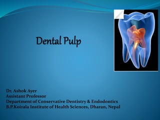
  • 1. Dr. Ashok Ayer Assistant Professor Department of Conservative Dentistry & Endodontics B.P.Koirala Institute of Health Sciences, Dharan, Nepal
  • 2. Contents:  Introduction  Coronal and radicular pulp  Apical foramen  Accessory canal  Functions of dental pulp  Components of dental pulp  Functions of pulpal extracellular matrix  Organization of cells in the pulp  The principle cells of the pulp  The pathways of collagen synthesis  Matrix and ground substances  Vasculature and lymphatic supply  Innervation of Dentin- pulp complex  Disorders of the dental pulp  Advances in pulp vitality testing  Conclusion
  • 3. Dental Pulp  Occupies the center of each tooth.  Soft connective tissue that supports the dentin.  Total 52 pulp organs; 32: Permanent, 20: Primary  Total Volume of all permanent teeth pulp organs is 0.38 cc.  Mean volume of a single adult human pulp is 0.02 cc.
  • 4. Maxillary (Cubic Centimeter) Mandibular (Cubic Centimeter) Central Incisor 0.012 0.006 Lateral Incisor 0.011 0.007 Canine 0.015 0.014 First Premolar 0.018 0.015 Second Premolar 0.017 0.015 First Molar 0.068 0.053 Second Molar 0.044 0.032 Third Molar 0.023 0.031 Orban’s Oral histology & embryology: Pulp; Department of Oral Surgery, Newcastle - Tyne, England
  • 5. Coronal Pulp:  Six surfaces  Pulp horns, depends on the cuspal number.
  • 6.  Radicular Pulp:  The radicular portion of the pulp organs are continuous with the periapical connective tissue through the apical foramen or foramina.  As growth proceeds, more dentin is formed, so that when the root of teeth are matured the radicular pulp is narrower.  The apical pulp canal becomes smaller also because of apical cementum deposition.
  • 7.  Apical foramen:  Average size of apical foramen of the maxillary teeth in the adult is 0.4 mm  Mandibular teeth 0.3 mm  Sometimes it is found on the lateral side of the apex although the root itself is not curved.  Frequently there are two or more foramina separated by a portion of dentin and cementum or by cementum only.
  • 8.  Accessory canal:  Leading from the radicular pulp laterally through the root dentin to the periodontal tissue.  May be seen anywhere along the root but are most numerous in the apical third of the root.  Clinically significant in spread of infection, either from the pulp to the periodontal ligament or vice versa.
  • 9.  Occur in areas where there is premature loss of root sheath cells; these cells induce the formation of odontoblasts which form dentin.  May also occur where the developing root encounters a blood vessel.
  • 11.  Inductive:  Interact with the oral epithelial cells  Differentiation of the dental lamina and enamel organ formation.  Cells of pulp + blood vessels & nerves provides the tooth vitality
  • 12.  Formative:  Produces dentin that surrounds and protects the pulp.  Pulpal odontoblasts develop the organic matrix and function in its calcification.
  • 13.  Nutritive:  Blood vascular system of the pulp; nourishes dentin through the odontoblasts and their processes.  Protective:  Sensory nerve respond to pain  Nerves initiate reflexes that control circulation in the pulp.
  • 14.  Defensive or reparative:  First line of defense to injuries and infection of dentine  Tertiary dentine  Immuno-competent  Clearance of toxic substances
  • 15. Components of dental pulp Cells + (extracellular) Matrix Fiber Ground substance • Collagen • Elastin Structural Adhesive • Fibronectin • Laminin • HS • DS • CS GAG Proteoglycan • Decorin • Versican
  • 16. Components of dental pulp  CELLS (odontoblast, fibroblast, undifferentiated cell, macrophage, dendritic cell)  FIBERS AND GLYCOPROTEIN (collagen type I, III, no elastic fiber, fibronectin)  GROUND SUBSTANCES (glycosaminoglycans, chondroitin sulfate proteoglycan)  BLOOD VESSELS, NERVES, LYMPH VESSELS
  • 17.  Maintain tissue’s physical properties and integrity  Control of growth and development and repairs  Control of cell migration  Control of diffusion of macromolecules Functions of pulpal extracellular matrix
  • 18. Collagen in dental pulp  Concentration varies from species to species, 32% in human pulp.  Higher content in the middle and apical pulp.  Total collagen decreases with age.  Interestingly high level of collagen type III. (43%) : vascular content, tissue extensibility (cf. Elastin)  Absence of elastin (except in b.v.).
  • 19. Adhesive glycoproteins in dental pulp  Fibronectin found in predentine NOT mature dentine.  Fibronectin present in pulp and dental papilla.  Fibroblasts synthesize pulpal fibronectin.  Fibronectin is expressed during reparative dentinogenesis. Immunoreactive fibronectin molecules detected along the border of predentine and between odontoblast (Yoshiba et al., 1994)
  • 20. Glycosaminoglycans in dental pulp  Chondroitin sulfate, dermatan sulfate, hyaluronic acid present  Amount of uronic acid decreases with age  Total GAG decreases with reduced dentinogenic activity  Decorin may involve in mineral nucleation at the mineralization front
  • 21. Organization of cells in the pulp tight junction nerve terminals
  • 22. Four distinct zones: 1. The odontoblastic zone at the pulp periphery 2. A cell free zone of Weil beneath the odontoblast; prominent in the coronal pulp 3. A cell rich zone; high cell density 4. The pulp core; major vessels and nerves
  • 23.
  • 24.  The principle cells of the pulp:  Odontoblasts  Fibroblast  Undifferentiated mesenchymal cells  Macrophages  Immunocompetent cells
  • 25.  Odontoblasts:  The most distinctive cells of the dental pulp  Form a layer lining the periphery of the pulp and have a process extending into the dentin  Arranged in palisade pattern of three to five cells deep  59,000 to 76,000 per square milimeter in coronal dentin, with a lesser number in root dentin.
  • 26.  Active cells:  Elongated, basal nucleus, much basophilic cytoplasm, promonent golgi zone.  Resting cell:  Stubby, little cytoplasm, more hematoxophilic nucleus.
  • 27.  Odontoblast process begins at the neck of the cells just above the apical junctional complex where the cell gradually begins to narrow as it enters predentin.  The process is devoid of major organelles but does display an abundance of microtubules and filaments arranged in a linear pattern along its length.
  • 28.  The pathways of collagen synthesis:  The spherical distensions contain free polypeptides that assemble as a triple helix in the cylindrical distensions to form the procollagen molecule.  The cylindrical distension bud off as secretory granules.  Secretory granules that are transported toward the odontoblast process, where their content is released.
  • 29. Synthesis of collagen and its assembly into fibrils and fiber
  • 30. Some types (of 15) of known collagen Type Molecular Tissue distribution Fibril-forming I [a1(I)]2 a2(I) bone, skin, tendon, ligaments (90%) of body collagen II [a1(II)]3 cartilage, intervertebral disc, notochord, vitreous humor of eye III [a1(III)]3 skin, blood vessels, internal organs V [a1(V]2 a2(V) as type I XI [a1(XI] a2(XI) a3(XI) as type II Fibril-associated IX [a1(IX] a2(IX) a3(IX) cartilage (with type II) XII [a1(XII)]3 tendon, ligaments (with some type I) Network-forming IV [a1(IV)]2 a2(IV) basal laminae VII [a1(VII)]3 anchoring fibrils beneath stratified squmous epithelia
  • 31. RGD = cell-binding domain The structure of a fibronectin dimer.
  • 32. Structure of a GAG Structure of proteoglycans
  • 33. Aggrecan mechanical support (cartilage) Betaglycan binds TGF-beta (cell surface*, matrix) Decorin binds type I and (CNT) TGF-beta Perlecan basal laminae (basal laminae) Syndecan-1 binds FGF (cell surface*) * = Integral membrane proteoglycan Some known proteoglycans:
  • 34.  Junctions occur between adjacent odontoblasts involving  Gap junctions  Occluding zones (Tight junctions)  Desmosomes  The actin filaments inserting into the adherent junction are prominent and form a terminal cell web.
  • 35.  This junctional complex does not form a zonula, completely encircling the cell, as occurs in epithelia; (it is focal, and there is some debate whether it can restrict the passage of molecules and ions from the pulp into the dentin layer)  Serum proteins seem to pass freely between odontoblasts and are found in dentin
  • 36. Fibroblasts:  Greatest number in the pulp  Numerous in coronal pulp where they form the cell- rich zone.  The function is to form and maintain pulp matrix.
  • 37. Undifferentiated Ectomesenchymal Cells:  Represents the pool from which the connective tissues of the pulp are derived.  Depending upon the stimulus these cells may give rise to odontoblasts and fibroblasts.  In older pulp they diminish, thereby reducing the regenerative potential of the pulp.
  • 38. Macrophages  Located throughout the pulp center.  Involved in the elimination of dead cells, the presence of which indicates that turnover of dental pulp fibroblast occurs.
  • 39. Lymphocytes  In normal pulp T lymphocytes are found, but B lymphocytes are scare.
  • 40. Dendritic Cells  Bone marrow derived, antigen presenting dendritic cells.  Beneath the odontoblast layer.  They capture and present foreign antigen to the T cells.
  • 41.  Cells participate in immunosurvillance and increase in number in carious teeth.  Infiltrate odontoblast and project their processes into the tubules.  8% of total cell population.
  • 42. Matrix and Ground Substance  Principally Type I and Type III collagen.  Composed of glycosaminoglycans, glycoproteins, and water.  Overall collagen content increases with age.
  • 43.  The greatest concentration of collagen generally occurs in the most apical portion of the pulp. Significance:  During pulpectomy; Engaging the pulp with a barbed broach in the region of apex affords a better opportunity to remove the tissue intact.
  • 44. Vasculature and Lymphatic Supply  Circulation establishes the tissue fluid pressure.  One or sometimes two vessels of arteriolar size (about 150µm) enter the apical foramen with the sensory and sympathetic nerve bundles.  Smaller vessels, without any accompanying nerve bundle, enter the pulp through the minor foramina.
  • 46. The arterioles occupy a central position within the pulp and, as they pass through the radicular portion of pulp, give off smaller lateral branches.  Occasionally U- looping of pulpal arterioles is seen, and this anatomic configuration is thought to be related to the regulation of blood flow.
  • 47. Pulp tissue is highlyvascularized. 40-50 ml/min/100g (Kim, 1985)
  • 48.  Some terminal capillary loops extend upward between the odontoblasts to abut the predentin if dentinogenesis is occurring.  Located on the periphery of the capillaries at random intervals are pericytes.  Pericytes are contractile cells capable of reducing the size of the vessel lumen.
  • 49.  Anastomosis are point of direct communication between the arterial and venous sides of the circulation.  Lymphatic vessels also occur in the pulp tissue, they exit via one or two large vessels through the apical foramen.
  • 50.  Sympathetic adrenergic nerves terminate in relation to the smooth muscle cells of the arteriolar walls.  Afferent free nerve endings terminate in relation to arterioles, capillaries and veins and serve as effectors by releasing various neuropeptides that exert an effect on the vascular system.
  • 51. Dental pulp interstitial fluid (ISF) and exchange of substances between plasma and ISF. (* values from Tonder and Kvinnsland, 1983; Ciucchi et al., 1995) (5.5-10.3 mm Hg*) (43 mm Hg) (20 mm Hg) (35 mm Hg) Hydrostatic pressure in dental pulp
  • 52. Innervation of Dentin- Pulp Complex  Nerve enter the pulp through apical foramen, along the afferent blood veessels, and together from the neurovascular bundle.  Each nerve fiber has been estimated to provide at least eight terminal branches.
  • 53. These branches ultimately contribute to an extensive plexus of nerves in the cell free zone just below the cell bodies of the odontoblasts in the crown portion of the tooth.
  • 54. Approx. 1800 non myelinated + 400 myelinated Intradentinal nerves are mostly found in pulpal horns.
  • 55.  This plexus of nerves, which is called the subodontoblastic plexus of Raschkow, occupies the cell- free zone of Weil and can be demonstrated in silver nitrate stained sections under the light microscope or by immunocytochemical techniques.
  • 56.  The nerve bundles that enter the tooth pulp consist principally of : Sensory afferent nerves of the trigeminal nerve and Sympathetic branches from the superior cervical ganglion.
  • 57. As the nerve bundle ascends coronally; The myelinated axons gradually loose their mylein coating, So that a proportional increase in the number of unmyelinated axons occurs in the more coronal aspect of the tooth.
  • 58. A-delta fibers  Conduction velocity 2-30 m/s  Lower threshold  Involved in fast, sharp pain  Stimulated by hydrodynamic stimuli  Sensitive to ischemia  Sharp pain C fibers  Conduction velocity 0-2 m/s  Higher threshold  Involved in slow, dull pain  Stimulated by direct pulp damage  Sensitive to anesthetics  Dull pain Types and properties of pulpal sensory nerve fibers A-beta fibers  Conduction velocity 30-70 m/s  Very low threshold, non-noxious sensation  50% of myelinated fibers in pulp  Functions not fully known Non-myelinated sympathetic fibers  Conduction velocity 0-2 m/s  Post-ganglionic fibers of superior cervical ganglion  Vasoconstriction
  • 59.  A small number of axons pass between the odontoblast cell bodies to enter the dentinal tubules in proximity to the odontoblast process.
  • 60.
  • 61. Possible mechanisms of dentine sensitivity Hydrodynamic mechanism (Gysi, 1900; Brannstrom, 1963)
  • 62. Pulp venules STIMULATION Increased pulp interstitial fluid Increased pulp pressure Increased tubular fluid flow Release of inflammatory agents? Increased blood viscosity and rbc congestion in capillary bed Increased A-V shunt blood flow Outward dentinal fluid flow and aspiration of odontoblasts CNS, Pain, Reflexes Vasodilation, Increased permeability Pulpal axonal reflex due to dentine stimulation Without infection, Vascular changes could be resolved. Axon reflex SP, CGRP Dentine
  • 63. Disorders of the Dental Pulp
  • 64. Pulp Stones  Pulp stones, or denticles, frequently are found in pulp tissue.  Discrete calcified masses that have calcium phosphorus ratios comparable to that of dentin.  More frequently at the orifice of the pulp chamber or within the root canal.
  • 65.  Concentric layers of mineralized tissue formed by surface accretion around blood thrombi, dying or dead cells, or collagen fibers.  Occasionally a pulp stone may contain tubules and be surrounded by cells resembling odontoblasts.
  • 66.  Such stones are rare and, if seen, occur close to the apex of the tooth. Such stones are referred to as ‘true’ pulp stones as opposed to ‘false’ stones having no cells associated with them.
  • 67.  If during the formation of a pulp stone, union occurs between it and the dentin wall, or if secondary dentin deposition surrounds the stone, the pulp stone is called an attached stone.
  • 68.  The presence of pulp stones is significant in that They reduce the overall number of cells within the pulp and Act as an impediment to debridement and enlargement of the root canal system during endodontic treatment.
  • 69.
  • 70. Age Changes  Decrease in the volume of pulp chamber and root canal brought about by continued dentin deposition.  On occasion can appear to be obliterated almost completely.  From about the age of 20 years, cells gradually decrease in number until age 70, when the cell density has decreased by about half.
  • 71.  Fibrosis is due to aging & Injury.  Increase in collagen fibers’ bundles which becomes more evident with the decrease in pulp size
  • 72.  Lose and a degeneration of myelinated and unmyelinated axons that correlate with an age- related reduction in sensitivity.  Irregular areas of dystrophic calcification, especially in central pulp.  Gradual reduction of tubule diameter.
  • 73.  The continued deposition often leads to complete closure of the tubule; as can be seen readily in a ground section of dentin, because the dentin becomes translucent (or sclerotic).  Sclerotic dentin is found frequently near the root apex in teeth from middle aged individuals.
  • 74. Pulpitis  Acute or chronic.  Partial or total.  Open or closed.  Exudative or suppurative.  Reversible or irreversible.
  • 75.  Pulpitis is a dynamic process and presents a continuous spectrum of changes reflecting interplay between cause and host defenses.  Poor correlation between microscopic changes & clinical symptoms.
  • 76. Pulpitis: Clinical Features  Presents as pain which patient may have difficulty in localizing to a particular tooth.  Pain may radiate to adjacent jaw, face, ear, or neck.  May be continuous for several days or may occur intermittently over a longer period.  Pulpitis is often described as acute or chronic based on duration and severity of symptoms.
  • 77. Acute pulpitis  Severe throbbing, lancinating pain on thermal stimulation or lying down, keeps patient awake.  Generally lasts 10-15 minutes but may be more or less continuous (reversible pulpitis).  With progression, may become spontaneous & continuous (irreversible pulpitis).
  • 78. Chronic pulpitis  Bouts of dull aching which can last for an hour or more.  Pain on thermal stimulation or spontaneously.
  • 79.  Pulpitis may be asymptomatic.  Most important decision clinically is whether pulpitis is reversible or irreversible.  Decision is made based on many factors including: 1. Severity of symptoms. 2. Duration of symptoms. 3. Size of carious lesion. 4. Pulp tests. 5. Direct observation during operative procedure. 6. Age of patient.
  • 80. Pulpitis: Etiology  Microbial:  Dental caries.  Traumatic exposure.  Marginal leakage.  Cracked tooth  Coronal fracture.  Attrition.
  • 81.  Abrasion.  Traumatic restorative procedure.  Invaginated odontome.  Advanced periodontitis (periodontal-endodontic lesion).
  • 82.  Pulpitis starts before leading organisms in carious dentin reach pulp.  Pulpitis is not usually seen histologically until organisms are within 1 mm of the pulp in permanent teeth, or 2 mm in deciduous teeth.
  • 83. Chemical and thermal injury  During restorative procedures: frictional heat, irritant substances.  May respond by reactionary dentin formation.
  • 84. Barotrauma (aerodontalgia)  Flying at high altitude in unpressurized aircraft, or rapid decompression in divers.  Attributed to formation of nitrogen bubbles in pulp tissue or vessels.  Thought not to be a direct cause, but rather an exacerbating cause in presence of caries.
  • 85. Pulpitis: Histopathology  Poor correlation between microscopic changes & clinical symptoms.  Inflammatory process may be modified by several factors:  Nature, severity and duration of insult.  Efficiency of host defenses.  Efficiency of pulpo-dentinal complex defenses.  Special anatomy of pulp: surrounded by hard tissue and cannot tolerate edema. 85
  • 86.  Reactionary dentin may continue to form after onset of pulpitis if odontoblasts and pulp have not been irreversibly damaged, and may protect pulp.  Pulpitis caused by caries starts as a localized area, but extends throughout pulp if caries is not treated. 86
  • 87. If inflammation is severe, local microcirculation may be compromised, leading to local necrosis and suppuration of pulp (pulp abscess), or diffuse suppuration and necrosis.
  • 88. Pulpitis: Chronic Hyperplastic Pulpitis (Pulp Polyp) Open pulpitis or chronic hyperplastic pulpitis (pulp polyp):  Large carious cavities.  Young molar teeth with wide apices and good blood supply. 88
  • 89.  Usually devoid of sensation on gentle probing.  Polyp consists of chronically inflamed hyperplastic granulation tissue protruding from pulp cavity.  May become epithelialized by spontaneous grafting of desquamated oral epithelial cells from saliva.
  • 90. Pulp Necrosis  May follow pulpitis or trauma to apical blood vessels.  Coagulative necrosis after ischemia. 90
  • 91.  Liquefactive necrosis after pulpitis; may become gangrenous with foul odor upon infection by putrefactive bacteria from caries.  Pulp necrosis in sickling crisis of sickle cell anemia.
  • 93. Effects of cavity Preparation:  Frictional heat  Desiccation  Exposure of dentinal tubules  Direct damage to odontoblast processes  Chemical treatment to exposed dentinal surface
  • 94.  Cavity preparation: speed, heat, pressure & coolant may all cause pulp irritation.  Aspiration or displacement of odontoblasts into dentinal tubules, with reduction of numbers. 94
  • 95. Factors associated with the restorative material & its placement  Material toxicity  Insertion pressure  Thermal effects  Induced stresses
  • 96. Effects subsequent to restoration  Marginal leakage  Cuspal fracture Effects of cavity preparation & restorative materials may further complicate pulpitis caused by caries or other causes. Thickness & nature of remaining dentine may affect pulp response to dental material.
  • 97. Advances in Pulp Vitality testing
  • 98.  Pulse Oximetry  Dental sensor (a modified finger probe) that can be successfully applied and adapted to the tooth and well suited to detect pulsatile absorbance.  The principle: relates the absorption of light, by a solute to its concentration and optical properties at a given light wavelength.
  • 99.  It also depends on the absorbance characteristics of haemoglobin in the red and infra-red range  In the red region, oxyhaemoglobin absorbs less light than deoxyhaemoglobin and vice versa in the infrared region.
  • 100. Hence one wavelength was sensitive to changes in oxygenation and the second was insensitive to compensate for changes in tissue thickness, haemoglobin content and light intensity.
  • 101.  The system consists of a probe containing a diode that emits light in two wavelengths: I. Red light of approximately 660 nm II. Infra-red light of approximately 850 nm  It is also useful in cases of impact injury where the blood supply remains intact but the nerve supply is damaged
  • 102. Dual Wavelength Spectrophotometry Dual wavelength spectrophotometry (DWLS) is a method independent of a pulsatile circulation. The presence of arterioles rather than arteries in the pulp and its rigid encapsulation by surrouding dentine and enamel make it difficult to detect a pulse in the pulp space.
  • 103. This method measures oxygenation changes in the capillary bed rather than in the supply vessels and hence does not depend on a pulsatile blood flow. A major advantage is that it uses visible light that is filtered and guided to the tooth by fibreoptics The test is noninvasive and yields objective results.
  • 104. Laser doppler flowmetry Laser Doppler Flowmetry (LDF) is a noninvasive, electro optical technique, Which allows the semi-quantitative recording of pulpal blood flow.  The Laser Doppler technique measures blood flow in the very small blood vessels of the microvasculature.
  • 105. The technique depends on the Doppler principle; whereby light from a laser diode incident on the tissue is scattered by moving RBC's and As a consequence, the frequency broadened.
  • 106. The primary issues in pulp-vitality testing as follows: A non-vital post-traumatized incisor has a better long-term prognosis; If root canal therapy is completed before the necrotic pulp gets infected.
  • 107. The best outcome for the post traumatized immature incisor is for it; To revascularize and, Continue normal root development, including increased root wall thickness.  Which is not possible to assess with conventional electrical and thermal testing
  • 108. Conclusion Thus the Preservation of Healthy Pulp during operative procedures and successful management in cases of disease are two of the most important challenges.
  • 109. References: 1. Seltzer and Bender's Dental Pulp; 2002 by Quintessence Publishing Co, Inc; Rev. ed. of: The dental pulp / Samuel Seltzer, I.B. Bender. 3rd ed. c1984. 2. Oral histology; Development, Structure and Function: A.R. Ten Cate: 7th Edition 3. Orban’s Oral Histology and Embryology 4. Shafer’s Textbook of Oral Pathology; 5th Edition 5. Yamada, Y., Ito, K., Nakamura, S., Ueda, M. & Nagasaka, T. (2010). Promising cell- based therapy for bone regeneration using stem cells from deciduous teeth, dental pulp, and bone marrow. Cell Transplantation. [Epub ahead of print], (October 2010) 6. Gronthos, S., Mangani, M., Brahim, J., Robey, PG. & Shi, S. (2000). Postnatal human dental pulp stem cells (DPSCs) in vitro and in vivo. Proceedings of the National Academy of Sciences of the United States of America, Vol.97, No.25, (December 2000), pp. 13625- 13630, ISSN 0027-8424 7. Miura, M., Gronthos, S., Zhao, M., Lu, B., Fisher, LW., Robey, PG. & Shi, S. (2003). SHED: stem cells from human exfoliated deciduous teeth. Proceedings of the National Academy of Sciences of the United Stases of America, Vol.100, No.10, (May 2003), pp.5807-5812, ISSN 0027-8424
  • 110. 8. Seo, BM., Miura, M., Gronthos, S., Bartold, PM., Batouli, S., Brahim, J., Young, M., Robey,PG., Wang, CY. & Shi, S. (2004). Investigation of multipotent postnatal stem cells from human periodontal ligament. Lancet, Vol.364, No.9429, (July 2004), pp.149-155, ISSN 0140-6736 9. Sonoyama, W., Liu, Y., Fang, D., Yamaza, T., Seo, BM., Zhang, C., Liu, H., Gronthos, S.,Wang, CY., Shi, S. & Wang, S. (2006). Mesenchymal stem cell-mediated functional tooth regeneration in swine. PLoS One.Vol.1, (December 2006), pp.e79 10. Morsczeck, C., Gotz, W., Schierholz, J., Zeilhofer, F., Kuhn, U., Mohl, C., Sippel, C. & Hoffmann, KH. (2005). Isolation of precursor cells (PCs) from human dental follicle of wisdom teeth. Matrix Biology, Vol.24, No.2, (April 2005), pp.155-165, ISSN 0945- 053X 11. Huang, GT., (2009). Pulp and dentin tissue engineering and regeneration: current progress. Regenerative Medicine, Vol.4, No.5, (September 2009), pp.697-707 ISSN 1746-076X
  • 111. 12. D'Aquino, R., De Rosa, A., Laino, G., Caruso, F., Guida, L., Rullo, R., Checchi, V., Laino, L., Tirino, V. & Papaccio, G. (2009). Human dental pulp stem cells: from biology to clinical applications. Journal of Experimental Zoology Part B: Molecular and Developmental Evolution, Vol. 312, No.5, (July 2009), pp. 408-15, ISSN 1552-5007 13. Batouli, S., Miura, M., Brahim, J., Tsutsui, TW., Fisher, LW., Gronthos, S., Robey, PG. & Shi, S. (2003). Comparison of stem cell- mediated osteogenesis and dentinogenesis. Journal of Dental Research, Vol.82, No.12, (December 2003), pp. 976–981, ISSN 0022- 0345 14. Laino, G., D'Aquino, R., Graziano, A., Lanza, V., Carinci, F., Naro, F., Pirozzi, G., & Papaccio, G. (2005). A new population of human adult dental pulp stem cells: a useful source of living autologous fibrous bone tissue (LAB). Journal of Bone and Mineral Research, Vol.20, No.8, (August 2005), pp.1394-1402, ISSN 0884-0431 15. Nakashima, M. (2005). Bone morphogenetic proteins in dentin regeneration for potential use in endodontic therapy. Cytokine & Growth Factor Reviews, Vol.16, No.3, (June 2005), pp.369-376 ISSN 1359- 6101
  • 112. 16. Sun, HH., Jin, T., Yu, Q. & Chen, FM. (2011). Biological approaches toward dental pulp regeneration by tissue engineering. Journal of Tissue Engineering and Regenerative Medicine. Vol.5, No.4, (April 2011), pp. e1-e16 17. Zavan Barbara et al. Dental pulp stem cells and tissue engineering strategies for clinical application of odontoiatric field. Journal of Biomaterial science and Engineering.