1. Introduction to Vitamins
Vitamins are organic molecules that perform a wide variety of functions in the body. The
most prominent function is as cofactors for enzymatic reactions. Vitamins generally cannot be
synthesized by mammalian cells and, therefore, must be supplied in the diet. The vitamins are
classified into two groups.
Water Soluble Vitamins Fat Soluble Vitamins
• Thiamin (B1)
• Riboflavin (B2)
• Niacin (B3)
• Cobalamin (B12)
• Pantothenic Acid (B5)
• Pyridoxal, Pyridoxamine,
Pyridoxine (B6)
• Biotin Folic Acid
• Ascorbic Acid
All letter vitamins except
Vitamin C
• Vitamin A
• Vitamin D
• Vitamin E
• Vitamin K
1. Vitamins serve as cofactors in enzyme catalysis
2. The cofactor nay be bound to the enzyme or free
3. When bound, the cofactor becomes the prosthetic group
of the enzyme
4. enzyme + cofactor is called the ‘Holoenzyme’
enzyme alone is called the ‘Apoenzyme
Thiamin (Vitamin B1)
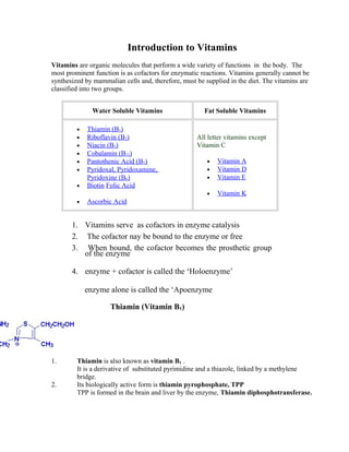
1. Thiamin is also known as vitamin B1 .
It is a derivative of substituted pyrimidine and a thiazole, linked by a methylene
bridge.
2. Its biologically active form is thiamin pyrophosphate, TPP
TPP is formed in the brain and liver by the enzyme, Thiamin diphosphotransferase.
2. Coenzyme Form: Thiamin pyrophosphate
3. TPP serves as a cofactor for the pyruvate and α-ketoglutarate dehydrogenase
reactions as well as the transketolase catalyzed reactions of the pentose phosphate
pathway.
4. Deficiency of thiamin leads to neurological conditions like ataxia, mental confusion,
peripheral neuropathy and a disease known as Beriberi
Riboflavin (Vitamin B2)
Riboflavin structure
1. Riboflavin is also known as vitamin B2.
2. The coenzymes forms of riboflavin are, flavin mononucleotide (FMN)
and flavin adenine inucleotide (FAD).
3. The enzymes that require FMN or FAD as cofactors are termed
flavoproteins.
4. Flavoproteins are involved in a wide range of redox reactions, e.g.
succinate dehydrogenase and xanthine oxidase.
5. The reduced forms of FMN and FAD are formed, FMNH2 and FADH2,
respectively.
3. Structure of FAD
Nitrogens 1 & 5 carry hydrogens in FADH2
Coenzyme Function:
1. Riboflavin acts as an integral component of two coenzymes: FAD
(flavin adenine dinucleotide) and FMN (flavin mononucleotide).
2. FAD and FMN are known as flavins since they are derived from
riboflavin. FMN and FAD serve as cofactors for a family of proteins
called flavoenzymes.
4. 3. Flavoenzymes catalyze a wide range of biochemical reactions,
typically redox reactions. They are key elements in cellular
respiration. In cellular respiration, FAD and FMN act as intermediate
hydrogen acceptors in the mitochondrial electron transport chain,
accepting hydrogens derived from foodstuffs, and transferring
electrons to the cytochrome system. During this process, ATP is
released (2 moles of ATP per mole of FADH2).
Clinical Significances of Flavin Deficiency
6. Riboflavin deficiency is often seen in chronic alcoholics due to their poor
dietetic habits.
7. Symptoms associated with riboflavin deficiency include, glossitis, seborrhea,
angular stomatitis.
8. Riboflavin is an orange powder, and water solutions have intense greenish
yellow fluorescence.
9. Riboflavin, is found naturally in the food we eat. Sources of riboflavin include
organ meats (liver, kidney, and heart) and certain plants such as almonds,
mushrooms, whole grain, soybeans, and green leafy vegetables.
Biotin (Vitamin H or B7)
1. Biotin, also known as vitamin H or B7.
2. It is a water-soluble B-complex vitamin which is composed of an ureido
(tetrahydroimidazalone) ring fused with a tetrahydrothiophene ring. A valeric acid
substituent is attached to one of the carbon atoms of the tetrahydrothiophene ring.
3. Biotin is important in the catalysis of essential metabolic reactions of (a) biosynthesis
of fatty acids, (b) gluconeogenesis, and (c) metabolism of Leucine.
4. Biotin is found in numerous foods and also is synthesized by intestinal bacteria and as
such deficiencies of the vitamin are rare. Deficiencies are generally seen only after long
antibiotic therapies which prevent intestinal absorption of the biotin.
5. Biotin is the cofactor (no coenzyme form, involved as it is) required for enzymes that
are involved in carboxylation reactions, e.g. Acetyl-CoA carboxylase and pyruvate
carboxylase.
5. Vitamin B6
Vitamin B6 is the name given to three related pyrimidine derivatives:
Pyridoxine Pyridoxal Pyridoxamine
1. Pyridoxine, pyridoxal and pyridoxamine are collectively known as vitamin B6
2. All three compounds are efficiently converted in the body to the coenzyme form
of vitamin B6, pyridoxal phosphate (PALP)
3. This conversion is catalyzed by the ATP requiring enzyme, pyridoxal kinase.
Coenzyme form of Vit-B6: Pyridoxal Phosphate
5. Pyridoxal phosphate functions as a cofactor for transamination, deacrboxylation and
recemase reactions
6. All these reactions involve the formation of a Schiff’s base linkage (-N=CH-)
The phenyl ring with positive charge on the nitrogen atom, delocalized over the ring
stabilizes the Schiff’s base.
7.. Deficiencies of vitamin B6 are rare and usually are related to an overall deficiency of
all the B-complex vitamins.
8. .Isoniazid and penicillamine (used to treat rheumatoid arthritis and cystinurias) are two
drugs that complex with pyridoxal and pyridoxal phosphate resulting in a deficiency in
this vitamin.
Niacin (Vitamin B3)
Nicotinamide Nicotinic Acid
6. 1. Niacin (nicotinic acid and nicotinamide) is also known as vitamin B3.
2. The coenzyme forms of niacin are nicotinamide adenine dinucleotide (NAD+
) and
nicotinamide adenine dinucleotide phosphate (NADP+
).
3. Both NAD+
and NADP+
function as cofactors for numerous
dehydrogenase, e.g.,
lactate and malate dehydrogenases.
Structure of NAD+
4. In the oxidized form of NAD (NAD+
) the pyridine ring is positively charged
Due to the delocalization of the positive charge on the nitrogen atom.
5. In the reduced form, this positive charge is removed and the C-atom at
Position 4 gains a H-atom forming –CH2 group as shown in the insert in the
box insert. The -OH phosphorylated in NADP+
is indicated by the red arrow.
6. Difficiency of Niacin leads to glossitis of the tongue, dermatitis, weight loss,
diarrhea, depression and dementia. When these symptoms are severe, the
condition is known as pellagra.
Biochemical Functions:
7. The coenzymes, NAD+
and NADP+
are involved in a variety of oxidation-reduction
reactions. They accept electrons in the form of hydride ion , H
-
(hydrogen atom + an
electron).
The reduction occurs in the pyridine ring, resulting in the neutralization of the
Positive charge. The overall reaction can be written as:
7. AH2 + NAD+
A + NADH + H+
(reduced substrate) (oxidized substrate)
NADP+
functions exactly like NAD+
There are nearly 40 different oxidoreductases which use either NAD+
or
NADP+
as cofactors. Some are specific to only NAD+
or only NADP+
,
while some use either one of them. These coenzymes are loosely bound to
enzymes and can be separated from the enzymes by dialysis.
In addition to its role in oxido-reduction reactions, NADH acts as carrier of
Reducing equivalents (electrons) from metabolic intermediates and delivers them
To the ETC in mitochondria where they are oxidized to produce ATP. 3 moles of
ATP are produced per mole of NADH oxidized.
While NADH is generally functions as a coenzyme if catabolic reactions, NADPH is
involved in anabolic reactions like the biosynthesis of fatty acids and some reactions of
PPP.
Pantothenic Acid (Vitamin B5)
Pantoic acid β-alanine
1. Pantothenic acid is also known as vitamin B5.
2. Pantothenic acid is formed from β-alanine and pantoic acid.
3. The coenzyme form of Pantothenate is coenzyme A.
8. 4. At least 70 enzymes require CoA for their action.
5. Deficiency of pantothenic acid is extremely rare due to its widespread
distribution in whole grain cereals, legumes and meat.
Coenzyme A
1. Coenzyme A is formed from pantothenic acid and 3 –moles of ATP
In a 4-step reaction.
2. Coenzyme A or CoA has a terminal thiol group which is the reactive part
of the coenzyme. Acyl groups (free fatty acids) are linked to CoA by a
thioester bond (-S-CoA) to give acyl CoA. Thus
Acetate forms Acetyl CoA
CH3-COO-
CH3-CO-S-CoA
Succinate forms Succinyl CoA
-
OOC-CH2CH2-COO- -
OOC-CH2-CH2-CO-S-CoA
Propionatate forms Propionyl CoA
CH3-CH2-COO- CH3-CH2-CO-S-CoA
3. Coenzyme A serves a carrier of activated acetyl or acyl groups as thiol
esters. This function is similar to ATP which acts as a carrier of activated
phosphoryl group (-PO3
2-
). Some of the reactions that involve coenzyme A
are:
CoASH
(a) Pyruvate Acetyl CoA
PDH
CH3-CO-COO- CH3-CO-S-CoA
9. Vitamin B12 Cobalamin
1. Cobalamin is more commonly known as vitamin B12.
2. It is composed of a complex tetrapyrrol ring structure (corrin ring)
and a cobalt ion in the center.
3. Vitamin B12 is synthesized exclusvely by microorganisms and is found in the
liver of animals, bound to proteins.
4. The vitamin must be hydrolyzed from protein in order to be active.
Hydrolysis occurs in the stomach by gastric acids or in the intestines by trypsin
digestion. After absorption, the vitamin is transported to the liver in the blood.
5. The Co2+
ion present at the center of the tetrahydropyrrole ring system is
usually coordinated to one of the following:
a. Cyanide (cyanocobalamine) (B12a)
b. Hydroxyl (hydroxycobalamine) (B12b)
c. Nitrite (nitrocobalamine) (B12c)
6. There are two coenzyme forms of Vitamine B12.
(a) 5-deoxyadenosyl cobalamine: -CN is replaced by 5’-deoxyadenosine
(b) Methylcobalamine: -CN is replaced by methyl group.
7. Biochemical Function: Vitamin B12 is a cofactor for two clinically
significant reactions in the body. In fatty acid synthesis, the enzyme
methylmalonyl-CoA mutase, requires vitamin B12 as a cofactor in the
10. conversion of methylmalonyl-CoA to succinyl-CoA.
The second reaction requiring vitamin B12 catalyzes the conversion of
homocysteine to methionine and is catalyzed by methionine synthase.
Folic Acid (folacin)
Pteridine PABA Glutamate
positions 7 & 8 carry hydrogens in dihydrofolate (DHF)
positions 5, 6, 7, & 8 carry hydrogens in tetrahydrofolate (THF)
1. Folic acid is abundantly found in green leaves (Latin, folium means leaf)
2. It is conjugated molecule consisting of a pteridine ring structure linked
to para-aminobenzoic acid (PABA) that forms pteroic acid, which is
conjugated to a glutamic acid residue.
3. Folic acid is obtained primarily from yeasts and leafy vegetables as well
as animal liver.
4. Animals cannot synthesize PABA nor attach glutamate residues to
pteroic acid, thus, requiring folate intake in the diet.
5. Folic acid is reduced within cells (principally in the liver where it is
stored) to tetrahydrofolate (THF also H4folate) through the action of
dihydrofolate reductase (DHFR), an NADPH-requiring enzyme.
6. The function of THF derivatives is to carry and transfer various forms of
one carbon units during biosynthetic reactions. The one carbon units
are methyl, methylene, methenyl, formyl or formimino groups.
7. Active center of tetrahydrofolate (THF): Note that the N5
position is
the site of attachment of methyl groups, the N10
the site for attachment of
formyl and formimino groups and that both N5
and N10
bridge the
methylene and methenyl groups.
11. 8. These one carbon transfer reactions are required in the biosynthesis of
serine, methionine, glycine, choline and the purine nucleotides and
dTMP.
Clinical Significance of Folate Deficiency
Folate deficiency results in complications nearly identical to those described for
vitamin B12 deficiency. The most pronounced effect of folate deficiency on
cellular processes is upon DNA synthesis.
Ascorbic Acid
Ascorbic Acid
1. Ascorbic acid is more commonly known as vitamin C.
2. Ascorbic acid is derived from glucose via the uronic acid pathway.
3. Ascaorbic acid has no coenzyme form. The active form of vitamin C is
ascorbate acid itself.
4. The main function of ascorbate is as a reducing agent in a number of different
reactions. Vitamin C has the potential to reduce cytochromes a and c of the
respiratory chain as well as molecular oxygen.
5. The most important reaction requiring ascorbate as a cofactor is the
hydroxylation of proline residues in collagen. Vitamin C is, therefore,
required for the maintenance of normal connective tissue as well as for
wound healing since synthesis of connective tissue is the first event in
wound tissue
12. NN
N
N
O
N
N
S S
HO
OH
HO
CH2
Vitamin D 1, 25-dihydroxycholecalciferol Calcium/phosphate metabolism
CHO
Vitamin A 11-cis-retinol Visual Cycle
Lipoic Acid Lipoyl Lysine Acyl Group Transfer
-CH2-CH2-CH2-CH2COO-
HOCH2
CH 3
CH 3
OHO
O
O
PO
-
Co
+
N-C-CH2-CH2-
CH3
CH
CH2
H3C
H3C
CH3
CH3
CH2
CH2-C-NH2
O
H
O
CH2
CH2-C-NH2
O
CH2-C-NH2
OCH3
CH3
H3C
H3C
CH2
CH2
C
NH2
O
H2C
CO
H2N
H2C
CO
H2N
Vitamin B12 Coenzyme B12 (Cobalamin) 1, 2 Prototropic Shift
13. O
Vitamin E α-Tocophoreol Antioxidant
HO
CH3
Vitamin K Phylloquinone γ-Carboxylation of Glutamate
Blood Clotting
O
O