
Recommended
More Related Content
What's hot
What's hot (20)
Viewers also liked
Similar to Lenses of slit lamp biomicroscope & indirect ophthalmoscope.
Similar to Lenses of slit lamp biomicroscope & indirect ophthalmoscope. (20)
Recently uploaded
Recently uploaded (20)
Lenses of slit lamp biomicroscope & indirect ophthalmoscope.
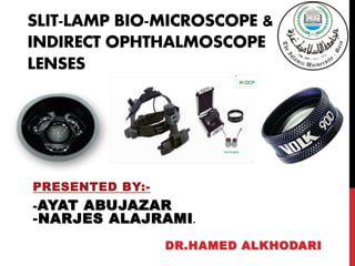
- 1. SLIT-LAMP BIO-MICROSCOPE & INDIRECT OPHTHALMOSCOPE LENSES PRESENTED BY:- -AYAT ABUJAZAR -NARJES ALAJRAMI. DR.HAMED ALKHODARI
- 2. COMPONENTS:- Volk double aspheric lenses Gold three mirror lens Quick review Introduction Principle of indirect ophthalmoscope 30D lens 20D lens 15D lens Indirect ophthalmoscopic lenses Slit lamp biomicroscopic lenses
- 3. SLIT LAMP BIOMICROSCOPY LENSES Quick Review Slit lamp biomicroscopy is used to illuminate and examine under magnification the anterior segment of the eye. There are many different attachments of the slit-lamp: 1.Video attachment . 2.Goldman tonometer . 3.Gold 3-mirror lens . 4.Volk double aspheric lens. (60D-78D-90D) 5.Direct contact goniolenses.
- 4. VOLK DOUBLE ASPHERIC LENS History -In 1956, aspheric ophthalmic lenses for subnormal vision were developed by Dr. David Volk. He found that an aspheric surface corrected the aberrations present in more common spherical lenses. -Several developments occurred through the years, leading up to 1982 when all Volk lenses for indirect ophthalmoscopy were redesigned with both surfaces aspheric, providing a substantial improvement in image quality.
- 5. VOLK DOUBLE ASPHERIC LENS Introduction Volk's 60D,78D and 90D fundus lenses have establishes slit lamp indirect ophthalmoscopy as a standard diagnostic procedure for comprehensive fundus evaluation. Examination of the retina by Slit lamp and Volk double aspheric lenses is called a Bio- microscopic Indirect Ophthalmoscope BIO.
- 6. VOLK DOUBLE ASPHERIC LENS 60D Primary Application :– • High Magnification Views of the Posterior Pole. • High magnification lens for detailed optic disc and macula imaging. • Ideal diameter for use in the orbital area. ( Its 31mm diameter allows a wide field of view and facilitates easy handling within the orbital area.) • Working distance from the cornea is 11mm.
- 7. VOLK DOUBLE ASPHERIC LENS 78D Primary application:- The Double Aspheric 78D is an excellent general diagnosis . Ideal balance of magnification and field of view. Working distance from the cornea is 7mm.
- 8. VOLK DOUBLE ASPHERIC LENS 90D Primary application: General diagnosis and small pupil examinations. The original Volk 90D lens started the slit lamp examination. It features a small 26mm diameter ring is ideal for dynamic fundoscopy. The Volk 90D has very good small pupil capabilities, making it ideal for a quick look at the posterior pole. Working distance from the cornea is 6.5mm.
- 9. VOLK DOUBLE ASPHERIC LENS Color of 90D volk lens and it’s case:- There are many of 90D VOLK’s colors , like green ,red, silver, blue, gold and violet.
- 10. +60D , +78D , +90D. For emmetropic eye, +60D lens with M(slit lamp)=X10 : D.P(eye)=60D. - Magnification = M.(retina) *M.(slit lamp). = 6060 * 10 =X10. For myopic eye by -8,+78D lens with M(slit lamp)= X10: D.P(eye)= 68. - Magnification = M.(retina) *M.(slit lamp). = 6878 * 10 =X8.7. For hyperopic eye by +10,+90D lens with M(slit lamp)=X16: D.P(eye)=50. - Magnification = M.(retina) *M.(slit lamp). = 5090 *10 =X5.5 .
- 11. BIOMICROSCOPIC INDIRECT OPHTHALMOSCOPE The patient’s pupil may be dilated and background lights dimmed as for direct ophthalmoscopy. Once the patient is positioned comfortably at the slit lamp. The slit lamp viewing piece and the light column are kept at an angle of 90 degrees. The intensity of the beam is kept to the minimum possible and the magnification preferably set at 10× initially. The slit beam is set around 1.5–2.5 mm wide and 5–10 mm long. The beam is focused onto the patient’s pupil and the condensing lens aligned at around 1 cm from the patient’s eye.
- 12. BIOMICROSCOPIC INDIRECT OPHTHALMOSCOPE The slit lamp is then pulled backwards gradually towards the examiner until it comes into focus with the aerial image of the fundus between the condensing lens and the slit lamp. Alternatively, the slit lamp could be drawn back completely towards the examiner and then gradually moved forwards until the image comes into focus As with indirect ophthalmoscopy, the image from a non- contact Volk Lens slit lamp biomicroscopic examination is inverted and laterally reversed.
- 14. VOLK DOUBLE ASPHERIC LENS Characteristics of volk lenses:- 1.Stereoscopic ,3 dimensional view of the retina: _Binocular viewing through the slit lamp. 2.Better image achieved when viewing through media opacities: _Cataract. 3.Allows for manipulation of image: _Slit lamp magnification& filters. 4.Image size less affected by patient refractive error.
- 15. GOLDMANN THREE MIRROR Primary Application:- Viewing and treatment of the Anterior Chamber and Central and Peripheral Fundus. Because the curvature of the contact surface of the lens is steeper than that of the cornea, a viscous coupling substance with the same refractive index as the cornea is required to bridge the gap between the cornea and the goniolens.
- 16. GOLDMANN THREE MIRROR It is important to be familiar with each part of the lens as follows: C-PEG 1. The central part provides a 30° upright view of the posterior pole. 2. The equatorial mirror (largest and oblong-shaped) enables visualization from 30° to the equator. 3. The peripheral mirror (intermediate in size and square- shaped) enables visualization between the equator and the ora serrata. 4. The gonioscopy mirror (smallest and dome-shaped) may be used for visualizing the extreme retinal periphery and pars plana.
- 19. GOLDMANN THREE MIRROR Characteristics:- 3- dimensional view obtained. Used both on undilated and dilated pupil. Mirror images can be confusing. Inadvertent pressure on cornea can lead to wide angle in AC.
- 20. INDIRECT OPHTHALMOSCOPE LENSES Introduction:- BIO condensing lenses (Hand held lenses ) are biconvex, aspheric designs with one surface more curved than the other. Less curved surface toward patient’s eye (silver).
- 21. INDIRECT OPHTHALMOSCOPE LENSES The hand-held lens acts both as:- 1. A condensing lens for the illuminating system. 2. A lens for forming an inverted image of the retina in space.
- 22. INDIRECT OPHTHALMOSCOPE LENSES The technique is called Indirect because the fundus is seen through a condensing lens. The image is formed close to the principle focus of the lens, between the lens and the observer.
- 23. INDIRECT OPHTHALMOSCOPE LENSES The condensing lens is a powerful convex lens (the usual power used is +14, + 20,and + 33 D ) The power of the condensing lens determines:- Retinal Magnification Field of view Stereopsis.
- 25. THE PRINCIPLE OF INDIRECT OPHTHALMOSCOPE
- 26. 1- magnification of a lens = dioptric power/ 4. 2- magnification of the retina = D.P. of the eye /D.P of the lens. 3- Stereopsis = magnification / 4 . 4- Field of view = (D.P. of the lens x 2 ).
- 27. The high dioptric power lens (30D) has the highest magnification :- It yields the least magnification of the retina, 60/30 = 2. Stereopsis is half that of the normal, 2/4= 1/2 Field of view is generally the largest = (60 degrees, 30 x 2 ).
- 28. 30D lens is used to obtain a panoramic view when detail and stereopsis are not as important , and used with small pupil.
- 29. The middle dioptric power lens of (20D). A- The retinal magnification = 60/20 = X3 B- The stereopsis is 3/4 that of the normal. C- The field of view is 40 degrees (20x2 ).
- 30. 20D lens most widely used, since it provides an adequate field of view, stereopsis and magnification.
- 31. The low dioptric power lens of 14 or 15D. A- The retinal magnification = 60/15 = X4. B- The stereopsis is full ( 4/4 ). C- The field of view is 30 degrees (15x2 ).
- 32. 15D lens, is most useful for detailed view of the macula or optic disc or for determining elevation of the retina in shallow retinal detachment.
- 33. INDIRECT OPHTHALMOSCOPE LENSES Working distance from cornea stereopsisField of view magnificati on Lens power (D 26mm1260 degree2x+30 47mm3440 degree3.25x+20 72mm130degree4.17 x+14+15
- 34. +30D , +20D , +15D . For emmetropic eye , +15 D lens :D.P(eye)= 60D. - stereopsis = 154 =3.75. -M(retina)= 6015 = X4. -field of view = 15*2 =30. For myopic eye by -8 , +20D lens :D.P(eye)=60- -8 =68d. M(retina)=6820=X3.4 . - stereopsis = 204 = 5 - field of view = 20*2 = 40. For hyperopic eye by +10, +30D lens: D.P(eye)=60-10=50d. -M(retina)=5030 = X1.6. -stereopsis = 304= 7.5 . -field of view = 30*2 =60 .