VIP Call Girls Pune Vrinda 9907093804 Short 1500 Night 6000 Best call girls S...
Chapter 15 - The Cardiovascular System - Part 1
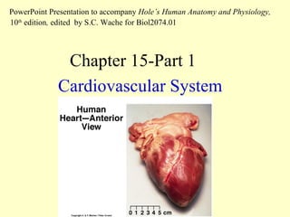
1. Cardiovascular System Chapter 15-Part 1 PowerPoint Presentation to accompany Hole’s Human Anatomy and Physiology, 10 th edition , edited by S.C. Wache for Biol2074.01
2. You are responsible for the following figures and topics: Part I. Structure and function of the heart. Fig. 15.1 – The cardiovascular system supplies lungs and body in separate pathways. Fig. 15.2 Anterior view of the human heart. Fig. 15.3 – Location of the heart in the mediastinum. Fig. 15.4 – It is enclosed by a double membrane pericardium. Fig. 15.5 - The heart wall. Fig. 15.10, 15.11 - Two circuits: pulmonary circuit; systemic or body circuit. Fig. 15.6, 15.9 - Name all the parts: 4 valves, 4 chambers, vessels going in and out of the heart. Fig. 15.4 , 15.13 - Anterior and posterior views of the heart. Fig. 15.15 - Coronary circuit. Pathway of blood flow Fig. 15.17 - The figure is made up of 5 subsets. Recordings during a cardiac cycle. Fig. 15.18 – Heart valves can be listened to at 2nd/3rd ribs and 5th/6th ribs. Fig. 15.19 - Cardiac conduction system. Fig. 15.22 - Study the phases of a cardiac cycle. Fig. 15.24 - control of cardiac output - [see table in the attached lecture handout] Read Clin. Appl. 15.5, p. 578, regarding hypertension. Fig. 15.37 – Vasoconstriction v. vasodilation control BP. Fig. 15.39 - When cardiac output increases and BP is high… Fig. 15.40 - Vasodilation in response to high BP.
18. Fig. 15.16 Note that the R and L atria contract together followed by the R and L ventricles contracting together. This results in the typical lub-dup sound in a stethoscope. Note that it takes two such sounds, lub-dup lub-dup, for one complete passage of one volume of blood through the R and L sides of the heart. Note: Following is a complex figure of the cardiac cycle !!!
![You are responsible for the following figures and topics: Part I. Structure and function of the heart. Fig. 15.1 – The cardiovascular system supplies lungs and body in separate pathways. Fig. 15.2 Anterior view of the human heart. Fig. 15.3 – Location of the heart in the mediastinum. Fig. 15.4 – It is enclosed by a double membrane pericardium. Fig. 15.5 - The heart wall. Fig. 15.10, 15.11 - Two circuits: pulmonary circuit; systemic or body circuit. Fig. 15.6, 15.9 - Name all the parts: 4 valves, 4 chambers, vessels going in and out of the heart. Fig. 15.4 , 15.13 - Anterior and posterior views of the heart. Fig. 15.15 - Coronary circuit. Pathway of blood flow Fig. 15.17 - The figure is made up of 5 subsets. Recordings during a cardiac cycle. Fig. 15.18 – Heart valves can be listened to at 2nd/3rd ribs and 5th/6th ribs. Fig. 15.19 - Cardiac conduction system. Fig. 15.22 - Study the phases of a cardiac cycle. Fig. 15.24 - control of cardiac output - [see table in the attached lecture handout] Read Clin. Appl. 15.5, p. 578, regarding hypertension. Fig. 15.37 – Vasoconstriction v. vasodilation control BP. Fig. 15.39 - When cardiac output increases and BP is high… Fig. 15.40 - Vasodilation in response to high BP.](data:image/gif;base64,R0lGODlhAQABAIAAAAAAAP///yH5BAEAAAAALAAAAAABAAEAAAIBRAA7)