SlideShare a Scribd company logo
1 of 5
Download to read offline
173
INTRODUCTION
Corneal anomalies are often present at birth. The cause can be genetic,
the result of a teratogen, or idiopathic. At the fifth week of gestation,
the lens vesicle separates from the surface ectoderm. Mesenchymal
neural crest cells migrate between the surface ectoderm and the optic
cup in what will become the anterior chamber.1
The first wave of mes-
enchyme becomes the corneal endothelium and trabecular meshwork,
the second becomes the corneal stromal keratocytes, and the third
becomes the anterior iris stroma. Separation of these mesenchyme lay-
ers forms the anterior chamber. Alterations in this process result in the
anomalies of corneal size, shape, and clarity, as described below.
Recent studies from large corneal referral practices have shown the
most common conditions requiring surgical intervention were: poste-
rior polymorphous dystrophy, Peters’ anomaly, congenital glaucoma,
and sclerocornea. After a mean follow-up of 40 months, 78% of corneal
grafts remained clear.2
However, medical management accounted for
the majority (52.7%) of eyes with congenital corneal opacities.3
SIZE AND SHAPE ANOMALIES
MICROCORNEA
The cornea’s horizontal diameter is normally 9.5–10 mm at birth and
10–12.5 mm in adulthood. An adult cornea less than 10 mm in hori-
zontal diameter is called microcornea and may occur in conjunction
with microphthalmos, which is a small disorganized eye often associ-
ated with anomalies such as coloboma of the iris, retina, choroid, and
even the optic nerve (Fig. 4-3-1). In contrast, nanophthalmos is a small
functional eye that has normal internal organization and proportions.
Epidemiology, Pathogenesis,
and Ocular Manifestations
Most cases are sporadic, although autosomal recessive and autosomal
dominant pedigrees have been reported.4
Microphthalmos in associa-
tion with dermal aplasia and sclerocornea is known as MIDAS syn-
drome (caused by deletion in Xp22).5
In microcornea, because the
remainder of the eye is normal in size, angle-closure glaucoma may
occur as the lens enlarges. An autosomal dominant variant of micro-
cornea associated with cataract and other anterior segment anomalies
has been described.6
Microcornea has been associated with numerous
syndromes, including the “micro” syndrome of microcornea, congenital
cataract, mental retardation, retinal dystrophy, optic atrophy, hypogeni-
talism, and microcephaly.7
Treatment
Treatment involves spectacle correction for hyperopia resulting from
the flat cornea. Other ocular pathology such as cataract and glaucoma
may require separate treatment.
MEGALOCORNEA
Epidemiology, Pathogenesis,
and Ocular Manifestations
Megalocornea is characterized by bilateral anterior segment enlarge-
ment with a corneal horizontal diameter of 12 mm or greater at birth
and 13 mm or greater after 2 years of age (Fig. 4-3-2). It is distinct from
buphthalmos, which manifests as elevated intraocular pressure and an
enlarged globe (which includes an enlarged cornea). The cause appears
to be related to defective growth of the optic cup, which leaves a larger
space for development of the cornea. In utero, elevated intraocular pres-
sure, which spontaneously arrests, may result in similar changes, but
presumably the endothelial cell density decreases and the entire globe
enlarges.
A number of variants of this disorder have been described. Auto-
somal dominant megalocornea without other ocular abnormalities is
the least common. X-linked recessive megalocornea is reported more
frequently and is associated with iris transillumination, pigment dis-
persion, lens subluxation, arcus, and central crocodile shagreen.8,9
Endothelial cell density is normal, which confirms that the enlarge-
ment does not arise from corneal stretching, and corneal clarity and
thickness are usually normal.10
The genetic locus for X-linked megalo-
cornea appears to be in the region Xq21–q22.6. Megalocornea has been
reported in association with congenital miosis,11
ectopia lentis, ectopia
pupillae, and mental retardation.12
Megalophthalmos is an enlarged cornea in an overall enlarged eye
that does not have glaucoma. This is most likely autosomal recessive
with findings similar to X-linked megalocornea, in addition to increased
axial length (often > 30 mm), juvenile cataract, and high myopia.8
Treatment
Treatment is not necessary except for spectacle correction for myopic
refractive error.
Joel Sugar, Hormuz P. Wadia, Roshni A. Vasaiwala 4.3Congenital Corneal Anomalies
SECTION 2 Congenital Abnormailities
PART 4 CORNEA AND OCULAR SURFACE DISEASES
Definition: Developmental abnormalities of the cornea present
at birth.
Key features
■ Not acquired
■ Generally involve defects in the migration of mesenchyme in the
anterior segment of the eye
■ Often associated with iris and lens anomalies
Fig. 4-3-1 Colobomatous microphthalmos. Right eye; note the small cornea and
typical coloboma.
4
174
CORNEAANDOCULARSURFACEDISEASES
Fig. 4-3-2 Megalocornea. (A) This patient has corneal diameters of 14 mm
bilaterally. (B) Gross specimen shows appearance of enlarged cornea and heavily
pigmented trabecular meshwork. (B, Courtesy of Dr. M.Yanoff)
A
B
Fig. 4-3-3 Posterior embryotoxon. Schwalbe’s line is evident nasally, superiorly, and
temporally in the eye of this patient.
CORNEAL ABSENCE
Isolated absence of the cornea does not occur. Concomitant and severe
developmental anomalies of the anterior segment or the entire eye are
seen because of the intimate relationship between the ocular structures
in the development of the cornea. Anophthalmos is the extreme;
absence of the cornea and other ocular structures may occur as part of
more extreme genetic developmental disorders. Cryptophthalmos
involves partial or complete failure of eyelid formation, dermoid trans-
formation of the cornea, an undeveloped anterior segment, or a
rudimentary cyst-like globe with an absent anterior segment. Cryp-
tophthalmos also may be associated with systemic anomalies such
as syndactyly and genitourinary anomalies. This syndrome, known
as Fraser syndrome, is inherited as an autosomal recessive trait.
Pseudocryptophthalmos occurs when the lids fail to separate and the
underlying globe is intact.
CONGENITAL CORNEAL ECTASIA
Keratoglobus (discussed in Chapter 4.18) is not congenital, but corneal
ectasia may be present at birth as part of a congenital anterior staphy-
loma, which is often seen with Peters’ anomaly. Congenital corneal
ectasia includes significantly more corneal thinning and bulging com-
pared to Peters’ anomaly (see Fig. 4-3-6), presumably as a consequence
of the same developmental abnormalities in the migration of mesen-
chyme. This anomaly is usually unilateral and is often associated with
iris developmental defects. Anterior staphyloma may also occur as a
result of inflammatory or infectious corneal thinning in utero.
ANOMALIES OF CORNEAL CLARITY
Changes in the first wave of neural crest mesenchyme migration result
in anomalies of the corneal endothelium and anterior chamber angle,
while those in the second wave lead to corneal stromal alterations, and
those in the third wave affect iris development. The factors that com-
monly result in maldevelopment impact more than just one phase in
this process of neural crest differentiation.
ANTERIOR EMBRYOTOXON
Anterior embryotoxon refers to a congenital broad limbus superiorly
with an otherwise normal anterior segment, representing merely a
broad transition from sclera to cornea. The term also is used to describe
an appearance similar to arcus senilis (arcus juvenilis) present at birth.
Though it is often sporadic, autosomal dominant and autosomal reces-
sive pedigrees have been described.
POSTERIOR EMBRYOTOXON
Posterior embryotoxon is likely the most frequently seen anomaly, with
the prevalence being reported as high as 24% in a random population.13
It consists of thickening and anterior displacement of Schwalbe’s line,
which is seen most readily at the slit lamp in the temporal cornea (Fig.
4-3-3). The term toxon is derived from the Greek word for bow, in refer-
ence to the crescent of Schwalbe’s line; when present alone, this has no
functional significance.
CORNEAL KELOIDS
Keloids are white, glistening, protuberant lesions that involve all or part
of the cornea. Although usually resulting from trauma or ocular inflam-
mation, they may be present at birth. Histopathologically, they repre-
sent an irregular array of collagen bundles, fibroblasts, and capillaries
arising in the corneal stroma. They are sometimes progressive and may
be associated with disorders that involve oculodigital manipulation, as
in Lowe’s syndrome. In otherwise healthy eyes, keratoplasty is appro-
priate.14
For lesions in which progressive growth causes discomfort,
dissection of the lesion from the cornea followed by covering with a
conjunctival flap may halt progression.
DERMOIDS
Dermoids (choristomas, or growths of tissue not normally present) may
be found on the cornea, usually at the inferotemporal limbus. At times
they may involve larger areas of the cornea, the entire limbus, the entire
cornea, or the interior of the eye. They usually are round, domed, and
pink to white to yellow in color. They may have hair, or in the lipoder-
moid variant, globules of lipid, as evident on slit-lamp examination.
Induced astigmatism, even amblyopia, may be present. Limbal dermoids
may be associated with other malformations, e.g., Goldenhar’s syn-
drome, which involves lid colobomas, hemifacial microsomia,
CongenitalCornealAnomalies
4.3
175
iridocorneal endothelial (ICE) syndrome (which is acquired and usually
unilateral), posterior polymorphous dystrophy with iridocorneal adhe-
sions, and the iridogoniodysgenesis syndrome.20
PETERS’ ANOMALY
Ocular Manifestations
Peters’ anomaly includes a variety of findings, not all of which need to
be present to make the diagnosis. Most cases are sporadic, but Peters’
anomaly can be recessively or occasionally dominantly inherited.
Eighty percent of cases are bilateral. The pathogenesis involves altera-
tion of the migration of neural crest cells. Type I consists of a central or
paracentral corneal opacity with iris strands that arise from the collar-
ette and attach to the periphery of the opacity. Initially, a defect in
corneal endothelium and Descemet’s membrane is present, often with
marked corneal edema, which may extend well beyond the defect (Fig.
4-3-6). Over time, the surrounding endothelium covers the defect and
produces new basement membrane, and the edema regresses leaving
the corneal opacity only.21
Posterior keratoconus and posterior ulcer of
von Hippel may be thought of as Peters’ anomaly without iris adhe-
sions. Peters’ anomaly type II has lens adherence to the posterior cor-
nea, failure of complete separation of the lens from the cornea, and/or
cataract. Type I usually is unilateral, while type II frequently is bilateral.
Peters’ anomaly may be associated with other ocular anomalies (Fig.
4-3-7), including microcornea, cornea plana, sclerocornea, chorioreti-
nal coloboma, iris coloboma, dysgenesis of the angle and iris, persistent
hyperplastic primary vitreous, microphthalmos, optic nerve hypoplasia
and foveal hypoplasia.22
The most frequent associated ocular abnormal-
ity is glaucoma in over 50% of cases.
Systemic Associations
The systemic associations of Peters’ anomaly include short stature, facial
dysmorphism, developmental delay, and delayed skeletal maturation,
preauricular skin tags, and other ear anomalies. Limbal dermoids also
may be associated with mandibular and other facial anomalies.
Histopathology confirms the presence of skin-like collagen with skin
adnexal appendages, which include hair follicles, sweat and sebaceous
glands, and fat. Treatment consists of simple excision, since a plane can
be developed between the lesion and normal sclera, limbus, and cornea,
and the dermoid can be readily removed. Infrequently, the lesion is of
sufficient depth to warrant concurrent lamellar keratoplasty to fill the
defect15
and very rarely may penetrate into the anterior of the eye. The
depth can be ascertained by ultrasound biomicroscopy.16
A rare case of
spontaneous partial regression of a congenital corneal dermoid has
been reported.17
AXENFELD’S ANOMALY AND
RIEGER’S SYNDROME
Axenfeld’s anomaly consists of bilateral posterior embryotoxon with iris
strands adherent to Schwalbe’s line (Fig. 4-3-4). Rieger’s syndrome
includes the changes of Axenfeld’s anomaly along with iris atrophy,
corectopia, and polycoria. Dental anomalies and a flattened midface and
nasal bridge are associated with the Axenfeld–Rieger syndrome (Fig. 4-3-
5). The term anomaly refers to the localized anatomical changes seen,
while the term syndrome refers to the more widespread ocular and sys-
temic findings. Glaucoma occurs in about half of the patients who have
Axenfeld–Rieger syndrome. It appears that Axenfeld’s anomaly and
Rieger’s syndrome arise from retention of neural crest remnants and
primordial endothelium on the iris and chamber angle.18
Defects in the
PITX 2 gene on chromosome 4q25 and in the FKHL7 gene on 6p25, as
well as other defects, have been found in different families with the
Axenfeld–Rieger phenotype.19
Differential diagnosis includes the
Fig. 4-3-4 Axenfeld’s anomaly (posterior embryotoxon). Histological section shows
an iris process attached to the anteriorly displaced Schwalbe’s ring. (CourtesyofDrR.Y.Foos.)
Fig. 4-3-5 Axenfeld–Rieger syndrome. This patient has bilateral glaucoma as well as
dental and facial anomalies.
Fig. 4-3-6 Peters’ anomaly in a neonate. (A) Typical Peters’anomaly type I. (B)
Severe Peters’involvement with corneal thinning and ectasia. This is called a
congenital anterior staphyloma.
A
B
4
176
CORNEAANDOCULARSURFACEDISEASES
Fig. 4-3-7 Severe Peters’ anomaly. This infant has bilateral glaucoma, Peters’
anomaly type II, and features of sclerocornea.
Fig. 4-3-8 Peters’ anomaly. Lens material is attached to the posterior cornea (LM).
Centrally, the endothelium, Descemet’s membrane, and Bowman’s membrane are
not present . The large space (arrow) is an artifact caused by fixation shrinkage.
(Courtesy of Dr. M.Yanoff)
LM
TABLE 4-3-1 RELATIONSHIP OF EMBRYONIC NEURAL CREST
MIGRATORY“WAVES”TO VARIOUS ANOMALIES
Anomaly Mesenchymal wave abnormality
1st 2nd 3rd
Posterior embryotoxon x
Axenfeld–Rieger syndrome x x
Peters’anomaly x x
Posterior keratoconus x
Sclerocornea x
which form the Krause–Kivlin syndrome (inheritance is autosomal reces-
sive). The Peters’-plus syndrome consists of Peters’ anomaly with syn-
dactyly, genitourinary anomalies, brachycephaly, central nervous system
anomalies, cardiac disease, or deafness.23,24
Peters’ anomaly also has
been reported as part of the fetal alcohol syndrome.25
Most interesting is
the finding of mutations at the PAX 6 locus on chromosome 11p13 in
some Peters’ anomaly patients.26
The PAX 6 gene appears to play a regu-
latory role in embryogenesis and also has been found to be abnormal in
aniridia and autosomal dominant keratitis.27
PAX 6 is normal, however,
in most patients with Peters’ anomaly.28
Increasing molecular evidence
has also been found with mutations in the CYP1B1 gene as a possible
causative role in Peters’ anomaly.29
This gene defect along with other
defects (such as in Axenfeld–Rieger, 4q25) have thus been implicated in
ocular development as well as glaucoma.30
Pathology
Pathology of Peters’ anomaly shows absence of Descemet’s membrane
and endothelium in the area of opacity initially (Fig. 4-3-8). As men-
tioned earlier, over time, the endothelial and Descemet’s membrane
defects are repaired by surrounding cells. Except for residual fibrosis in
the posterior stroma, the remainder of the cornea, except for a usually
absent central Bowman’s membrane, is normal.
Treatment and Outcome
Treatment of Peters’ anomaly includes treatment of the associated glau-
coma. Typically, penetrating keratoplasty is considered when corneal
opacification is bilateral. A recent study, however, examined results of
penetrating keratoplasty for unilateral corneal opacification in Peters’
anomaly.31
Of 14 eyes, 11 (78.6%) had clear grafts at 30 months’ follow-
up. Ten of 14 children gained seemingly useful vision after strict
amblyopia therapy. The Boston Keratoprosthesis has become an alter-
native to penetrating keratoplasty in children with Peters’ anomaly who
are at high risk for graft failure.22
Although visual outcomes often are
not ideal, the establishment of useful vision is worthwhile.32
SCLEROCORNEA
Sclerocornea refers to a non-progressive, non-inflammatory scleral-like
clouding of the cornea, which may be peripheral or diffuse. It results
from a disorder of the second wave mesenchyme migration and may be
associated with corneal flattening or cornea plana because of the par-
ticipation in the formation of the limbus. Peripheral forms need to be
differentiated from a congenital broad limbus or anterior embryotoxon.
Sclerocornea may be associated with other anomalies of anterior seg-
ment development, such as Peters’ anomaly; indeed, the entire anterior
segment developmental abnormalities discussed here may be consid-
ered part of a spectrum and often have many overlapping findings.
Glaucoma is common. Associations with systemic anomalies and
other ocular anomalies may occur, as noted earlier. Inheritance may be
Access the complete reference list online at
autosomal dominant, recessive, or X-linked. Most cases, however, are
sporadic and usually bilateral. Treatment consists of keratoplasty, or
most recently the Boston Keratoprosthesis, once the glaucoma has been
controlled. Outcomes are poor, usually because of the difficulty in con-
trolling glaucoma or secondary to optic nerve anomalies.
A review of the relationship of the embryonic neural crest migratory
waves to the anomalies discussed is given in Table 4-3-1.
KEY REFERENCES
Alward WL. Axenfeld–Rieger syndrome in the age of molecular genetics. Am J Ophthalmol
2000;130:107–15.
Churchill AJ, Booth AP, Anwar R, et al. PAX 6 is normal in most cases of Peters’anomaly.
Eye 1998;12:299–303.
Cook CS. Experimental models of anterior segment dysgenesis. Ophthalmic Pediatr Genet
1989;10:33–46.
Dana MR, Schaumberg DA, Moyes AL, et al. Corneal transplantation in children with Peters’
anomaly and mesenchymal dysgenesis. Multicenter Pediatric Keratoplasty Study.
Ophthalmology 1997;104:1580–6.
Hanson IM, Fletcher JM, Jordan T, et al. Mutations at the PAX 6 locus are found in heterogeneous
anterior segment malformations including Peters’anomaly. Nat Genet 1994;6:168–73.
Harissi-Dagher M, Colby K. Anterior segment dysgenesis: Peters anomaly and sclerocornea.
Int Ophthalmol Clin 2008;48:35–42.
Heon E, Barsoum-Homsy M, Cevrette L, et al. Peters’anomaly, the spectrum of associated ocular
malformations. Ophthalmic Pediatr Genet 1992;13:137–43.
Mader TH, Stulting D. Technique for the removal of limbal dermoids. Cornea 1998;17:66–7.
Mayer UM. Peters’anomaly and combination with other malformations. Ophthalmic Pediatr
Genet 1992;13:131–5.
Meire FM. Megalocornea, clinical and genetic aspects. Doc Ophthalmol 1994;87:1–1121.
Mejia LF, Acosta C, Santamaria JP. Clinical, surgical, and histopathologic characteristics of corneal
keloid. Cornea 2001;20:421–4.
Michaeli A, Markovich A, Rootman DS. Corneal transplants for the treatment of congenital
corneal opacities. J Pediatr Ophthalmol Strabismus 2005;42:34–44.
Miller MT, Epstein RJ, Sugar J, et al. Anterior segment anomalies associated with the fetal alcohol
syndrome. J Pediatr Ophthalmol Strabismus 1984;21:8–18.
Rezende RA, Uchoa UB, Uchoa R, et al. Congenital corneal opacities in a cornea referral practice.
Cornea 2004;23:565–70.
Shields MB, Buckley E, Klintworth GK, et al. Axenfeld–Rieger syndrome: a spectrum of
developmental disorders. Surv Ophthalmol 1985;29:387–409.
CongenitalCornealAnomalies
4.3
176.e1
REFERENCES
1. Cook CS. Experimental models of anterior segment dysgenesis. Ophthalmic Pediatr Genet
1989;10:33–46.
2. Michaeli A, Markovich A, Rootman DS. Corneal transplants for the treatment of congenital
corneal opacities. J Pediatr Ophthalmol Strabismus 2005;42:34–44.
3. Rezende RA, Uchoa UB, Uchoa R, et al. Congenital corneal opacities in a cornea referral
practice. Cornea 2004;23:565–70.
4. Vingolo EM, Steindl K, Forte R, et al. Autosomal dominant simple microphthalmos. J Med
Genet 1994;31:721–5.
5. Zvulunov A, Kachko L, Manor E, et al. Reticulolinear aplasia cutis congenita of the face and
neck: a distinctive cutaneous manifestation in several syndromes linked to Xp22. Br J
Dermatol 1998;138:104–52.
6. Salmon JF, Wallis CE, Murray ADN. Variable expressivity of autosomal dominant microcornea
with cataract. Arch Ophthalmol 1988;106:505–10.
7. Warburg M, Sjo O, Fledelius HC, et al. Autosomal recessive microcephaly, microcornea,
congenital cataract, mental retardation, optic atrophy and hypogenitalism. Am J Dis Child
1993;147:1309–12.
8. Meire FM. Megalocornea, clinical and genetic aspects. Doc Ophthalmol 1994;87:1–1121.
9. Pletz C, Hentsch R. Hereditary anterior megalophthalmos – a genealogical study of 12
patients in 4 generations. Klin Monatsbl Augenheilkd 2000;217:284–8.
10. Skuta GL, Sugar J, Ericson ES. Corneal endothelial cell measurements in megalocornea.
Arch Ophthalmol 1983;101:51–3.
11. Meire FM, Delleman JW. Autosomal dominant congenital miosis with megalocornea.
Ophthalmic Paediatr Genet 1992;13:123–9.
12. Antinolo G, Rufo M, Borrego S, et al. Megalocornea–mental retardation syndrome: an
additional case. Am J Med Genet 1994;52:196–7.
13. Ozeki H, Shirai S, Majima A, et al. Clinical evaluation of posterior embryotoxon in one
institution. Jpn J Ophthalmol 1997;41:422–5.
14. Mejia LF, Acosta C, Santamaria JP. Clinical, surgical, and histopathologic characteristics of
corneal keloid. Cornea 2001;20:421–4.
15. Mader TH, Stulting D. Technique for the removal of limbal dermoids. Cornea 1998;17:66–7.
16. Lanzl M, Augsburger JJ, Hertle RW, et al. The role of ultrasound biomicroscopy in surgical
planning for limbal dermoids. Cornea 1998;17:604–6.
17. Roncevic MB, Doresic JP, Md LD. Large congenital corneal dermoid with spontaneous partial
regression: the first report. Cornea 2011;30:219–21.
18. Shields MB, Buckley E, Klintworth GK, et al. Axenfeld–Rieger syndrome: a spectrum of
developmental disorders. Surv Ophthalmol 1985;29:387–409.
19. Alward WL. Axenfeld–Rieger syndrome in the age of molecular genetics. Am J Ophthalmol
2000;130:107–15.
20. Walter MA, Mirzayans F, Means AJ, et al. Autosomal-dominant iridogoniodysgenesis and
Axenfeld–Rieger syndrome are genetically distinct. Ophthalmology 1996;103:1907–15.
21. Townsend WM, Font RL, Zimmerman LE. Congenital corneal leukomas II. Histopathologic
findings in 19 eyes with central defect in Descemet’s membrane. Am J Ophthalmol
1974;77:192–206.
22. Harissi-Dagher M, Colby K. Anterior segment dysgenesis: Peters anomaly and sclerocornea.
Int Ophthalmol Clin 2008;48:35–42.
23. Heon E, Barsoum-Homsy M, Cevrette L, et al. Peters’anomaly, the spectrum of associated
ocular malformations. Ophthalmic Pediatr Genet 1992;13:137–43.
24. Mayer UM. Peters’anomaly and combination with other malformations. Ophthalmic Pediatr
Genet 1992;13:131–5.
25. Miller MT, Epstein RJ, Sugar J, et al. Anterior segment anomalies associated with the fetal
alcohol syndrome. J Pediatr Ophthalmol Strabismus 1984;21:8–18.
26. Hanson IM, Fletcher JM, Jordan T, et al. Mutations at the PAX 6 locus are found in
heterogeneous anterior segment malformations including Peters’anomaly. Nat Genet
1994;6:168–73.
27. Mirzayans F, Pearce WG, MacDonald IM, et al. Mutation of the PAX 6 gene in patients with
autosomal dominant keratitis. Am J Hum Genet 1995;57:539–48.
28. Churchill AJ, Booth AP, Anwar R, et al. PAX 6 is normal in most cases of Peters’anomaly.
Eye 1998;12:299–303.
29. Vincent A, Billingsley G, Priston M, et al. Further support of the role of CYP1B1 in patients
with Peters’anomaly. Mol Vis 2006;12:506–10.
30. Doward W, Periseen R, Lloyd GC. A mutation in the RIEG 1 gene associated with Peters’
anomaly. J Med Genet 1999;36:152–5.
31. Basdekidou C, Dureau P, Edelson C, et al. Should unilateral congenital corneal opacities in
Peters’anomaly be grafted? Eur J Ophthalmol 2011;21:695–9.
32. Dana MR, Schaumberg DA, Moyes AL, et al. Corneal transplantation in children with Peters’
anomaly and mesenchymal dysgenesis. Multicenter Pediatric Keratoplasty Study.
Ophthalmology 1997;104:1580–6.

More Related Content

What's hot

What's hot (20)

PERSISTENT HYPERPLASTIC PRIMARY VITREOUS
PERSISTENT HYPERPLASTIC PRIMARY VITREOUS  PERSISTENT HYPERPLASTIC PRIMARY VITREOUS
PERSISTENT HYPERPLASTIC PRIMARY VITREOUS
 
Ac/a ratio
Ac/a ratio Ac/a ratio
Ac/a ratio
 
Disc Damage Likelihod Scale (DDLS)
Disc Damage Likelihod Scale (DDLS)Disc Damage Likelihod Scale (DDLS)
Disc Damage Likelihod Scale (DDLS)
 
Corneal degeneration dystrophies
Corneal degeneration dystrophiesCorneal degeneration dystrophies
Corneal degeneration dystrophies
 
Intermitent exotropia
Intermitent exotropiaIntermitent exotropia
Intermitent exotropia
 
Disorders of cornea: Ectatic Disorders, Corneal Dystrophy and Degeneration, I...
Disorders of cornea: Ectatic Disorders, Corneal Dystrophy and Degeneration, I...Disorders of cornea: Ectatic Disorders, Corneal Dystrophy and Degeneration, I...
Disorders of cornea: Ectatic Disorders, Corneal Dystrophy and Degeneration, I...
 
Optic atrophy and low vision
Optic atrophy and low visionOptic atrophy and low vision
Optic atrophy and low vision
 
ANATOMY OF IRIS AND ITS CONGENITAL ANOMALIES
ANATOMY OF IRIS AND ITS CONGENITAL ANOMALIESANATOMY OF IRIS AND ITS CONGENITAL ANOMALIES
ANATOMY OF IRIS AND ITS CONGENITAL ANOMALIES
 
Anatomy & physiology of cornea
Anatomy & physiology of corneaAnatomy & physiology of cornea
Anatomy & physiology of cornea
 
Anomalies of accommodation, convergence & its management
Anomalies of accommodation, convergence & its managementAnomalies of accommodation, convergence & its management
Anomalies of accommodation, convergence & its management
 
Coloboma
ColobomaColoboma
Coloboma
 
Keratoconus
Keratoconus Keratoconus
Keratoconus
 
Corneal dystrophy and degeneration
Corneal  dystrophy and degenerationCorneal  dystrophy and degeneration
Corneal dystrophy and degeneration
 
Macular hole
Macular holeMacular hole
Macular hole
 
Multifocal iols
Multifocal iolsMultifocal iols
Multifocal iols
 
DUOCHROM TEST.pptx
DUOCHROM TEST.pptxDUOCHROM TEST.pptx
DUOCHROM TEST.pptx
 
Measuring interpupillary distance
Measuring interpupillary distanceMeasuring interpupillary distance
Measuring interpupillary distance
 
binocular single vision
binocular single visionbinocular single vision
binocular single vision
 
Iol power calculation in pediatric patients
Iol power calculation in pediatric patientsIol power calculation in pediatric patients
Iol power calculation in pediatric patients
 
Pediatric contact lens
Pediatric contact lensPediatric contact lens
Pediatric contact lens
 

Similar to Congenital corneal anomalies

DR SONAL Myopia and astigmatism.pptx
DR SONAL Myopia and astigmatism.pptxDR SONAL Myopia and astigmatism.pptx
DR SONAL Myopia and astigmatism.pptx
ssuser637864
 
Approach to cases of congenital glaucoma, developmental
Approach to cases of congenital glaucoma, developmentalApproach to cases of congenital glaucoma, developmental
Approach to cases of congenital glaucoma, developmental
Bipin Bista
 
Glaucoma and cataract include treatment
Glaucoma and cataract include treatmentGlaucoma and cataract include treatment
Glaucoma and cataract include treatment
vaisakhgopakumar
 
Abnormalities of Shape of Cornea and Corneal Opacity.pptx
Abnormalities of Shape of Cornea and Corneal Opacity.pptxAbnormalities of Shape of Cornea and Corneal Opacity.pptx
Abnormalities of Shape of Cornea and Corneal Opacity.pptx
KAJAYKIRAN41
 
Congenital optic disc anomalies
Congenital optic disc anomaliesCongenital optic disc anomalies
Congenital optic disc anomalies
Jagdish Dukre
 
Posterior segment manifestations of blunt trauma
Posterior segment manifestations of blunt traumaPosterior segment manifestations of blunt trauma
Posterior segment manifestations of blunt trauma
SSSIHMS-PG
 

Similar to Congenital corneal anomalies (20)

DR SONAL Myopia and astigmatism.pptx
DR SONAL Myopia and astigmatism.pptxDR SONAL Myopia and astigmatism.pptx
DR SONAL Myopia and astigmatism.pptx
 
Approach to cases of congenital glaucoma, developmental
Approach to cases of congenital glaucoma, developmentalApproach to cases of congenital glaucoma, developmental
Approach to cases of congenital glaucoma, developmental
 
Approach to cases of congenital glaucoma, developmental
Approach to cases of congenital glaucoma, developmentalApproach to cases of congenital glaucoma, developmental
Approach to cases of congenital glaucoma, developmental
 
myopia & hypermetropia.pptx
myopia & hypermetropia.pptxmyopia & hypermetropia.pptx
myopia & hypermetropia.pptx
 
Congenital glaucomas
Congenital glaucomasCongenital glaucomas
Congenital glaucomas
 
Childhood gaucoma 2
Childhood gaucoma 2Childhood gaucoma 2
Childhood gaucoma 2
 
MYOPIA
MYOPIAMYOPIA
MYOPIA
 
Ophthalmology 5th year, 4th lecture (Dr. Bakhtyar)
Ophthalmology 5th year, 4th lecture (Dr. Bakhtyar)Ophthalmology 5th year, 4th lecture (Dr. Bakhtyar)
Ophthalmology 5th year, 4th lecture (Dr. Bakhtyar)
 
CORNEAL DEGENERATION AND DYSTROPHIES.pptx
CORNEAL DEGENERATION AND DYSTROPHIES.pptxCORNEAL DEGENERATION AND DYSTROPHIES.pptx
CORNEAL DEGENERATION AND DYSTROPHIES.pptx
 
Congenital glaucoma.pptx
Congenital glaucoma.pptxCongenital glaucoma.pptx
Congenital glaucoma.pptx
 
Common Cases: Skin conditions and the Eye
Common Cases: Skin conditions and the Eye Common Cases: Skin conditions and the Eye
Common Cases: Skin conditions and the Eye
 
Glaucoma and cataract include treatment
Glaucoma and cataract include treatmentGlaucoma and cataract include treatment
Glaucoma and cataract include treatment
 
Abnormalities of Shape of Cornea and Corneal Opacity.pptx
Abnormalities of Shape of Cornea and Corneal Opacity.pptxAbnormalities of Shape of Cornea and Corneal Opacity.pptx
Abnormalities of Shape of Cornea and Corneal Opacity.pptx
 
Ophthalmology 5th year, 2nd lecture (Dr. Tara)
Ophthalmology 5th year, 2nd lecture (Dr. Tara)Ophthalmology 5th year, 2nd lecture (Dr. Tara)
Ophthalmology 5th year, 2nd lecture (Dr. Tara)
 
Myopic maculopathy in Pathological Myopia
Myopic maculopathy in Pathological MyopiaMyopic maculopathy in Pathological Myopia
Myopic maculopathy in Pathological Myopia
 
Corneal Ectasias
Corneal Ectasias Corneal Ectasias
Corneal Ectasias
 
Congenital glaucomas
Congenital glaucomasCongenital glaucomas
Congenital glaucomas
 
Congenital optic disc anomalies
Congenital optic disc anomaliesCongenital optic disc anomalies
Congenital optic disc anomalies
 
Cataracts
CataractsCataracts
Cataracts
 
Posterior segment manifestations of blunt trauma
Posterior segment manifestations of blunt traumaPosterior segment manifestations of blunt trauma
Posterior segment manifestations of blunt trauma
 

More from Socrates Narvaez

Aqueous tear deficiency, sjögren syndrome and mucin deficiency moc - 2014 ...
Aqueous tear deficiency, sjögren syndrome and mucin deficiency   moc - 2014 ...Aqueous tear deficiency, sjögren syndrome and mucin deficiency   moc - 2014 ...
Aqueous tear deficiency, sjögren syndrome and mucin deficiency moc - 2014 ...
Socrates Narvaez
 
Slit lamp biomicroscopy performance and record-keeping - moc - 2014 - page 16-18
Slit lamp biomicroscopy performance and record-keeping - moc - 2014 - page 16-18Slit lamp biomicroscopy performance and record-keeping - moc - 2014 - page 16-18
Slit lamp biomicroscopy performance and record-keeping - moc - 2014 - page 16-18
Socrates Narvaez
 

More from Socrates Narvaez (20)

Ojos prehistóricos el oftalmosaurio y el tuátara
Ojos prehistóricos  el oftalmosaurio y el tuátaraOjos prehistóricos  el oftalmosaurio y el tuátara
Ojos prehistóricos el oftalmosaurio y el tuátara
 
El monóculo de anna dorothea therbusch
El monóculo de anna dorothea therbuschEl monóculo de anna dorothea therbusch
El monóculo de anna dorothea therbusch
 
Patología ocular en la obra de duccio di buoninsegna. jesús devuelve la vis...
Patología ocular en la obra de duccio di buoninsegna. jesús devuelve la vis...Patología ocular en la obra de duccio di buoninsegna. jesús devuelve la vis...
Patología ocular en la obra de duccio di buoninsegna. jesús devuelve la vis...
 
Patología ocular en la obra de piero della francesca
Patología ocular en la obra de piero della francescaPatología ocular en la obra de piero della francesca
Patología ocular en la obra de piero della francesca
 
La ceguera de galileo galilei
La ceguera de galileo galileiLa ceguera de galileo galilei
La ceguera de galileo galilei
 
Herpes zoster ophthalmicus
Herpes zoster ophthalmicusHerpes zoster ophthalmicus
Herpes zoster ophthalmicus
 
Blepharitis
BlepharitisBlepharitis
Blepharitis
 
Corneal topography and wavefront imaging
Corneal topography and wavefront imagingCorneal topography and wavefront imaging
Corneal topography and wavefront imaging
 
Corneal anatomy physiology and wound healing
Corneal anatomy physiology and wound healingCorneal anatomy physiology and wound healing
Corneal anatomy physiology and wound healing
 
Dry eye
Dry eyeDry eye
Dry eye
 
Corneal endothelium
Corneal endotheliumCorneal endothelium
Corneal endothelium
 
Clinical anatomy of the eyelids
Clinical anatomy of the eyelidsClinical anatomy of the eyelids
Clinical anatomy of the eyelids
 
La ceguera de galileo galilei
La ceguera de galileo galileiLa ceguera de galileo galilei
La ceguera de galileo galilei
 
Cuidados oftalmologicos en la primera guerra mundia, 100 años despues
Cuidados oftalmologicos en la primera guerra mundia, 100 años despuesCuidados oftalmologicos en la primera guerra mundia, 100 años despues
Cuidados oftalmologicos en la primera guerra mundia, 100 años despues
 
Filamentary keratopathy - moc - 2014, pages 63-65
Filamentary keratopathy - moc - 2014, pages 63-65Filamentary keratopathy - moc - 2014, pages 63-65
Filamentary keratopathy - moc - 2014, pages 63-65
 
Aqueous tear deficiency, sjögren syndrome and mucin deficiency moc - 2014 ...
Aqueous tear deficiency, sjögren syndrome and mucin deficiency   moc - 2014 ...Aqueous tear deficiency, sjögren syndrome and mucin deficiency   moc - 2014 ...
Aqueous tear deficiency, sjögren syndrome and mucin deficiency moc - 2014 ...
 
Conjunctivochalasis moc - cornea 2014 - pages 55-57
Conjunctivochalasis   moc - cornea 2014 - pages 55-57Conjunctivochalasis   moc - cornea 2014 - pages 55-57
Conjunctivochalasis moc - cornea 2014 - pages 55-57
 
Tear film evaluation static and dynamic assessments tear break up time schirm...
Tear film evaluation static and dynamic assessments tear break up time schirm...Tear film evaluation static and dynamic assessments tear break up time schirm...
Tear film evaluation static and dynamic assessments tear break up time schirm...
 
Slit lamp biomicroscopy performance and record-keeping - moc - 2014 - page 16-18
Slit lamp biomicroscopy performance and record-keeping - moc - 2014 - page 16-18Slit lamp biomicroscopy performance and record-keeping - moc - 2014 - page 16-18
Slit lamp biomicroscopy performance and record-keeping - moc - 2014 - page 16-18
 
Patologia ocular en la obra de rembrandt sanson cegado por los filisteos
Patologia ocular en la obra de rembrandt sanson cegado por los filisteosPatologia ocular en la obra de rembrandt sanson cegado por los filisteos
Patologia ocular en la obra de rembrandt sanson cegado por los filisteos
 

Recently uploaded

Russian Escort Service in Delhi 11k Hotel Foreigner Russian Call Girls in Delhi
Russian Escort Service in Delhi 11k Hotel Foreigner Russian Call Girls in DelhiRussian Escort Service in Delhi 11k Hotel Foreigner Russian Call Girls in Delhi
Russian Escort Service in Delhi 11k Hotel Foreigner Russian Call Girls in Delhi
kauryashika82
 
The basics of sentences session 3pptx.pptx
The basics of sentences session 3pptx.pptxThe basics of sentences session 3pptx.pptx
The basics of sentences session 3pptx.pptx
heathfieldcps1
 
Activity 01 - Artificial Culture (1).pdf
Activity 01 - Artificial Culture (1).pdfActivity 01 - Artificial Culture (1).pdf
Activity 01 - Artificial Culture (1).pdf
ciinovamais
 

Recently uploaded (20)

Russian Escort Service in Delhi 11k Hotel Foreigner Russian Call Girls in Delhi
Russian Escort Service in Delhi 11k Hotel Foreigner Russian Call Girls in DelhiRussian Escort Service in Delhi 11k Hotel Foreigner Russian Call Girls in Delhi
Russian Escort Service in Delhi 11k Hotel Foreigner Russian Call Girls in Delhi
 
Magic bus Group work1and 2 (Team 3).pptx
Magic bus Group work1and 2 (Team 3).pptxMagic bus Group work1and 2 (Team 3).pptx
Magic bus Group work1and 2 (Team 3).pptx
 
The basics of sentences session 3pptx.pptx
The basics of sentences session 3pptx.pptxThe basics of sentences session 3pptx.pptx
The basics of sentences session 3pptx.pptx
 
On National Teacher Day, meet the 2024-25 Kenan Fellows
On National Teacher Day, meet the 2024-25 Kenan FellowsOn National Teacher Day, meet the 2024-25 Kenan Fellows
On National Teacher Day, meet the 2024-25 Kenan Fellows
 
Sociology 101 Demonstration of Learning Exhibit
Sociology 101 Demonstration of Learning ExhibitSociology 101 Demonstration of Learning Exhibit
Sociology 101 Demonstration of Learning Exhibit
 
Holdier Curriculum Vitae (April 2024).pdf
Holdier Curriculum Vitae (April 2024).pdfHoldier Curriculum Vitae (April 2024).pdf
Holdier Curriculum Vitae (April 2024).pdf
 
TỔNG ÔN TẬP THI VÀO LỚP 10 MÔN TIẾNG ANH NĂM HỌC 2023 - 2024 CÓ ĐÁP ÁN (NGỮ Â...
TỔNG ÔN TẬP THI VÀO LỚP 10 MÔN TIẾNG ANH NĂM HỌC 2023 - 2024 CÓ ĐÁP ÁN (NGỮ Â...TỔNG ÔN TẬP THI VÀO LỚP 10 MÔN TIẾNG ANH NĂM HỌC 2023 - 2024 CÓ ĐÁP ÁN (NGỮ Â...
TỔNG ÔN TẬP THI VÀO LỚP 10 MÔN TIẾNG ANH NĂM HỌC 2023 - 2024 CÓ ĐÁP ÁN (NGỮ Â...
 
Mixin Classes in Odoo 17 How to Extend Models Using Mixin Classes
Mixin Classes in Odoo 17  How to Extend Models Using Mixin ClassesMixin Classes in Odoo 17  How to Extend Models Using Mixin Classes
Mixin Classes in Odoo 17 How to Extend Models Using Mixin Classes
 
Application orientated numerical on hev.ppt
Application orientated numerical on hev.pptApplication orientated numerical on hev.ppt
Application orientated numerical on hev.ppt
 
SKILL OF INTRODUCING THE LESSON MICRO SKILLS.pptx
SKILL OF INTRODUCING THE LESSON MICRO SKILLS.pptxSKILL OF INTRODUCING THE LESSON MICRO SKILLS.pptx
SKILL OF INTRODUCING THE LESSON MICRO SKILLS.pptx
 
Introduction to Nonprofit Accounting: The Basics
Introduction to Nonprofit Accounting: The BasicsIntroduction to Nonprofit Accounting: The Basics
Introduction to Nonprofit Accounting: The Basics
 
How to Create and Manage Wizard in Odoo 17
How to Create and Manage Wizard in Odoo 17How to Create and Manage Wizard in Odoo 17
How to Create and Manage Wizard in Odoo 17
 
Unit-IV- Pharma. Marketing Channels.pptx
Unit-IV- Pharma. Marketing Channels.pptxUnit-IV- Pharma. Marketing Channels.pptx
Unit-IV- Pharma. Marketing Channels.pptx
 
Grant Readiness 101 TechSoup and Remy Consulting
Grant Readiness 101 TechSoup and Remy ConsultingGrant Readiness 101 TechSoup and Remy Consulting
Grant Readiness 101 TechSoup and Remy Consulting
 
UGC NET Paper 1 Mathematical Reasoning & Aptitude.pdf
UGC NET Paper 1 Mathematical Reasoning & Aptitude.pdfUGC NET Paper 1 Mathematical Reasoning & Aptitude.pdf
UGC NET Paper 1 Mathematical Reasoning & Aptitude.pdf
 
Activity 01 - Artificial Culture (1).pdf
Activity 01 - Artificial Culture (1).pdfActivity 01 - Artificial Culture (1).pdf
Activity 01 - Artificial Culture (1).pdf
 
Explore beautiful and ugly buildings. Mathematics helps us create beautiful d...
Explore beautiful and ugly buildings. Mathematics helps us create beautiful d...Explore beautiful and ugly buildings. Mathematics helps us create beautiful d...
Explore beautiful and ugly buildings. Mathematics helps us create beautiful d...
 
ICT Role in 21st Century Education & its Challenges.pptx
ICT Role in 21st Century Education & its Challenges.pptxICT Role in 21st Century Education & its Challenges.pptx
ICT Role in 21st Century Education & its Challenges.pptx
 
ICT role in 21st century education and it's challenges.
ICT role in 21st century education and it's challenges.ICT role in 21st century education and it's challenges.
ICT role in 21st century education and it's challenges.
 
Key note speaker Neum_Admir Softic_ENG.pdf
Key note speaker Neum_Admir Softic_ENG.pdfKey note speaker Neum_Admir Softic_ENG.pdf
Key note speaker Neum_Admir Softic_ENG.pdf
 

Congenital corneal anomalies

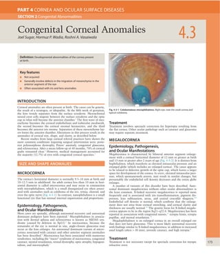
  • 1. 173 INTRODUCTION Corneal anomalies are often present at birth. The cause can be genetic, the result of a teratogen, or idiopathic. At the fifth week of gestation, the lens vesicle separates from the surface ectoderm. Mesenchymal neural crest cells migrate between the surface ectoderm and the optic cup in what will become the anterior chamber.1 The first wave of mes- enchyme becomes the corneal endothelium and trabecular meshwork, the second becomes the corneal stromal keratocytes, and the third becomes the anterior iris stroma. Separation of these mesenchyme lay- ers forms the anterior chamber. Alterations in this process result in the anomalies of corneal size, shape, and clarity, as described below. Recent studies from large corneal referral practices have shown the most common conditions requiring surgical intervention were: poste- rior polymorphous dystrophy, Peters’ anomaly, congenital glaucoma, and sclerocornea. After a mean follow-up of 40 months, 78% of corneal grafts remained clear.2 However, medical management accounted for the majority (52.7%) of eyes with congenital corneal opacities.3 SIZE AND SHAPE ANOMALIES MICROCORNEA The cornea’s horizontal diameter is normally 9.5–10 mm at birth and 10–12.5 mm in adulthood. An adult cornea less than 10 mm in hori- zontal diameter is called microcornea and may occur in conjunction with microphthalmos, which is a small disorganized eye often associ- ated with anomalies such as coloboma of the iris, retina, choroid, and even the optic nerve (Fig. 4-3-1). In contrast, nanophthalmos is a small functional eye that has normal internal organization and proportions. Epidemiology, Pathogenesis, and Ocular Manifestations Most cases are sporadic, although autosomal recessive and autosomal dominant pedigrees have been reported.4 Microphthalmos in associa- tion with dermal aplasia and sclerocornea is known as MIDAS syn- drome (caused by deletion in Xp22).5 In microcornea, because the remainder of the eye is normal in size, angle-closure glaucoma may occur as the lens enlarges. An autosomal dominant variant of micro- cornea associated with cataract and other anterior segment anomalies has been described.6 Microcornea has been associated with numerous syndromes, including the “micro” syndrome of microcornea, congenital cataract, mental retardation, retinal dystrophy, optic atrophy, hypogeni- talism, and microcephaly.7 Treatment Treatment involves spectacle correction for hyperopia resulting from the flat cornea. Other ocular pathology such as cataract and glaucoma may require separate treatment. MEGALOCORNEA Epidemiology, Pathogenesis, and Ocular Manifestations Megalocornea is characterized by bilateral anterior segment enlarge- ment with a corneal horizontal diameter of 12 mm or greater at birth and 13 mm or greater after 2 years of age (Fig. 4-3-2). It is distinct from buphthalmos, which manifests as elevated intraocular pressure and an enlarged globe (which includes an enlarged cornea). The cause appears to be related to defective growth of the optic cup, which leaves a larger space for development of the cornea. In utero, elevated intraocular pres- sure, which spontaneously arrests, may result in similar changes, but presumably the endothelial cell density decreases and the entire globe enlarges. A number of variants of this disorder have been described. Auto- somal dominant megalocornea without other ocular abnormalities is the least common. X-linked recessive megalocornea is reported more frequently and is associated with iris transillumination, pigment dis- persion, lens subluxation, arcus, and central crocodile shagreen.8,9 Endothelial cell density is normal, which confirms that the enlarge- ment does not arise from corneal stretching, and corneal clarity and thickness are usually normal.10 The genetic locus for X-linked megalo- cornea appears to be in the region Xq21–q22.6. Megalocornea has been reported in association with congenital miosis,11 ectopia lentis, ectopia pupillae, and mental retardation.12 Megalophthalmos is an enlarged cornea in an overall enlarged eye that does not have glaucoma. This is most likely autosomal recessive with findings similar to X-linked megalocornea, in addition to increased axial length (often > 30 mm), juvenile cataract, and high myopia.8 Treatment Treatment is not necessary except for spectacle correction for myopic refractive error. Joel Sugar, Hormuz P. Wadia, Roshni A. Vasaiwala 4.3Congenital Corneal Anomalies SECTION 2 Congenital Abnormailities PART 4 CORNEA AND OCULAR SURFACE DISEASES Definition: Developmental abnormalities of the cornea present at birth. Key features ■ Not acquired ■ Generally involve defects in the migration of mesenchyme in the anterior segment of the eye ■ Often associated with iris and lens anomalies Fig. 4-3-1 Colobomatous microphthalmos. Right eye; note the small cornea and typical coloboma.
  • 2. 4 174 CORNEAANDOCULARSURFACEDISEASES Fig. 4-3-2 Megalocornea. (A) This patient has corneal diameters of 14 mm bilaterally. (B) Gross specimen shows appearance of enlarged cornea and heavily pigmented trabecular meshwork. (B, Courtesy of Dr. M.Yanoff) A B Fig. 4-3-3 Posterior embryotoxon. Schwalbe’s line is evident nasally, superiorly, and temporally in the eye of this patient. CORNEAL ABSENCE Isolated absence of the cornea does not occur. Concomitant and severe developmental anomalies of the anterior segment or the entire eye are seen because of the intimate relationship between the ocular structures in the development of the cornea. Anophthalmos is the extreme; absence of the cornea and other ocular structures may occur as part of more extreme genetic developmental disorders. Cryptophthalmos involves partial or complete failure of eyelid formation, dermoid trans- formation of the cornea, an undeveloped anterior segment, or a rudimentary cyst-like globe with an absent anterior segment. Cryp- tophthalmos also may be associated with systemic anomalies such as syndactyly and genitourinary anomalies. This syndrome, known as Fraser syndrome, is inherited as an autosomal recessive trait. Pseudocryptophthalmos occurs when the lids fail to separate and the underlying globe is intact. CONGENITAL CORNEAL ECTASIA Keratoglobus (discussed in Chapter 4.18) is not congenital, but corneal ectasia may be present at birth as part of a congenital anterior staphy- loma, which is often seen with Peters’ anomaly. Congenital corneal ectasia includes significantly more corneal thinning and bulging com- pared to Peters’ anomaly (see Fig. 4-3-6), presumably as a consequence of the same developmental abnormalities in the migration of mesen- chyme. This anomaly is usually unilateral and is often associated with iris developmental defects. Anterior staphyloma may also occur as a result of inflammatory or infectious corneal thinning in utero. ANOMALIES OF CORNEAL CLARITY Changes in the first wave of neural crest mesenchyme migration result in anomalies of the corneal endothelium and anterior chamber angle, while those in the second wave lead to corneal stromal alterations, and those in the third wave affect iris development. The factors that com- monly result in maldevelopment impact more than just one phase in this process of neural crest differentiation. ANTERIOR EMBRYOTOXON Anterior embryotoxon refers to a congenital broad limbus superiorly with an otherwise normal anterior segment, representing merely a broad transition from sclera to cornea. The term also is used to describe an appearance similar to arcus senilis (arcus juvenilis) present at birth. Though it is often sporadic, autosomal dominant and autosomal reces- sive pedigrees have been described. POSTERIOR EMBRYOTOXON Posterior embryotoxon is likely the most frequently seen anomaly, with the prevalence being reported as high as 24% in a random population.13 It consists of thickening and anterior displacement of Schwalbe’s line, which is seen most readily at the slit lamp in the temporal cornea (Fig. 4-3-3). The term toxon is derived from the Greek word for bow, in refer- ence to the crescent of Schwalbe’s line; when present alone, this has no functional significance. CORNEAL KELOIDS Keloids are white, glistening, protuberant lesions that involve all or part of the cornea. Although usually resulting from trauma or ocular inflam- mation, they may be present at birth. Histopathologically, they repre- sent an irregular array of collagen bundles, fibroblasts, and capillaries arising in the corneal stroma. They are sometimes progressive and may be associated with disorders that involve oculodigital manipulation, as in Lowe’s syndrome. In otherwise healthy eyes, keratoplasty is appro- priate.14 For lesions in which progressive growth causes discomfort, dissection of the lesion from the cornea followed by covering with a conjunctival flap may halt progression. DERMOIDS Dermoids (choristomas, or growths of tissue not normally present) may be found on the cornea, usually at the inferotemporal limbus. At times they may involve larger areas of the cornea, the entire limbus, the entire cornea, or the interior of the eye. They usually are round, domed, and pink to white to yellow in color. They may have hair, or in the lipoder- moid variant, globules of lipid, as evident on slit-lamp examination. Induced astigmatism, even amblyopia, may be present. Limbal dermoids may be associated with other malformations, e.g., Goldenhar’s syn- drome, which involves lid colobomas, hemifacial microsomia,
  • 3. CongenitalCornealAnomalies 4.3 175 iridocorneal endothelial (ICE) syndrome (which is acquired and usually unilateral), posterior polymorphous dystrophy with iridocorneal adhe- sions, and the iridogoniodysgenesis syndrome.20 PETERS’ ANOMALY Ocular Manifestations Peters’ anomaly includes a variety of findings, not all of which need to be present to make the diagnosis. Most cases are sporadic, but Peters’ anomaly can be recessively or occasionally dominantly inherited. Eighty percent of cases are bilateral. The pathogenesis involves altera- tion of the migration of neural crest cells. Type I consists of a central or paracentral corneal opacity with iris strands that arise from the collar- ette and attach to the periphery of the opacity. Initially, a defect in corneal endothelium and Descemet’s membrane is present, often with marked corneal edema, which may extend well beyond the defect (Fig. 4-3-6). Over time, the surrounding endothelium covers the defect and produces new basement membrane, and the edema regresses leaving the corneal opacity only.21 Posterior keratoconus and posterior ulcer of von Hippel may be thought of as Peters’ anomaly without iris adhe- sions. Peters’ anomaly type II has lens adherence to the posterior cor- nea, failure of complete separation of the lens from the cornea, and/or cataract. Type I usually is unilateral, while type II frequently is bilateral. Peters’ anomaly may be associated with other ocular anomalies (Fig. 4-3-7), including microcornea, cornea plana, sclerocornea, chorioreti- nal coloboma, iris coloboma, dysgenesis of the angle and iris, persistent hyperplastic primary vitreous, microphthalmos, optic nerve hypoplasia and foveal hypoplasia.22 The most frequent associated ocular abnormal- ity is glaucoma in over 50% of cases. Systemic Associations The systemic associations of Peters’ anomaly include short stature, facial dysmorphism, developmental delay, and delayed skeletal maturation, preauricular skin tags, and other ear anomalies. Limbal dermoids also may be associated with mandibular and other facial anomalies. Histopathology confirms the presence of skin-like collagen with skin adnexal appendages, which include hair follicles, sweat and sebaceous glands, and fat. Treatment consists of simple excision, since a plane can be developed between the lesion and normal sclera, limbus, and cornea, and the dermoid can be readily removed. Infrequently, the lesion is of sufficient depth to warrant concurrent lamellar keratoplasty to fill the defect15 and very rarely may penetrate into the anterior of the eye. The depth can be ascertained by ultrasound biomicroscopy.16 A rare case of spontaneous partial regression of a congenital corneal dermoid has been reported.17 AXENFELD’S ANOMALY AND RIEGER’S SYNDROME Axenfeld’s anomaly consists of bilateral posterior embryotoxon with iris strands adherent to Schwalbe’s line (Fig. 4-3-4). Rieger’s syndrome includes the changes of Axenfeld’s anomaly along with iris atrophy, corectopia, and polycoria. Dental anomalies and a flattened midface and nasal bridge are associated with the Axenfeld–Rieger syndrome (Fig. 4-3- 5). The term anomaly refers to the localized anatomical changes seen, while the term syndrome refers to the more widespread ocular and sys- temic findings. Glaucoma occurs in about half of the patients who have Axenfeld–Rieger syndrome. It appears that Axenfeld’s anomaly and Rieger’s syndrome arise from retention of neural crest remnants and primordial endothelium on the iris and chamber angle.18 Defects in the PITX 2 gene on chromosome 4q25 and in the FKHL7 gene on 6p25, as well as other defects, have been found in different families with the Axenfeld–Rieger phenotype.19 Differential diagnosis includes the Fig. 4-3-4 Axenfeld’s anomaly (posterior embryotoxon). Histological section shows an iris process attached to the anteriorly displaced Schwalbe’s ring. (CourtesyofDrR.Y.Foos.) Fig. 4-3-5 Axenfeld–Rieger syndrome. This patient has bilateral glaucoma as well as dental and facial anomalies. Fig. 4-3-6 Peters’ anomaly in a neonate. (A) Typical Peters’anomaly type I. (B) Severe Peters’involvement with corneal thinning and ectasia. This is called a congenital anterior staphyloma. A B
  • 4. 4 176 CORNEAANDOCULARSURFACEDISEASES Fig. 4-3-7 Severe Peters’ anomaly. This infant has bilateral glaucoma, Peters’ anomaly type II, and features of sclerocornea. Fig. 4-3-8 Peters’ anomaly. Lens material is attached to the posterior cornea (LM). Centrally, the endothelium, Descemet’s membrane, and Bowman’s membrane are not present . The large space (arrow) is an artifact caused by fixation shrinkage. (Courtesy of Dr. M.Yanoff) LM TABLE 4-3-1 RELATIONSHIP OF EMBRYONIC NEURAL CREST MIGRATORY“WAVES”TO VARIOUS ANOMALIES Anomaly Mesenchymal wave abnormality 1st 2nd 3rd Posterior embryotoxon x Axenfeld–Rieger syndrome x x Peters’anomaly x x Posterior keratoconus x Sclerocornea x which form the Krause–Kivlin syndrome (inheritance is autosomal reces- sive). The Peters’-plus syndrome consists of Peters’ anomaly with syn- dactyly, genitourinary anomalies, brachycephaly, central nervous system anomalies, cardiac disease, or deafness.23,24 Peters’ anomaly also has been reported as part of the fetal alcohol syndrome.25 Most interesting is the finding of mutations at the PAX 6 locus on chromosome 11p13 in some Peters’ anomaly patients.26 The PAX 6 gene appears to play a regu- latory role in embryogenesis and also has been found to be abnormal in aniridia and autosomal dominant keratitis.27 PAX 6 is normal, however, in most patients with Peters’ anomaly.28 Increasing molecular evidence has also been found with mutations in the CYP1B1 gene as a possible causative role in Peters’ anomaly.29 This gene defect along with other defects (such as in Axenfeld–Rieger, 4q25) have thus been implicated in ocular development as well as glaucoma.30 Pathology Pathology of Peters’ anomaly shows absence of Descemet’s membrane and endothelium in the area of opacity initially (Fig. 4-3-8). As men- tioned earlier, over time, the endothelial and Descemet’s membrane defects are repaired by surrounding cells. Except for residual fibrosis in the posterior stroma, the remainder of the cornea, except for a usually absent central Bowman’s membrane, is normal. Treatment and Outcome Treatment of Peters’ anomaly includes treatment of the associated glau- coma. Typically, penetrating keratoplasty is considered when corneal opacification is bilateral. A recent study, however, examined results of penetrating keratoplasty for unilateral corneal opacification in Peters’ anomaly.31 Of 14 eyes, 11 (78.6%) had clear grafts at 30 months’ follow- up. Ten of 14 children gained seemingly useful vision after strict amblyopia therapy. The Boston Keratoprosthesis has become an alter- native to penetrating keratoplasty in children with Peters’ anomaly who are at high risk for graft failure.22 Although visual outcomes often are not ideal, the establishment of useful vision is worthwhile.32 SCLEROCORNEA Sclerocornea refers to a non-progressive, non-inflammatory scleral-like clouding of the cornea, which may be peripheral or diffuse. It results from a disorder of the second wave mesenchyme migration and may be associated with corneal flattening or cornea plana because of the par- ticipation in the formation of the limbus. Peripheral forms need to be differentiated from a congenital broad limbus or anterior embryotoxon. Sclerocornea may be associated with other anomalies of anterior seg- ment development, such as Peters’ anomaly; indeed, the entire anterior segment developmental abnormalities discussed here may be consid- ered part of a spectrum and often have many overlapping findings. Glaucoma is common. Associations with systemic anomalies and other ocular anomalies may occur, as noted earlier. Inheritance may be Access the complete reference list online at autosomal dominant, recessive, or X-linked. Most cases, however, are sporadic and usually bilateral. Treatment consists of keratoplasty, or most recently the Boston Keratoprosthesis, once the glaucoma has been controlled. Outcomes are poor, usually because of the difficulty in con- trolling glaucoma or secondary to optic nerve anomalies. A review of the relationship of the embryonic neural crest migratory waves to the anomalies discussed is given in Table 4-3-1. KEY REFERENCES Alward WL. Axenfeld–Rieger syndrome in the age of molecular genetics. Am J Ophthalmol 2000;130:107–15. Churchill AJ, Booth AP, Anwar R, et al. PAX 6 is normal in most cases of Peters’anomaly. Eye 1998;12:299–303. Cook CS. Experimental models of anterior segment dysgenesis. Ophthalmic Pediatr Genet 1989;10:33–46. Dana MR, Schaumberg DA, Moyes AL, et al. Corneal transplantation in children with Peters’ anomaly and mesenchymal dysgenesis. Multicenter Pediatric Keratoplasty Study. Ophthalmology 1997;104:1580–6. Hanson IM, Fletcher JM, Jordan T, et al. Mutations at the PAX 6 locus are found in heterogeneous anterior segment malformations including Peters’anomaly. Nat Genet 1994;6:168–73. Harissi-Dagher M, Colby K. Anterior segment dysgenesis: Peters anomaly and sclerocornea. Int Ophthalmol Clin 2008;48:35–42. Heon E, Barsoum-Homsy M, Cevrette L, et al. Peters’anomaly, the spectrum of associated ocular malformations. Ophthalmic Pediatr Genet 1992;13:137–43. Mader TH, Stulting D. Technique for the removal of limbal dermoids. Cornea 1998;17:66–7. Mayer UM. Peters’anomaly and combination with other malformations. Ophthalmic Pediatr Genet 1992;13:131–5. Meire FM. Megalocornea, clinical and genetic aspects. Doc Ophthalmol 1994;87:1–1121. Mejia LF, Acosta C, Santamaria JP. Clinical, surgical, and histopathologic characteristics of corneal keloid. Cornea 2001;20:421–4. Michaeli A, Markovich A, Rootman DS. Corneal transplants for the treatment of congenital corneal opacities. J Pediatr Ophthalmol Strabismus 2005;42:34–44. Miller MT, Epstein RJ, Sugar J, et al. Anterior segment anomalies associated with the fetal alcohol syndrome. J Pediatr Ophthalmol Strabismus 1984;21:8–18. Rezende RA, Uchoa UB, Uchoa R, et al. Congenital corneal opacities in a cornea referral practice. Cornea 2004;23:565–70. Shields MB, Buckley E, Klintworth GK, et al. Axenfeld–Rieger syndrome: a spectrum of developmental disorders. Surv Ophthalmol 1985;29:387–409.
  • 5. CongenitalCornealAnomalies 4.3 176.e1 REFERENCES 1. Cook CS. Experimental models of anterior segment dysgenesis. Ophthalmic Pediatr Genet 1989;10:33–46. 2. Michaeli A, Markovich A, Rootman DS. Corneal transplants for the treatment of congenital corneal opacities. J Pediatr Ophthalmol Strabismus 2005;42:34–44. 3. Rezende RA, Uchoa UB, Uchoa R, et al. Congenital corneal opacities in a cornea referral practice. Cornea 2004;23:565–70. 4. Vingolo EM, Steindl K, Forte R, et al. Autosomal dominant simple microphthalmos. J Med Genet 1994;31:721–5. 5. Zvulunov A, Kachko L, Manor E, et al. Reticulolinear aplasia cutis congenita of the face and neck: a distinctive cutaneous manifestation in several syndromes linked to Xp22. Br J Dermatol 1998;138:104–52. 6. Salmon JF, Wallis CE, Murray ADN. Variable expressivity of autosomal dominant microcornea with cataract. Arch Ophthalmol 1988;106:505–10. 7. Warburg M, Sjo O, Fledelius HC, et al. Autosomal recessive microcephaly, microcornea, congenital cataract, mental retardation, optic atrophy and hypogenitalism. Am J Dis Child 1993;147:1309–12. 8. Meire FM. Megalocornea, clinical and genetic aspects. Doc Ophthalmol 1994;87:1–1121. 9. Pletz C, Hentsch R. Hereditary anterior megalophthalmos – a genealogical study of 12 patients in 4 generations. Klin Monatsbl Augenheilkd 2000;217:284–8. 10. Skuta GL, Sugar J, Ericson ES. Corneal endothelial cell measurements in megalocornea. Arch Ophthalmol 1983;101:51–3. 11. Meire FM, Delleman JW. Autosomal dominant congenital miosis with megalocornea. Ophthalmic Paediatr Genet 1992;13:123–9. 12. Antinolo G, Rufo M, Borrego S, et al. Megalocornea–mental retardation syndrome: an additional case. Am J Med Genet 1994;52:196–7. 13. Ozeki H, Shirai S, Majima A, et al. Clinical evaluation of posterior embryotoxon in one institution. Jpn J Ophthalmol 1997;41:422–5. 14. Mejia LF, Acosta C, Santamaria JP. Clinical, surgical, and histopathologic characteristics of corneal keloid. Cornea 2001;20:421–4. 15. Mader TH, Stulting D. Technique for the removal of limbal dermoids. Cornea 1998;17:66–7. 16. Lanzl M, Augsburger JJ, Hertle RW, et al. The role of ultrasound biomicroscopy in surgical planning for limbal dermoids. Cornea 1998;17:604–6. 17. Roncevic MB, Doresic JP, Md LD. Large congenital corneal dermoid with spontaneous partial regression: the first report. Cornea 2011;30:219–21. 18. Shields MB, Buckley E, Klintworth GK, et al. Axenfeld–Rieger syndrome: a spectrum of developmental disorders. Surv Ophthalmol 1985;29:387–409. 19. Alward WL. Axenfeld–Rieger syndrome in the age of molecular genetics. Am J Ophthalmol 2000;130:107–15. 20. Walter MA, Mirzayans F, Means AJ, et al. Autosomal-dominant iridogoniodysgenesis and Axenfeld–Rieger syndrome are genetically distinct. Ophthalmology 1996;103:1907–15. 21. Townsend WM, Font RL, Zimmerman LE. Congenital corneal leukomas II. Histopathologic findings in 19 eyes with central defect in Descemet’s membrane. Am J Ophthalmol 1974;77:192–206. 22. Harissi-Dagher M, Colby K. Anterior segment dysgenesis: Peters anomaly and sclerocornea. Int Ophthalmol Clin 2008;48:35–42. 23. Heon E, Barsoum-Homsy M, Cevrette L, et al. Peters’anomaly, the spectrum of associated ocular malformations. Ophthalmic Pediatr Genet 1992;13:137–43. 24. Mayer UM. Peters’anomaly and combination with other malformations. Ophthalmic Pediatr Genet 1992;13:131–5. 25. Miller MT, Epstein RJ, Sugar J, et al. Anterior segment anomalies associated with the fetal alcohol syndrome. J Pediatr Ophthalmol Strabismus 1984;21:8–18. 26. Hanson IM, Fletcher JM, Jordan T, et al. Mutations at the PAX 6 locus are found in heterogeneous anterior segment malformations including Peters’anomaly. Nat Genet 1994;6:168–73. 27. Mirzayans F, Pearce WG, MacDonald IM, et al. Mutation of the PAX 6 gene in patients with autosomal dominant keratitis. Am J Hum Genet 1995;57:539–48. 28. Churchill AJ, Booth AP, Anwar R, et al. PAX 6 is normal in most cases of Peters’anomaly. Eye 1998;12:299–303. 29. Vincent A, Billingsley G, Priston M, et al. Further support of the role of CYP1B1 in patients with Peters’anomaly. Mol Vis 2006;12:506–10. 30. Doward W, Periseen R, Lloyd GC. A mutation in the RIEG 1 gene associated with Peters’ anomaly. J Med Genet 1999;36:152–5. 31. Basdekidou C, Dureau P, Edelson C, et al. Should unilateral congenital corneal opacities in Peters’anomaly be grafted? Eur J Ophthalmol 2011;21:695–9. 32. Dana MR, Schaumberg DA, Moyes AL, et al. Corneal transplantation in children with Peters’ anomaly and mesenchymal dysgenesis. Multicenter Pediatric Keratoplasty Study. Ophthalmology 1997;104:1580–6.