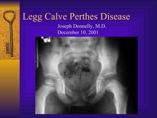
3. History
♦ Late 19th century: “hip infections” that
resolved without surgery
♦ First described in 1910
♦ Early path studies: cartilaginous islands in
the epiphysis
4. Epidemiology
♦ Disorder of the hip in young children
♦ Usually ages 4-8yo
♦ As early as 2yo, as late as teens
♦ Boys:Girls= 4-5:1
♦ Bilateral 10-12%
♦ No evidence of inheritance
5. Etiology
♦ Unknown
♦ Past theories: infection, inflammation,
trauma, congenital
♦ Most current theories involve vascular
compromise
– Sanches 1973: “second infarction theory”
7. Pathogenesis
♦ Histologic changes described by 1913
♦ Secondary ossification center= covered by
cartilage of 3 zones:
– Superficial
– Epiphyseal
– Thin cartilage zone
♦ Capillaries penetrate thin zone from below
9. Pathogenesis
♦ Epiphyseal cartilage in LCP disease:
– Superficial zone is normal but thickened
– Middle zone has 1)areas of extreme
hypercellularity in clusters and 2)areas of loose
fibrocartilaginous matrix
♦ Superficial and middle layers nourished by
synovial fluid
♦ Deep layer relies on blood supply
10.
11. Pathogenesis
♦ Physeal plate: cleft formation, amorphis
debris, blood extravasation
♦ Metaphyseal region: normal bone
separated by cartilaginous matrix
♦ Epiphyseal changes can be seen also in
greater trochanter, acetabulum
22. Healed Stage
♦ Left with residual deformity from disease
and repair process
♦ Differs from AVN following Fx or
dislocation
23. Presentation
♦ Often insidious onset of a limp
♦ C/O pain in groin, thigh, knee
♦ 17% relate trauma hx
♦ Can have an acute onset
24. Physical Exam
♦ Decreased ROM, especially abduction and
internal rotation
♦ Trendelenburg test often positive
♦ Adductor contracture
♦ Muscular atrophy of thigh/buttock/calf
♦ Limb length discrepency
25. Imaging
♦ AP pelvis
♦ Frog leg lateral
♦ Key= view films
sequentially over
course of dz
♦ Arthrography
♦ MRI role undefined
26. Differential Diagnosis
♦ Important to rule out infectious etiology
(septic arthritis, toxic synovitis)
♦ Others:
– Chondrolysis -Neoplasm
– JRA -Sickle Cell
– Osteomyelitis -Traumatic AVN
– Lymphoma -Medication
27. Radiographic Classifications
♦ Describe extent of epiphyseal disease
♦ Catterall classification= most commonly
used
– 4 groups based on amount of femoral head
involvement
– Also presence of sequestrum, metaphyseal rxn,
subchondral fx
32. Lateral Pillar Classification
♦ 3 groups:
– A) no lateral pillar
involvment
– B) >50% lat height
intact
– C) <50% lat height
intact
33. Salter-Thompson Classification
♦ Simplification of Catterall
♦ Based on status of lateral margin of capital
femoral epiphysis
♦ Group A (Catterall I & II equivalent)
♦ Group B (Catterall III & IV equivalent)
34. Prognosis
♦ 60% of kids do well without tx
♦ AGE is key prognostic factor:
– <6yo= good outcome regardless of tx
– 6-8yo= not always good results with just
containment
– >9yo= containment option is questionable,
poorer prognosis, significant residual defect
35. Prognosis
♦ Flat femoral head incongruent with
acetabulum= worst prognosis
♦ Do not treat in reossification stage
(>15mos)
36. Non-operative Tx
♦ Improve ROM 1st
♦ Bracing:
– Removable abduction orthosis
– Pietrie casts
– Hips abducted and internally rotated
♦ Wean from brace when improved x-ray
healing signs
38. Non-operative Tx
♦ Check serial radiographs
– Q3-4 mos with ROM testing
♦ Continue bracing until:
– Lateral column ossifies
– Sclerotic areas in epiphysis gone
♦ Cast/brace uninvolved side
39. Operative Tx
♦ If non-op tx cannot maintain containment
♦ Surgically ideal pt:
– 6-9yo
– Catterral II-III
– Good ROM
– <12mos sx
– In collapsing phase
40. Surgical Tx
♦ Surgical options:
– Excise lat extruding head portion to stop
hinging abduction
– Acetabular (innominate) osteotomy to cover
head
– Varus femoral osteotomy
– Arthrodesis
-described 1910 independently by Legg, Calve, Perthes, and Waldenstrom -Legg= “obscure affectation of the hip”, thought secondary to pressure injury flattening the femoral head -Calve: same year, 10 cases non inflamm/self limiting disease due to delayed osteogenesis. Also reported coxa vara and increased femoral head size -Perthes- “arthritis deformans juveniles, possible inflammatory condition -Waldenstrom: thought it was TB
-incidence of positive family hx ranges from 1.6% to 20%, but no hard evidence of predisposition -is more common in certain geographic areas (urban>rural)=nutritional?, later born children, strong association with ADHD (33%)
-Phemister- thought it was infectious but cx neg -Axhausen- thought bacillary embolism with weak infection which healed quickly -1975 Matsoukas showed association with prenatal rubella -1973 Sanches, infarcted animal femoral heads, unable to produce typical histologic picture of LCPdz with one infarction, could do it with a second. Supported by Inoue using human histologic material
-blood supply description (terminology)varies -3 main sources to the proximal femur: 1)extracapsular ring, 2)ascending cervical (retinacular branches) vessels 3)artery of the ligamentum teres -extracapsular ring: med and lat fem circumflex, gives rise to ascending cervical branches (extracapsular) which give of metaphyseal and epiphyseal branches -ant portion=mainly lat fem circ, post/lat/med=med fem circ -Chung found that greatest volume of flow= from lat ascending cervical (end of med fem circ)
-few human specimens have been studied, each showing only a stage of the dz and usually from sample of just one part of the involved head. Histologically not well illucidated
-Superficial zone=like adult articular cartilage -epiphyseal (middle) cartilage zone= becomes thinner as skeleton matures, epiphyseal bone enlarges -Deep thin zone= small clusters of cartilage cells that hypertrophy and degenerate -capillaries from below
-changes in zone 2 are abnormal, have different histochemical and US properties vs normal, also see small 2ndary ossification centers directly on the abnormal cartilage matrix -synovial fluid nourishes 2 superficial layers, continue to proliferate -deep layer affected by ischemic process
-a) superficial zone: area of disorganized cartilage -c) junction btwn normal and abnormal epiphyseal cartilage, note hypercellularity -d) extensive abnormal cartilage, bone forms directly on abnormal cartilage
-physeal plate: thinner than normal, irregular cell columns and cartilage masses -metaphysis: cartilage does not ossify, proliferates with bone, causes tongues of cartilage extending into metaphysis -skeletal surveys shows contour irregularities in 48% of normal contralateral capital epiphysis, suggesting it is a generalized disorder, more appropriately named a syndrome
-growth failure due to lack of blood supply -affected femoral ossific nucleus appears radiodense (relative osteopenia of surrounding bone vs. increased mass in that area?) -affected femoral head appears smaller vs. other side -wide med joint space due to: synovitis? Decreased head volume from necrosis and collapse? Due to increased blood flow to soft tissues (eg. Lig teres) causing lateral displacement? Most likely due to epiphyseal cartilage hypertrophy (x-ray phenomenon) -crescent sign= subchondral radiolucent zone, likely results from a subchondral stress fracture and the extent of this zone determines the extent of the necrotic fragment
-note smaller, denser ossific nucleus on L hip -crescent (fracture) sign -irregular physeal plate -blurry, radiolucent metaphysis
-same on frog
-increased radiodensity due to new bone forming on old bone
-fragmented epiphyseal bone, radiodense and lucent areas
-note shape deformity on R vs L
-AVN process after fx/dislocation does not undergo fragmentation
-must recognize thigh & knee pain as possible hip pathology -pain usually mild and relieved by rest, often present late due to mild sx
-early decreased abduction due to synovitis/spasm, may become permanent after development of femoral head deformity -adduction contracture due to long standing spasm -atrophy due to disuse due to pain, shows long standing nature -short limb due to head collapse= poor prognosis
-frog leg= better for crescent sign -compare films with previous to determine change -arthrography can show status of cartilage not shown on x-ray, check ROM to r/o hinging abduction -hinging abduction due to large femoral head extruding laterally & hinging over edge of acetabulum
-25% of anterocentral head involved -no sequestrum, no subchondral fx’s, normal metaphysis
-50% of anterolateral region involved -evidence of sequestrum/ subchondral (anterior) fx, med/lat pillars intact
-75% of head involved -large sequestrum, lat pillar (column) involved, sclerotic junction btwn normal/abnormal -subchondral fx line extends into post ½ of epiphysis
-whole head involved, widespread epiphyseal collapse -diffuse or central metaphyseal lesion -Posterior remodeling of ephiphysis -poor prognosis
-Catterral 1 and usually 2 do well without treatment
-to late to treat after 15mos
-abduction usually affected most of ROM -use PT to regain abduction (overcome spasm) and internal rotation -may require several weeks of abduction traction -don’t start bracing until abd/int rot restored to normal -arthrography pre-bracing to determine congruency throughout ROM -head collapse is independent of weight bearing, not necessarily NWB -hips braced in abd/ int rotation to transmit weight over wide area of acetabulum, prevents head collapse
-Study by Futami and Suzuki 1997, 6% of uninvolved contralat hips develop LCP when unbraced, 0 when braced -length of casting usually 6 weeks to start out
-coxa magna due to ossiffication of hypertrophied articular cartilage -OD=rare, occurs with the late onset of dz and with prolonged ineffectual repair stage