SlideShare a Scribd company logo
1 of 72
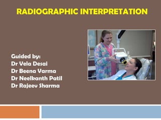
RADIOGRAPHIC INTERPRETATION


Guided by:
Dr Vela Desai
Dr Beena Varma
Dr Neelkanth Patil
Dr Rajeev Sharma
Radiograph
2


       Photographic image
       Radiosensitive surface
       Radiation – X rays/ Gamma rays
       Radiogram/shadowgram/roentgenogram


                                  11/15/2011
Role of radiographs
3


       Clinical examination phase
       Diagnosis( confirm/exclude)
       Treatment planning
       During treatment
       Follow up
       Blind screening tool-justify
       Limitations-replace clinical examination
       Need for further investigation

                                        11/15/2011
Radiographs in Diagnosis
4


     Diagnostic imaging is an integral part of the
      diagnostic process in clinical dentistry.
     Radiographs are often obtained as part of a

      complete examination.
     Appropriate radiographic interpretation is used
      along with clinical information and other tests
      to formulate a differential diagnosis

            Free PowerPoint Template from www.brainybetty.com   11/15/2011
Uses of radiographs
5


       Loss of tooth structure
       Caries(occlusal/proximal)
       Non carious(attrition,fracture)
       Periodontal diseases
       Endodontic diseases
       Impacted teeth
       Trauma
       Other bone pathologies
       Implants
              Free PowerPoint Template from www.brainybetty.com   11/15/2011
6


     Technique             Radiography
     Interpretation         Radiology
     Interpretation:

      Step by step analytical process that provides
      an exact idea of the clinical problem and helps
      to achieve the final diagnosis of any particular
      lesion.


                                      11/15/2011
Interpretation
7


       Three steps:
       Visualization
        Perception
       Integration of information
       Other diagnostic tools-vitality/mobility
       Pulp tester


                                          11/15/2011
8


       Clinical examination        Quality assurance
       Type of radiograph          Inadequate quality
       Number of                   Inadequate number
        radiographs                 Extraoral radiology
       Aids in interpretation      Biopsy/treatment-
                                     aids in site selection


                                           11/15/2011
FULL MOUTH INTRAORAL RADIOGRAPHS-IOPA & BITEWING

9                                        11/15/2011
10


     Ideal radiograph:
      Visual : density & contrast

      Geometric : sharpness/detail,

       resolution/definition, magnification, distortion
      Anatomical accuracy of radiographic images

      Adequate coverage of anatomical region of
       interest.


                                         11/15/2011
Viewing Conditions
11


       This should be done in a quiet, darkened room
       At least two good, evenly-lit viewing boxes are
        required
       A bright light illuminator is required for relatively
        over-exposed areas
       Mounted in holder
       Appropriate size of viewbox to accommodate film
       Magnifying glass-detailed examination of small
        regions

                                           11/15/2011
12


        A radiograph is a two dimensional image of a
         three dimensional object.
        Clark’s rule: The most distant object from the
         cone(lingual) moves towards the direction of
         the cone


                                        11/15/2011
Three-dimensional concept
13


       The  radiographic image is simply a
        Two-dimensional shadowgram of the patient
       The third dimension must be reconstructed
        mentally, preferably from two radiographic
        projections made at right angles (orthogonal
        projections) to each other
       Oblique projections may be required to assess
        anatomically complicated areas


                                        11/15/2011
Contrast perception:
14


        Ability to distinguish b/w two areas of
         radiographic image of diff densities-Weber’s
         law
        Minimum perceptible difference in gray level is
         proportional to the brightness level to which
         the subject is adapted.
        All areas on a radiograph represented as:
        Black
        Grey
        White
                                         11/15/2011
MACH BAND EFFECT
15

        Illusion consists of light or dark stripes that are
         perceived next to the boundary between two regions of
         an image that have different lightness gradients

        Spatial high-boost filtering performed by the human
         visual system on the luminance channel of the image
         captured by the retina.

        Mach bands are independent of orientation.

        This occurs when two circles of uniform brightness are
         placed side by side, separated by a sharp edge. Just
         along the edge one colour looks darker than it really is,
                                               11/15/2011
         while the other looks lighter.
16
     MACH BAND EFFECT   11/15/2011
17


         False-positive radiological diagnosis of dental
         caries
         Manifest adjacent to metal restorations or
         appliances, between enamel and dentin
         Misdiagnosis of horizontal root fractures
         because of the differing radiographic
         intensities of tooth and bone.


                                         11/15/2011
   RADIOLUSCENT-the capability of a substance
         with a relatively small atomic number to let a large
18
         amount of x-rays pass through it, thus producing
         darkened images on x-ray films.


                                               RADIOOPAQUE


                                                 RADIOLUSCENT


        RADIOOPACITY-the capability of a substance to
         hinder or completely stop the passage of x-rays,
         display as white/light areas on an exposed x-ray
         film.
                                            11/15/2011
Properties
19


        Atomic number
          The  higher the atomic number, the more
           radiopaque the tissue/object:
        Physical opacity
          Air, fluid and soft tissue have approximately the
           same atomic number, but the specific gravity of
           air is only 0.001, whereas that of fluid and soft
           tissue is 1
          Therefore air will appear black on a radiograph,
           compared with fluid and soft tissue, which appear
           more grey                          11/15/2011
20


        Thickness
          The thicker the tissue/object, the greater the
          attenuation of X-Rays and the more white the
          image .

          When  two tissues/objects are superimposed, the
          composite shadow formed by these will appear
          more opaque than either of the two separate
          tissues
        Bone(14;1.8)
                 Free PowerPoint Template from www.brainybetty.com   11/15/2011
Image analysis
21


        Identify normal anatomic landmarks
        Knowledge of normal v/s abnormal
        Attention to all regions on the film
         systematically
        Three circuits


                                        11/15/2011
First visual circuit: intraoral
22
     images
      Periapical before bitewing images
      Right maxilla to left; left mandible to right

      One anatomic structure at a time

      Eg: posterior maxilla-maxillary

       sinus,tuberosity,zygomatic process
      Normal anatomy

          bones, canals, foramina
     Check for symmetry

                                          11/15/2011
Use a systematic process
23


      Go back to the first quadrant and look at the
       trabecular pattern. Is it:
      Normal

      Symmetrical when compared to the
       contralateral
      side
      Sparse

      Dense

      In the direction of anatomical stress

      Altered
                                       11/15/2011
Fish net
     Step ladder


      Granular

24   TRABECULAR PATTERN   11/15/2011
Second visual circuit
25


        Examination of bone:
        Height of alveolar bone
        Crest relative to teeth
        Loss of height-more than 1.5 mm-periodontal
         disease
        Cortication
        Lamina dura + PDL space + tooth roots
        Carcinoma-erosion of alveolar crest+ ill defined
         borders.
               Free PowerPoint Template from www.brainybetty.com   11/15/2011
26   Free PowerPoint Template from www.brainybetty.com   11/15/2011
Third visual circuit
27


      Examination of dentition & associated
       structures
      Number, Sequence, appearance, root

       structure
      Crowns –defective enamel, caries

      Intreproximal areas & restorations

      Pulp chambers-size, contentRestoration
      Dentin
                                   Proximal caries
      Bone-radioluscent/radioopaque lesions
                                    Pulp


                                           11/15/2011
Check individual teeth
28


        Enamel, [amelogenesis imperfecta, mulberry molar, etc.]

        The dentin, [dens invaginatus or evaginatus, denticles etc.] T

        Pulp chamber [dentinogenesis imperfecta, odontogenesis
         imperfecta, odontodysplasia, taurodontism, individual obliteration of
         nerve canals, etc.]

        Apical area [root resorption, lucencies or opacities]

        periodontal ligament space [widened in early osteosarcoma
         (localized), scleroderma ( generalized) [ absent in
         hyperparathyroidism]

        Amount of bone support.
                  Free PowerPoint Template from www.brainybetty.com   11/15/2011
Routine assessment of
     radiographs
29


         Ensure that the radiograph is the one of the patient being
          examined, check the date, opd/no.
         Ensure two orthogonal projections are available.
         The radiographic views are named according to the direction the
          primary beam enters and leaves the tissue and the body part
          being examined
         The position of the patient during exposure should be known,
          and left/right markers should be identified
         The radiograph should be of high technical quality with respect to
          positioning, centring, collimation, exposure and development,
          and should be free from artefacts.


                                                     11/15/2011
30


        Every shadow visible must be evaluated to
         determine whether it is:
         A  feature of normal anatomy
          A composite structure formed by superimposition
           of structures
          An artefact produced by inaccurate positioning

          A pathologic lesion: must be ruled out first


               Free PowerPoint Template from www.brainybetty.com   11/15/2011
Interpretation is an orderly process


                Normal                                     Abnormal
                variation


                                   Developmental
                                                           Acquired
                                   abnormalities
                                                           abnormalities


Cyst    Benign Malignant        Inflammatory     Bone       Vascular Metabolic
                                                                               Trauma
       neoplasia neoplasia          lesion     dysplasia    analomy
31
Why describe the lesion?
32


        The radiographic description can give us
         indications of:
         Tissue of origin
         Biological behavior
        Prognosis
        Treatment concerns
        Diagnosis or a Differential Diagnosis


               Free PowerPoint Template from www.brainybetty.com   11/15/2011
Describing the Lesion
33


     1. Size
     2. Shape
     3. Location
     4. Density
     5. Borders
     6. Internal Architecture
     7. Effect on adjacent structures


             Free PowerPoint Template from www.brainybetty.com   11/15/2011
Aunty Minnie Approach
34


       Aunt Minny represents an abnormality which
        looks like one that the evaluator has seen
        before, or been told about.
       It would be difficult to recognise new findings
        using this approach
      Cousin Harry represents an abnormality which
        the evaluator has not seen for a long time, but
        would like to see
      Uncle Fred represents an abnormality which is
        often present

                                       11/15/2011
35


     Free PowerPoint Template from www.brainybetty.com   11/15/2011
Size
36


        Measure the lesion with a ruler. If you must
         estimate, use surrounding structures as
         guide
        Measure in two dimensions, width and
         height in mm or cm


                                       11/15/2011
Shape
37


     Regular shapes like Round, Triangular,
        Rhomboid etc.
     Irregular shape like circular, fluid
        filled(hydraulic)-cyst
     Scalloped-multilocular app.


               Odontogenic keratocyst   11/15/2011
38


     Scalloped/Multilocular-
         Ameloblastoma
                               11/15/2011
Location
39


      Is the lesion localized or generalized?
      Unilateral or bilateral

     (submandibular fossa), fibrous dysplasia
      Where is the lesion in relation to other
       structures and anatomic landmarks?
      Use terms such as:

      Mesial, Distal

      Inferior, Superior

      Posterior, Anterior


                                       11/15/2011
Soft tissues or jaws:
40


        Epicentre-coronal to tooth-odontogenic
         epithelium
         Epicenter of the lesion is above the
         mandibular canal->odontogenic in origin
        Epicentre->below IAC->non
         odontogenic(likely)
        Cartilaginous lesions, osteochondromas -
         >condyles
        If the epicenter of the lesion is in the sinus, not
         odontogenic in origin-alveolar process of
                                            11/15/2011
         maxilla
Density
41


        Is the lesion Radiopaque, Radiolucent, or
         Mixed Density
        Remember that opacity is relative to the
         adjacent structures.
        If the lesion is of mixed density, describe the
         appearance


                                          11/15/2011
Radioluscent to radioopaque
42
     structures
        Air,fat,gas
        Fluid
        Soft tissue
        Bone marrow
        Trabecular bone
        Cortical bone
        Enamel
        Metal

                           11/15/2011
Internal architecture
43


        Is the lesion uniform?
        Internal structures such as septae or
         loculations
        Septae –residual bone-long strands/walls
        Loculations are individual compartments(2)
        Soap bubble app- OKC
        Giant cell granuloma-wispy, granular
        Odontogenic myxoma-straight, thin
        Tooth-like elements-cementum
               Free PowerPoint Template from www.brainybetty.com   11/15/2011
Fibrous dysplasia
44


        More in number
        Shorter
        Aligned in response to stress
        Randomly oriented
        Ground glass/orange peel app


                                         11/15/2011
45   Free PowerPoint Template from www.brainybetty.com   11/15/2011
46
        Inflammatory lesion-new bone formation-thick
         trabeculae-more radioopaque
        Dystrophic calcifications-damaged soft tissue
         masses- calcified lymph nodes-cauliflower like
         masses
        Ewing’s sarcoma-onion skin app


                                                 11/15/2011
            Calcified lymph nodes-tuberculosis
Borders
47
        Well or poorly demarcated
        Punched out-sharp- (no bony reaction)-

                   multiple myeloma
        Corticated-uniform-periphery- (thin opaque border)

                           cyst
        Sclerotic (wide, uneven opaque border)

             Periapical cemental dysplasia

        Radioluscent(periphery)+ corticated

             Odontoma, cementoblastoma
                                                  11/15/2011
Periapical cemento                              Residual cyst
     osseous dysplasia


                                  Well defined borders


48            Free PowerPoint Template from www.brainybetty.com   11/15/2011
Ill defined borders
49


        Gradual transition-normal app bone &
         abnormal app trabaculae- sclerosing osteitis


        Invasive border-bone destruction-malignancy


               Free PowerPoint Template from www.brainybetty.com   11/15/2011
Jaw – examine the lesion in the
     jaw:
50


     · Site – location, extent, solitary, multi-focal or
       generalised
     · Size and shape – measure and describe. This
       may require one or more views.
     · Symmetry – examine contralateral site. Bilateral
       symmetry is suggestive of a normal variant
     · Border – sclerosis, resorption, lack of continuity
     · Contents – lucent or opaque. Homogenous or
       varying density
     · Association with other structures. Teeth
       displaced or resorbing
             Free PowerPoint Template from www.brainybetty.com   11/15/2011
Effect on adjacent structures
51


        Lesions behaviour & impact on surrounding
         structures-identification of disease
        Inflammatory disease-bone
         resorption/formation.
        A Space Occupying lesion creates its own
         space by displacing other structures, such as
         teeth, maxillary sinus, inferior alveolar canal,
         etc.
               Free PowerPoint Template from www.brainybetty.com   11/15/2011
52


        Epicentre above crown of teeth-follicular cysts-
         teeth apically
        Lesion-ramus of mandible-cherubism-anterior
         direction
        Papilla of developing tooth-lymphoma
        Widening of PDL, broken lamina dura-
         periapical/periodontal abscess
        Root resoption-periodontitis, trauma, tumors
        Reactive bone-periphery of lesion-benign slow
         growth
               Free PowerPoint Template from www.brainybetty.com   11/15/2011
53


        Inferior alveolar canal
        Superior displacement-fibrous dysplasia
        Widening of IAN-cortical boundary intact-
         benign vascular/neural lesion
        Irregular widening with cortical destruction,
         complete length of canal-malignant neoplasm


               Free PowerPoint Template from www.brainybetty.com   11/15/2011
Outer cortical bone/periosteal
54
                     reactions
        Slow growing-new bone-expanding lesion-
         outer cortical bone maintained
        Rapidly growing-periosteum does not respond-
         missing cortical plate
        Exudate from inflammatory lesion-lift
         periosteum off surface of the surface of cortical
         bone-periosteum lay down new bone.
        Onion skin app-leukaemia, langerhan’s cell
         histiocytosis
        Spiculated new bone-osteogenic sarcoma
                                          11/15/2011
Formulation of radiographic
55
                  interpretation
        Organised fashion
        Single observation
        Diagnosis


               Free PowerPoint Template from www.brainybetty.com   11/15/2011
56


        Decision 1: Normal V/S Abnormal
        Decision2: Developmental V/S Acquired
        Decision 3: Classification
        Decision 4: Ways To Proceed


                                      11/15/2011
Decision 1: Normal V/S Abnormal
57


        Structure of interest
        Variation of normal/represents abnormality


               Free PowerPoint Template from www.brainybetty.com   11/15/2011
Decision 2: Developmental V/S
     Acquired
58


        Area of interest: abnormal
        Radiographic characterstics: location,
         periphery, shape, internal structure, effect on
         surrounding structures
        Indicates developmental/acquired-external
         root resorption


               Free PowerPoint Template from www.brainybetty.com   11/15/2011
Decision 3: Classification
59


        Abnormality
        Appropriate category
        Treatment plan


               Free PowerPoint Template from www.brainybetty.com   11/15/2011
Decision 4: Ways To Proceed
60


        Analyse images
        Further imaging like CT, MRI
        Biopsy
        Treatment


               Free PowerPoint Template from www.brainybetty.com   11/15/2011
SOFT TISSUE.
61


        The examination of the radiographic
         appearance of soft tissue is all too often
         overlooked.
        This is particularly true on panoramic
         radiographs.
        If the clinical examination determines that soft
         tissue requires radiographic examination, kVp
         be reduced when the patient is exposed. Soft
         tissue structures in the maxillofacial region are
         often tongue, soft palate, tip and ala of the
         nose Free PowerPoint Template from www.brainybetty.com 11/15/2011
Correct terminology
62

        One examines a radiograph and NOT an X-ray. Bear in mind that

         an X-ray can not be seen. An X-ray is a photon / beam of energy.

        One does not see infection at the apex of a tooth. What one does

         see is the well / poorly demarcated radiolucency/opacity, x mm

         by y mms in size at the apex of tooth number X.

     For the same reason one does not speak about a PAP in radiology.


                                                      11/15/2011
63


        Periodontal bone loss is not periodontitis per
         se.

        Stay away from brand names. We do not
         have a panorex machine here. Use the word
         PANORAMIC radiograph or PAN.

         In radiologic terminology, a PA is a postero-
         anterior PowerPoint Template from www.brainybetty.com
               Free view.                                        11/15/2011
EXISTING DIAGNOSTIC
     RADIOGRAPHS
64


        An effective way to reduce unnecessary
         radiation to the patient is to avoid retaking
         [recent] radiographs that already exist. It is the
         clinician's responsibility to obtain these records
         from earlier health providers where possible.


               Free PowerPoint Template from www.brainybetty.com   11/15/2011
The diagnostic process is far from infallible. In any
     diagnostic procedure there are four possible outcomes:-
65


     1. True positive: The disease is present and
       correctly identified.
     2. False positive: The disease was absent but
       something on the radiograph convinced the
       clinician that it was present.
     3. True negative: No disease present and
       correctly determined.
     4. False negative: Disease is present but not
       detected. Occurs much too often
              Free PowerPoint Template from www.brainybetty.com   11/15/2011
RADIOGRAPHIC RECORDS
66

        The value of radiographs as a part of the integral
         records of a patient cannot be overstated.
         Good radiograph is difficult to match with written
         records and the radiograph is more indisputable than a
         written statement in a court of law provided the name of
         the patient is indicated as well as the date.
         However, this is not a call to expose the patient to
         ionizing radiation merely for the sake of documentation.
        One may not retake radiographs for the sake of
         improving one's grades. Radiographs legally must be
         kept for at least 5 years; some authorities state 7 years.

                                                 11/15/2011
DOCUMENTATION
67


        Clear medico-legal requirement for
         documentation of interpretation.
         Signed and dated radiographic report must be
         written with patient's record.
         Clinically useful in treatment planning and
         case presentation.


               Free PowerPoint Template from www.brainybetty.com   11/15/2011
Radiographic report
68


        Patient & general information
        Imaging procedure
        Clinical information
        Findings
        Radiographic interpretation


               Free PowerPoint Template from www.brainybetty.com   11/15/2011
RADIOGRAPHIC
69
     PRESCRIPTION

        Licensed dentist may prescribe radiographs
        Examination appropriate      radiographic
         views

          Maximum amount of information

         Minimum amount of ionizing radiation.

               Free PowerPoint Template from www.brainybetty.com   11/15/2011
70   CONCLUSION


            11/15/2011
References
71


        White and pharoah,principles and
         interpretation.IV edition,pg281-296
        W&P. Ch.14. Oral and Maxillofacial Imaging.
         Farman and NortjeNeill Serman.2000
        Dr. Parish P. Sedghizadeh. Radiographic
         pathology of the head and neck.
        Brocklebank L, Dental Radiology, Oxford
         University Press 1997.
         Deforge DH and Colmery BH, An Atlas of
         Dental Radiology, Iowa State University Press
         2000
               Free PowerPoint Template from www.brainybetty.com   11/15/2011
THANK YOU
72


     ...when you have eliminated the impossible,
        whatever remains, however improbable, must
        be the
     truth.
         Sir Arthur Conan Doyle, (Sherlock Holmes)
      British mystery author & physician (1859 - 1930)


                                      11/15/2011

More Related Content

What's hot

Multilocular radiolucencies
Multilocular radiolucenciesMultilocular radiolucencies
Multilocular radiolucenciesSelva Arockiam
 
Chairside investigations
Chairside investigationsChairside investigations
Chairside investigationsrumelaghoshbds
 
object Localization in intraoral radiographies
object Localization in intraoral radiographiesobject Localization in intraoral radiographies
object Localization in intraoral radiographieszohre rafi
 
Pigmented lesions of oral cavity
Pigmented lesions of oral cavityPigmented lesions of oral cavity
Pigmented lesions of oral cavityPraveena Veena
 
Radiographic techniques
Radiographic techniquesRadiographic techniques
Radiographic techniquesanusushanth
 
Radiographic Differential Diagnosis 2009
Radiographic Differential Diagnosis 2009Radiographic Differential Diagnosis 2009
Radiographic Differential Diagnosis 2009IAU Dent
 
Periapical radiograph
Periapical radiograph Periapical radiograph
Periapical radiograph UE
 
Paralleling and bisecting radiographic techniques
Paralleling and bisecting  radiographic techniquesParalleling and bisecting  radiographic techniques
Paralleling and bisecting radiographic techniquesDr. Ritu Gupta
 
panoramic-techique errors
panoramic-techique errorspanoramic-techique errors
panoramic-techique errorsParth Thakkar
 
Bisecting angle vs paralleling technique /orthodontic courses by Indian denta...
Bisecting angle vs paralleling technique /orthodontic courses by Indian denta...Bisecting angle vs paralleling technique /orthodontic courses by Indian denta...
Bisecting angle vs paralleling technique /orthodontic courses by Indian denta...Indian dental academy
 
Radiopaque Lesions
Radiopaque LesionsRadiopaque Lesions
Radiopaque LesionsMaryam Arbab
 
Factors controlling the beam of x-ray
Factors controlling the beam of x-rayFactors controlling the beam of x-ray
Factors controlling the beam of x-rayAnu Jose
 
mixed radiolucent radiopaque lesions of oral cavity
mixed radiolucent radiopaque lesions of oral cavitymixed radiolucent radiopaque lesions of oral cavity
mixed radiolucent radiopaque lesions of oral cavityStephanie Chahrouk
 
Anatomic landmarks seen in a IOPA
Anatomic landmarks seen in a IOPAAnatomic landmarks seen in a IOPA
Anatomic landmarks seen in a IOPAdrsundaram95
 
Intra oral radiograph techniques
Intra oral radiograph techniquesIntra oral radiograph techniques
Intra oral radiograph techniquessamar noby
 
PULP AND PERIAPICAL LESIONS OF THE TOOTH ppt
PULP AND PERIAPICAL LESIONS OF THE TOOTH pptPULP AND PERIAPICAL LESIONS OF THE TOOTH ppt
PULP AND PERIAPICAL LESIONS OF THE TOOTH pptK BHATTACHARJEE
 
Effects of radiation on oral tissues
Effects of radiation on oral tissuesEffects of radiation on oral tissues
Effects of radiation on oral tissuesAdwiti Vidushi
 
periapical radiolucencies
 periapical radiolucencies periapical radiolucencies
periapical radiolucenciesvidushiKhanna1
 

What's hot (20)

Multilocular radiolucencies
Multilocular radiolucenciesMultilocular radiolucencies
Multilocular radiolucencies
 
Chairside investigations
Chairside investigationsChairside investigations
Chairside investigations
 
object Localization in intraoral radiographies
object Localization in intraoral radiographiesobject Localization in intraoral radiographies
object Localization in intraoral radiographies
 
Panoramic radiography OPG
Panoramic radiography OPGPanoramic radiography OPG
Panoramic radiography OPG
 
Pigmented lesions of oral cavity
Pigmented lesions of oral cavityPigmented lesions of oral cavity
Pigmented lesions of oral cavity
 
Radiographic techniques
Radiographic techniquesRadiographic techniques
Radiographic techniques
 
Radiographic Differential Diagnosis 2009
Radiographic Differential Diagnosis 2009Radiographic Differential Diagnosis 2009
Radiographic Differential Diagnosis 2009
 
red and white lesions of oral cavity
red and white lesions of oral cavityred and white lesions of oral cavity
red and white lesions of oral cavity
 
Periapical radiograph
Periapical radiograph Periapical radiograph
Periapical radiograph
 
Paralleling and bisecting radiographic techniques
Paralleling and bisecting  radiographic techniquesParalleling and bisecting  radiographic techniques
Paralleling and bisecting radiographic techniques
 
panoramic-techique errors
panoramic-techique errorspanoramic-techique errors
panoramic-techique errors
 
Bisecting angle vs paralleling technique /orthodontic courses by Indian denta...
Bisecting angle vs paralleling technique /orthodontic courses by Indian denta...Bisecting angle vs paralleling technique /orthodontic courses by Indian denta...
Bisecting angle vs paralleling technique /orthodontic courses by Indian denta...
 
Radiopaque Lesions
Radiopaque LesionsRadiopaque Lesions
Radiopaque Lesions
 
Factors controlling the beam of x-ray
Factors controlling the beam of x-rayFactors controlling the beam of x-ray
Factors controlling the beam of x-ray
 
mixed radiolucent radiopaque lesions of oral cavity
mixed radiolucent radiopaque lesions of oral cavitymixed radiolucent radiopaque lesions of oral cavity
mixed radiolucent radiopaque lesions of oral cavity
 
Anatomic landmarks seen in a IOPA
Anatomic landmarks seen in a IOPAAnatomic landmarks seen in a IOPA
Anatomic landmarks seen in a IOPA
 
Intra oral radiograph techniques
Intra oral radiograph techniquesIntra oral radiograph techniques
Intra oral radiograph techniques
 
PULP AND PERIAPICAL LESIONS OF THE TOOTH ppt
PULP AND PERIAPICAL LESIONS OF THE TOOTH pptPULP AND PERIAPICAL LESIONS OF THE TOOTH ppt
PULP AND PERIAPICAL LESIONS OF THE TOOTH ppt
 
Effects of radiation on oral tissues
Effects of radiation on oral tissuesEffects of radiation on oral tissues
Effects of radiation on oral tissues
 
periapical radiolucencies
 periapical radiolucencies periapical radiolucencies
periapical radiolucencies
 

Viewers also liked

Periodontal management of pt. with diabetes mellitus, hypertension, iinfectiv...
Periodontal management of pt. with diabetes mellitus, hypertension, iinfectiv...Periodontal management of pt. with diabetes mellitus, hypertension, iinfectiv...
Periodontal management of pt. with diabetes mellitus, hypertension, iinfectiv...Ujwal Gautam
 
Dental trauma to primary teeth
Dental trauma to primary teethDental trauma to primary teeth
Dental trauma to primary teethFahimeh Vaziri
 
young permanent tooth
young permanent toothyoung permanent tooth
young permanent toothJeena Paul
 
Normal Radiographic Anatomical Landmarks
Normal Radiographic Anatomical LandmarksNormal Radiographic Anatomical Landmarks
Normal Radiographic Anatomical LandmarksDivya Rana
 
Radiographic errors and artifacts
Radiographic errors and artifactsRadiographic errors and artifacts
Radiographic errors and artifactsWAlid Salem
 
Radiographs in endodontic diagnosis
Radiographs in endodontic diagnosisRadiographs in endodontic diagnosis
Radiographs in endodontic diagnosisliya thomas
 
Endodontic radiograph
Endodontic radiographEndodontic radiograph
Endodontic radiographmstfa mgdy
 
Radiography in endodontics
Radiography in endodonticsRadiography in endodontics
Radiography in endodonticsJuny handA
 
Recent advances in imaging techniques/ /certified fixed orthodontic courses b...
Recent advances in imaging techniques/ /certified fixed orthodontic courses b...Recent advances in imaging techniques/ /certified fixed orthodontic courses b...
Recent advances in imaging techniques/ /certified fixed orthodontic courses b...Indian dental academy
 
Dental Digital Radiography in Easy to Understand Steps
Dental Digital  Radiography in Easy to Understand StepsDental Digital  Radiography in Easy to Understand Steps
Dental Digital Radiography in Easy to Understand StepsTheresa Nielsen
 
Radiographs in endodontics
Radiographs in endodonticsRadiographs in endodontics
Radiographs in endodonticsIAU Dent
 
Oral squamous cell carcinoma
Oral squamous cell carcinomaOral squamous cell carcinoma
Oral squamous cell carcinomapreethi95
 
radiology-image-characteristics
 radiology-image-characteristics radiology-image-characteristics
radiology-image-characteristicsParth Thakkar
 
Radiology in Endodontics
Radiology in EndodonticsRadiology in Endodontics
Radiology in EndodonticsJean Michael
 
Digital Radiography
Digital RadiographyDigital Radiography
Digital Radiographyricksw78
 

Viewers also liked (20)

Periodontal management of pt. with diabetes mellitus, hypertension, iinfectiv...
Periodontal management of pt. with diabetes mellitus, hypertension, iinfectiv...Periodontal management of pt. with diabetes mellitus, hypertension, iinfectiv...
Periodontal management of pt. with diabetes mellitus, hypertension, iinfectiv...
 
Dental trauma to primary teeth
Dental trauma to primary teethDental trauma to primary teeth
Dental trauma to primary teeth
 
young permanent tooth
young permanent toothyoung permanent tooth
young permanent tooth
 
Pulpotomy
Pulpotomy Pulpotomy
Pulpotomy
 
Normal Radiographic Anatomical Landmarks
Normal Radiographic Anatomical LandmarksNormal Radiographic Anatomical Landmarks
Normal Radiographic Anatomical Landmarks
 
Radiographic errors and artifacts
Radiographic errors and artifactsRadiographic errors and artifacts
Radiographic errors and artifacts
 
Radiographs in endodontic diagnosis
Radiographs in endodontic diagnosisRadiographs in endodontic diagnosis
Radiographs in endodontic diagnosis
 
Endodontic radiograph
Endodontic radiographEndodontic radiograph
Endodontic radiograph
 
Radiography in endodontics
Radiography in endodonticsRadiography in endodontics
Radiography in endodontics
 
Recent advances in imaging techniques/ /certified fixed orthodontic courses b...
Recent advances in imaging techniques/ /certified fixed orthodontic courses b...Recent advances in imaging techniques/ /certified fixed orthodontic courses b...
Recent advances in imaging techniques/ /certified fixed orthodontic courses b...
 
Dental Digital Radiography in Easy to Understand Steps
Dental Digital  Radiography in Easy to Understand StepsDental Digital  Radiography in Easy to Understand Steps
Dental Digital Radiography in Easy to Understand Steps
 
Radiographs in endodontics
Radiographs in endodonticsRadiographs in endodontics
Radiographs in endodontics
 
Oral squamous cell carcinoma
Oral squamous cell carcinomaOral squamous cell carcinoma
Oral squamous cell carcinoma
 
Digital imaging
Digital imagingDigital imaging
Digital imaging
 
radiology-image-characteristics
 radiology-image-characteristics radiology-image-characteristics
radiology-image-characteristics
 
Radiology in Endodontics
Radiology in EndodonticsRadiology in Endodontics
Radiology in Endodontics
 
Digital Radiography
Digital RadiographyDigital Radiography
Digital Radiography
 
Digital Radiography
Digital RadiographyDigital Radiography
Digital Radiography
 
Digital Radiography
Digital RadiographyDigital Radiography
Digital Radiography
 
digital radiography
digital radiographydigital radiography
digital radiography
 

Similar to Radiographic interpretation

Radiographic Interpretation2.pptx
Radiographic Interpretation2.pptxRadiographic Interpretation2.pptx
Radiographic Interpretation2.pptxDrCarlosIICapitan
 
Image Characteristic & Interpretation
Image Characteristic & InterpretationImage Characteristic & Interpretation
Image Characteristic & InterpretationWAlid Salem
 
x-ray films by Dr Sanjana Ravindra
 x-ray films by Dr Sanjana Ravindra x-ray films by Dr Sanjana Ravindra
x-ray films by Dr Sanjana RavindraDr. Sanjana Ravindra
 
Role of oct in ophthalmology
Role of oct in ophthalmologyRole of oct in ophthalmology
Role of oct in ophthalmologyShyam Kumar Sah
 
Radiology in dentistry
Radiology in dentistry Radiology in dentistry
Radiology in dentistry Masuma Ryzvee
 
Optical Coherence Tomography(OCT) in posterior segment diseases
Optical Coherence Tomography(OCT) in posterior segment diseasesOptical Coherence Tomography(OCT) in posterior segment diseases
Optical Coherence Tomography(OCT) in posterior segment diseasesShagil Khan
 
Optical coherence-tomography-120421562843883-4
Optical coherence-tomography-120421562843883-4Optical coherence-tomography-120421562843883-4
Optical coherence-tomography-120421562843883-4kebaplik
 
Optical coherence-tomography-120421562843883-4
Optical coherence-tomography-120421562843883-4Optical coherence-tomography-120421562843883-4
Optical coherence-tomography-120421562843883-4kebaplik
 
Optical Coherence Tomography - principle and uses in ophthalmology
Optical Coherence Tomography - principle and uses in ophthalmologyOptical Coherence Tomography - principle and uses in ophthalmology
Optical Coherence Tomography - principle and uses in ophthalmologytapan_jakkal
 
Medical Imaging.ppt
Medical Imaging.pptMedical Imaging.ppt
Medical Imaging.pptKevWatts
 
Diagnostic imaging of bones and joints (1)
Diagnostic imaging of bones and joints (1)Diagnostic imaging of bones and joints (1)
Diagnostic imaging of bones and joints (1)Vijaya Krishna
 
Diagnostic Imaging of Bones and Joints.ppt
Diagnostic Imaging of Bones and Joints.pptDiagnostic Imaging of Bones and Joints.ppt
Diagnostic Imaging of Bones and Joints.pptRaj Harshwal
 
Diagnostic Imaging of Bones and Joints.ppt
Diagnostic Imaging of Bones and Joints.pptDiagnostic Imaging of Bones and Joints.ppt
Diagnostic Imaging of Bones and Joints.pptGurumurthy B R
 
Radiological anatomy
Radiological anatomyRadiological anatomy
Radiological anatomyABU ZARIA
 
Diagnostic Imaging of Bones and Joints.pptx
Diagnostic Imaging of Bones and Joints.pptxDiagnostic Imaging of Bones and Joints.pptx
Diagnostic Imaging of Bones and Joints.pptxHarshad645155
 
Orthodontic radiograph..
Orthodontic radiograph.. Orthodontic radiograph..
Orthodontic radiograph.. nehal albelasy
 
Color and shade selection
Color and shade selectionColor and shade selection
Color and shade selectionMarisol Virola
 

Similar to Radiographic interpretation (20)

Radiographic Interpretation2.pptx
Radiographic Interpretation2.pptxRadiographic Interpretation2.pptx
Radiographic Interpretation2.pptx
 
Image Characteristic & Interpretation
Image Characteristic & InterpretationImage Characteristic & Interpretation
Image Characteristic & Interpretation
 
Imaging in periodontics
Imaging in periodonticsImaging in periodontics
Imaging in periodontics
 
B SCAN
B SCAN B SCAN
B SCAN
 
x-ray films by Dr Sanjana Ravindra
 x-ray films by Dr Sanjana Ravindra x-ray films by Dr Sanjana Ravindra
x-ray films by Dr Sanjana Ravindra
 
Role of oct in ophthalmology
Role of oct in ophthalmologyRole of oct in ophthalmology
Role of oct in ophthalmology
 
Radiology in dentistry
Radiology in dentistry Radiology in dentistry
Radiology in dentistry
 
Optical Coherence Tomography(OCT) in posterior segment diseases
Optical Coherence Tomography(OCT) in posterior segment diseasesOptical Coherence Tomography(OCT) in posterior segment diseases
Optical Coherence Tomography(OCT) in posterior segment diseases
 
Optical coherence-tomography-120421562843883-4
Optical coherence-tomography-120421562843883-4Optical coherence-tomography-120421562843883-4
Optical coherence-tomography-120421562843883-4
 
Optical coherence-tomography-120421562843883-4
Optical coherence-tomography-120421562843883-4Optical coherence-tomography-120421562843883-4
Optical coherence-tomography-120421562843883-4
 
Optical Coherence Tomography - principle and uses in ophthalmology
Optical Coherence Tomography - principle and uses in ophthalmologyOptical Coherence Tomography - principle and uses in ophthalmology
Optical Coherence Tomography - principle and uses in ophthalmology
 
Medical Imaging.ppt
Medical Imaging.pptMedical Imaging.ppt
Medical Imaging.ppt
 
Diagnostic imaging of bones and joints (1)
Diagnostic imaging of bones and joints (1)Diagnostic imaging of bones and joints (1)
Diagnostic imaging of bones and joints (1)
 
Diagnostic Imaging of Bones and Joints.ppt
Diagnostic Imaging of Bones and Joints.pptDiagnostic Imaging of Bones and Joints.ppt
Diagnostic Imaging of Bones and Joints.ppt
 
Diagnostic Imaging of Bones and Joints.ppt
Diagnostic Imaging of Bones and Joints.pptDiagnostic Imaging of Bones and Joints.ppt
Diagnostic Imaging of Bones and Joints.ppt
 
Radiological anatomy
Radiological anatomyRadiological anatomy
Radiological anatomy
 
Diagnostic Imaging of Bones and Joints.pptx
Diagnostic Imaging of Bones and Joints.pptxDiagnostic Imaging of Bones and Joints.pptx
Diagnostic Imaging of Bones and Joints.pptx
 
Orthodontic radiograph..
Orthodontic radiograph.. Orthodontic radiograph..
Orthodontic radiograph..
 
Color and shade selection
Color and shade selectionColor and shade selection
Color and shade selection
 
Cone beam CT: concepts and applications.pptx
Cone beam CT: concepts and applications.pptxCone beam CT: concepts and applications.pptx
Cone beam CT: concepts and applications.pptx
 

More from Dipika Bumb, Indian Cancer Society, New Delhi (7)

Banned drugs
Banned drugsBanned drugs
Banned drugs
 
Pigmented lesions
Pigmented lesionsPigmented lesions
Pigmented lesions
 
Digital Imaging
Digital ImagingDigital Imaging
Digital Imaging
 
TMJ Imaging
TMJ ImagingTMJ Imaging
TMJ Imaging
 
Bitemarks
BitemarksBitemarks
Bitemarks
 
Lasers in dentistry
Lasers in dentistryLasers in dentistry
Lasers in dentistry
 
Normal Radiographic Anatomy
Normal Radiographic AnatomyNormal Radiographic Anatomy
Normal Radiographic Anatomy
 

Recently uploaded

High Profile Call Girls Jaipur Vani 8445551418 Independent Escort Service Jaipur
High Profile Call Girls Jaipur Vani 8445551418 Independent Escort Service JaipurHigh Profile Call Girls Jaipur Vani 8445551418 Independent Escort Service Jaipur
High Profile Call Girls Jaipur Vani 8445551418 Independent Escort Service Jaipurparulsinha
 
Call Girls Whitefield Just Call 7001305949 Top Class Call Girl Service Available
Call Girls Whitefield Just Call 7001305949 Top Class Call Girl Service AvailableCall Girls Whitefield Just Call 7001305949 Top Class Call Girl Service Available
Call Girls Whitefield Just Call 7001305949 Top Class Call Girl Service Availablenarwatsonia7
 
Call Girls Frazer Town Just Call 7001305949 Top Class Call Girl Service Avail...
Call Girls Frazer Town Just Call 7001305949 Top Class Call Girl Service Avail...Call Girls Frazer Town Just Call 7001305949 Top Class Call Girl Service Avail...
Call Girls Frazer Town Just Call 7001305949 Top Class Call Girl Service Avail...narwatsonia7
 
Call Girls In Andheri East Call 9920874524 Book Hot And Sexy Girls
Call Girls In Andheri East Call 9920874524 Book Hot And Sexy GirlsCall Girls In Andheri East Call 9920874524 Book Hot And Sexy Girls
Call Girls In Andheri East Call 9920874524 Book Hot And Sexy Girlsnehamumbai
 
call girls in Connaught Place DELHI 🔝 >༒9540349809 🔝 genuine Escort Service ...
call girls in Connaught Place  DELHI 🔝 >༒9540349809 🔝 genuine Escort Service ...call girls in Connaught Place  DELHI 🔝 >༒9540349809 🔝 genuine Escort Service ...
call girls in Connaught Place DELHI 🔝 >༒9540349809 🔝 genuine Escort Service ...saminamagar
 
Call Girl Nagpur Sia 7001305949 Independent Escort Service Nagpur
Call Girl Nagpur Sia 7001305949 Independent Escort Service NagpurCall Girl Nagpur Sia 7001305949 Independent Escort Service Nagpur
Call Girl Nagpur Sia 7001305949 Independent Escort Service NagpurRiya Pathan
 
Call Girls Hebbal Just Call 7001305949 Top Class Call Girl Service Available
Call Girls Hebbal Just Call 7001305949 Top Class Call Girl Service AvailableCall Girls Hebbal Just Call 7001305949 Top Class Call Girl Service Available
Call Girls Hebbal Just Call 7001305949 Top Class Call Girl Service Availablenarwatsonia7
 
Call Girls Service Chennai Jiya 7001305949 Independent Escort Service Chennai
Call Girls Service Chennai Jiya 7001305949 Independent Escort Service ChennaiCall Girls Service Chennai Jiya 7001305949 Independent Escort Service Chennai
Call Girls Service Chennai Jiya 7001305949 Independent Escort Service ChennaiNehru place Escorts
 
Call Girls Service Nandiambakkam | 7001305949 At Low Cost Cash Payment Booking
Call Girls Service Nandiambakkam | 7001305949 At Low Cost Cash Payment BookingCall Girls Service Nandiambakkam | 7001305949 At Low Cost Cash Payment Booking
Call Girls Service Nandiambakkam | 7001305949 At Low Cost Cash Payment BookingNehru place Escorts
 
Call Girls Hosur Just Call 7001305949 Top Class Call Girl Service Available
Call Girls Hosur Just Call 7001305949 Top Class Call Girl Service AvailableCall Girls Hosur Just Call 7001305949 Top Class Call Girl Service Available
Call Girls Hosur Just Call 7001305949 Top Class Call Girl Service Availablenarwatsonia7
 
Mumbai Call Girls Service 9910780858 Real Russian Girls Looking Models
Mumbai Call Girls Service 9910780858 Real Russian Girls Looking ModelsMumbai Call Girls Service 9910780858 Real Russian Girls Looking Models
Mumbai Call Girls Service 9910780858 Real Russian Girls Looking Modelssonalikaur4
 
Call Girls Thane Just Call 9910780858 Get High Class Call Girls Service
Call Girls Thane Just Call 9910780858 Get High Class Call Girls ServiceCall Girls Thane Just Call 9910780858 Get High Class Call Girls Service
Call Girls Thane Just Call 9910780858 Get High Class Call Girls Servicesonalikaur4
 
Call Girl Lucknow Mallika 7001305949 Independent Escort Service Lucknow
Call Girl Lucknow Mallika 7001305949 Independent Escort Service LucknowCall Girl Lucknow Mallika 7001305949 Independent Escort Service Lucknow
Call Girl Lucknow Mallika 7001305949 Independent Escort Service Lucknownarwatsonia7
 
Hemostasis Physiology and Clinical correlations by Dr Faiza.pdf
Hemostasis Physiology and Clinical correlations by Dr Faiza.pdfHemostasis Physiology and Clinical correlations by Dr Faiza.pdf
Hemostasis Physiology and Clinical correlations by Dr Faiza.pdfMedicoseAcademics
 
Call Girls Electronic City Just Call 7001305949 Top Class Call Girl Service A...
Call Girls Electronic City Just Call 7001305949 Top Class Call Girl Service A...Call Girls Electronic City Just Call 7001305949 Top Class Call Girl Service A...
Call Girls Electronic City Just Call 7001305949 Top Class Call Girl Service A...narwatsonia7
 
College Call Girls Vyasarpadi Whatsapp 7001305949 Independent Escort Service
College Call Girls Vyasarpadi Whatsapp 7001305949 Independent Escort ServiceCollege Call Girls Vyasarpadi Whatsapp 7001305949 Independent Escort Service
College Call Girls Vyasarpadi Whatsapp 7001305949 Independent Escort ServiceNehru place Escorts
 
call girls in munirka DELHI 🔝 >༒9540349809 🔝 genuine Escort Service 🔝✔️✔️
call girls in munirka  DELHI 🔝 >༒9540349809 🔝 genuine Escort Service 🔝✔️✔️call girls in munirka  DELHI 🔝 >༒9540349809 🔝 genuine Escort Service 🔝✔️✔️
call girls in munirka DELHI 🔝 >༒9540349809 🔝 genuine Escort Service 🔝✔️✔️saminamagar
 
Housewife Call Girls Bangalore - Call 7001305949 Rs-3500 with A/C Room Cash o...
Housewife Call Girls Bangalore - Call 7001305949 Rs-3500 with A/C Room Cash o...Housewife Call Girls Bangalore - Call 7001305949 Rs-3500 with A/C Room Cash o...
Housewife Call Girls Bangalore - Call 7001305949 Rs-3500 with A/C Room Cash o...narwatsonia7
 
Noida Sector 135 Call Girls ( 9873940964 ) Book Hot And Sexy Girls In A Few C...
Noida Sector 135 Call Girls ( 9873940964 ) Book Hot And Sexy Girls In A Few C...Noida Sector 135 Call Girls ( 9873940964 ) Book Hot And Sexy Girls In A Few C...
Noida Sector 135 Call Girls ( 9873940964 ) Book Hot And Sexy Girls In A Few C...rajnisinghkjn
 
Kolkata Call Girls Services 9907093804 @24x7 High Class Babes Here Call Now
Kolkata Call Girls Services 9907093804 @24x7 High Class Babes Here Call NowKolkata Call Girls Services 9907093804 @24x7 High Class Babes Here Call Now
Kolkata Call Girls Services 9907093804 @24x7 High Class Babes Here Call NowNehru place Escorts
 

Recently uploaded (20)

High Profile Call Girls Jaipur Vani 8445551418 Independent Escort Service Jaipur
High Profile Call Girls Jaipur Vani 8445551418 Independent Escort Service JaipurHigh Profile Call Girls Jaipur Vani 8445551418 Independent Escort Service Jaipur
High Profile Call Girls Jaipur Vani 8445551418 Independent Escort Service Jaipur
 
Call Girls Whitefield Just Call 7001305949 Top Class Call Girl Service Available
Call Girls Whitefield Just Call 7001305949 Top Class Call Girl Service AvailableCall Girls Whitefield Just Call 7001305949 Top Class Call Girl Service Available
Call Girls Whitefield Just Call 7001305949 Top Class Call Girl Service Available
 
Call Girls Frazer Town Just Call 7001305949 Top Class Call Girl Service Avail...
Call Girls Frazer Town Just Call 7001305949 Top Class Call Girl Service Avail...Call Girls Frazer Town Just Call 7001305949 Top Class Call Girl Service Avail...
Call Girls Frazer Town Just Call 7001305949 Top Class Call Girl Service Avail...
 
Call Girls In Andheri East Call 9920874524 Book Hot And Sexy Girls
Call Girls In Andheri East Call 9920874524 Book Hot And Sexy GirlsCall Girls In Andheri East Call 9920874524 Book Hot And Sexy Girls
Call Girls In Andheri East Call 9920874524 Book Hot And Sexy Girls
 
call girls in Connaught Place DELHI 🔝 >༒9540349809 🔝 genuine Escort Service ...
call girls in Connaught Place  DELHI 🔝 >༒9540349809 🔝 genuine Escort Service ...call girls in Connaught Place  DELHI 🔝 >༒9540349809 🔝 genuine Escort Service ...
call girls in Connaught Place DELHI 🔝 >༒9540349809 🔝 genuine Escort Service ...
 
Call Girl Nagpur Sia 7001305949 Independent Escort Service Nagpur
Call Girl Nagpur Sia 7001305949 Independent Escort Service NagpurCall Girl Nagpur Sia 7001305949 Independent Escort Service Nagpur
Call Girl Nagpur Sia 7001305949 Independent Escort Service Nagpur
 
Call Girls Hebbal Just Call 7001305949 Top Class Call Girl Service Available
Call Girls Hebbal Just Call 7001305949 Top Class Call Girl Service AvailableCall Girls Hebbal Just Call 7001305949 Top Class Call Girl Service Available
Call Girls Hebbal Just Call 7001305949 Top Class Call Girl Service Available
 
Call Girls Service Chennai Jiya 7001305949 Independent Escort Service Chennai
Call Girls Service Chennai Jiya 7001305949 Independent Escort Service ChennaiCall Girls Service Chennai Jiya 7001305949 Independent Escort Service Chennai
Call Girls Service Chennai Jiya 7001305949 Independent Escort Service Chennai
 
Call Girls Service Nandiambakkam | 7001305949 At Low Cost Cash Payment Booking
Call Girls Service Nandiambakkam | 7001305949 At Low Cost Cash Payment BookingCall Girls Service Nandiambakkam | 7001305949 At Low Cost Cash Payment Booking
Call Girls Service Nandiambakkam | 7001305949 At Low Cost Cash Payment Booking
 
Call Girls Hosur Just Call 7001305949 Top Class Call Girl Service Available
Call Girls Hosur Just Call 7001305949 Top Class Call Girl Service AvailableCall Girls Hosur Just Call 7001305949 Top Class Call Girl Service Available
Call Girls Hosur Just Call 7001305949 Top Class Call Girl Service Available
 
Mumbai Call Girls Service 9910780858 Real Russian Girls Looking Models
Mumbai Call Girls Service 9910780858 Real Russian Girls Looking ModelsMumbai Call Girls Service 9910780858 Real Russian Girls Looking Models
Mumbai Call Girls Service 9910780858 Real Russian Girls Looking Models
 
Call Girls Thane Just Call 9910780858 Get High Class Call Girls Service
Call Girls Thane Just Call 9910780858 Get High Class Call Girls ServiceCall Girls Thane Just Call 9910780858 Get High Class Call Girls Service
Call Girls Thane Just Call 9910780858 Get High Class Call Girls Service
 
Call Girl Lucknow Mallika 7001305949 Independent Escort Service Lucknow
Call Girl Lucknow Mallika 7001305949 Independent Escort Service LucknowCall Girl Lucknow Mallika 7001305949 Independent Escort Service Lucknow
Call Girl Lucknow Mallika 7001305949 Independent Escort Service Lucknow
 
Hemostasis Physiology and Clinical correlations by Dr Faiza.pdf
Hemostasis Physiology and Clinical correlations by Dr Faiza.pdfHemostasis Physiology and Clinical correlations by Dr Faiza.pdf
Hemostasis Physiology and Clinical correlations by Dr Faiza.pdf
 
Call Girls Electronic City Just Call 7001305949 Top Class Call Girl Service A...
Call Girls Electronic City Just Call 7001305949 Top Class Call Girl Service A...Call Girls Electronic City Just Call 7001305949 Top Class Call Girl Service A...
Call Girls Electronic City Just Call 7001305949 Top Class Call Girl Service A...
 
College Call Girls Vyasarpadi Whatsapp 7001305949 Independent Escort Service
College Call Girls Vyasarpadi Whatsapp 7001305949 Independent Escort ServiceCollege Call Girls Vyasarpadi Whatsapp 7001305949 Independent Escort Service
College Call Girls Vyasarpadi Whatsapp 7001305949 Independent Escort Service
 
call girls in munirka DELHI 🔝 >༒9540349809 🔝 genuine Escort Service 🔝✔️✔️
call girls in munirka  DELHI 🔝 >༒9540349809 🔝 genuine Escort Service 🔝✔️✔️call girls in munirka  DELHI 🔝 >༒9540349809 🔝 genuine Escort Service 🔝✔️✔️
call girls in munirka DELHI 🔝 >༒9540349809 🔝 genuine Escort Service 🔝✔️✔️
 
Housewife Call Girls Bangalore - Call 7001305949 Rs-3500 with A/C Room Cash o...
Housewife Call Girls Bangalore - Call 7001305949 Rs-3500 with A/C Room Cash o...Housewife Call Girls Bangalore - Call 7001305949 Rs-3500 with A/C Room Cash o...
Housewife Call Girls Bangalore - Call 7001305949 Rs-3500 with A/C Room Cash o...
 
Noida Sector 135 Call Girls ( 9873940964 ) Book Hot And Sexy Girls In A Few C...
Noida Sector 135 Call Girls ( 9873940964 ) Book Hot And Sexy Girls In A Few C...Noida Sector 135 Call Girls ( 9873940964 ) Book Hot And Sexy Girls In A Few C...
Noida Sector 135 Call Girls ( 9873940964 ) Book Hot And Sexy Girls In A Few C...
 
Kolkata Call Girls Services 9907093804 @24x7 High Class Babes Here Call Now
Kolkata Call Girls Services 9907093804 @24x7 High Class Babes Here Call NowKolkata Call Girls Services 9907093804 @24x7 High Class Babes Here Call Now
Kolkata Call Girls Services 9907093804 @24x7 High Class Babes Here Call Now
 

Radiographic interpretation

  • 1. RADIOGRAPHIC INTERPRETATION Guided by: Dr Vela Desai Dr Beena Varma Dr Neelkanth Patil Dr Rajeev Sharma
  • 2. Radiograph 2  Photographic image  Radiosensitive surface  Radiation – X rays/ Gamma rays  Radiogram/shadowgram/roentgenogram 11/15/2011
  • 3. Role of radiographs 3  Clinical examination phase  Diagnosis( confirm/exclude)  Treatment planning  During treatment  Follow up  Blind screening tool-justify  Limitations-replace clinical examination  Need for further investigation 11/15/2011
  • 4. Radiographs in Diagnosis 4  Diagnostic imaging is an integral part of the diagnostic process in clinical dentistry.  Radiographs are often obtained as part of a complete examination.  Appropriate radiographic interpretation is used along with clinical information and other tests to formulate a differential diagnosis Free PowerPoint Template from www.brainybetty.com 11/15/2011
  • 5. Uses of radiographs 5  Loss of tooth structure  Caries(occlusal/proximal)  Non carious(attrition,fracture)  Periodontal diseases  Endodontic diseases  Impacted teeth  Trauma  Other bone pathologies  Implants Free PowerPoint Template from www.brainybetty.com 11/15/2011
  • 6. 6  Technique Radiography  Interpretation Radiology  Interpretation: Step by step analytical process that provides an exact idea of the clinical problem and helps to achieve the final diagnosis of any particular lesion. 11/15/2011
  • 7. Interpretation 7  Three steps:  Visualization  Perception  Integration of information  Other diagnostic tools-vitality/mobility  Pulp tester 11/15/2011
  • 8. 8  Clinical examination  Quality assurance  Type of radiograph  Inadequate quality  Number of  Inadequate number radiographs  Extraoral radiology  Aids in interpretation  Biopsy/treatment- aids in site selection 11/15/2011
  • 9. FULL MOUTH INTRAORAL RADIOGRAPHS-IOPA & BITEWING 9 11/15/2011
  • 10. 10 Ideal radiograph:  Visual : density & contrast  Geometric : sharpness/detail, resolution/definition, magnification, distortion  Anatomical accuracy of radiographic images  Adequate coverage of anatomical region of interest. 11/15/2011
  • 11. Viewing Conditions 11  This should be done in a quiet, darkened room  At least two good, evenly-lit viewing boxes are required  A bright light illuminator is required for relatively over-exposed areas  Mounted in holder  Appropriate size of viewbox to accommodate film  Magnifying glass-detailed examination of small regions 11/15/2011
  • 12. 12  A radiograph is a two dimensional image of a three dimensional object.  Clark’s rule: The most distant object from the cone(lingual) moves towards the direction of the cone 11/15/2011
  • 13. Three-dimensional concept 13  The radiographic image is simply a Two-dimensional shadowgram of the patient  The third dimension must be reconstructed mentally, preferably from two radiographic projections made at right angles (orthogonal projections) to each other  Oblique projections may be required to assess anatomically complicated areas 11/15/2011
  • 14. Contrast perception: 14  Ability to distinguish b/w two areas of radiographic image of diff densities-Weber’s law  Minimum perceptible difference in gray level is proportional to the brightness level to which the subject is adapted.  All areas on a radiograph represented as:  Black  Grey  White 11/15/2011
  • 15. MACH BAND EFFECT 15  Illusion consists of light or dark stripes that are perceived next to the boundary between two regions of an image that have different lightness gradients  Spatial high-boost filtering performed by the human visual system on the luminance channel of the image captured by the retina.  Mach bands are independent of orientation.  This occurs when two circles of uniform brightness are placed side by side, separated by a sharp edge. Just along the edge one colour looks darker than it really is, 11/15/2011 while the other looks lighter.
  • 16. 16 MACH BAND EFFECT 11/15/2011
  • 17. 17  False-positive radiological diagnosis of dental caries  Manifest adjacent to metal restorations or appliances, between enamel and dentin  Misdiagnosis of horizontal root fractures because of the differing radiographic intensities of tooth and bone. 11/15/2011
  • 18. RADIOLUSCENT-the capability of a substance with a relatively small atomic number to let a large 18 amount of x-rays pass through it, thus producing darkened images on x-ray films. RADIOOPAQUE RADIOLUSCENT  RADIOOPACITY-the capability of a substance to hinder or completely stop the passage of x-rays, display as white/light areas on an exposed x-ray film. 11/15/2011
  • 19. Properties 19  Atomic number  The higher the atomic number, the more radiopaque the tissue/object:  Physical opacity  Air, fluid and soft tissue have approximately the same atomic number, but the specific gravity of air is only 0.001, whereas that of fluid and soft tissue is 1  Therefore air will appear black on a radiograph, compared with fluid and soft tissue, which appear more grey 11/15/2011
  • 20. 20  Thickness  The thicker the tissue/object, the greater the attenuation of X-Rays and the more white the image .  When two tissues/objects are superimposed, the composite shadow formed by these will appear more opaque than either of the two separate tissues  Bone(14;1.8) Free PowerPoint Template from www.brainybetty.com 11/15/2011
  • 21. Image analysis 21  Identify normal anatomic landmarks  Knowledge of normal v/s abnormal  Attention to all regions on the film systematically  Three circuits 11/15/2011
  • 22. First visual circuit: intraoral 22 images  Periapical before bitewing images  Right maxilla to left; left mandible to right  One anatomic structure at a time  Eg: posterior maxilla-maxillary sinus,tuberosity,zygomatic process  Normal anatomy bones, canals, foramina Check for symmetry 11/15/2011
  • 23. Use a systematic process 23  Go back to the first quadrant and look at the trabecular pattern. Is it:  Normal  Symmetrical when compared to the contralateral side  Sparse  Dense  In the direction of anatomical stress  Altered 11/15/2011
  • 24. Fish net Step ladder Granular 24 TRABECULAR PATTERN 11/15/2011
  • 25. Second visual circuit 25  Examination of bone:  Height of alveolar bone  Crest relative to teeth  Loss of height-more than 1.5 mm-periodontal disease  Cortication  Lamina dura + PDL space + tooth roots  Carcinoma-erosion of alveolar crest+ ill defined borders. Free PowerPoint Template from www.brainybetty.com 11/15/2011
  • 26. 26 Free PowerPoint Template from www.brainybetty.com 11/15/2011
  • 27. Third visual circuit 27  Examination of dentition & associated structures  Number, Sequence, appearance, root structure  Crowns –defective enamel, caries  Intreproximal areas & restorations  Pulp chambers-size, contentRestoration Dentin Proximal caries  Bone-radioluscent/radioopaque lesions Pulp 11/15/2011
  • 28. Check individual teeth 28  Enamel, [amelogenesis imperfecta, mulberry molar, etc.]  The dentin, [dens invaginatus or evaginatus, denticles etc.] T  Pulp chamber [dentinogenesis imperfecta, odontogenesis imperfecta, odontodysplasia, taurodontism, individual obliteration of nerve canals, etc.]  Apical area [root resorption, lucencies or opacities]  periodontal ligament space [widened in early osteosarcoma (localized), scleroderma ( generalized) [ absent in hyperparathyroidism]  Amount of bone support. Free PowerPoint Template from www.brainybetty.com 11/15/2011
  • 29. Routine assessment of radiographs 29  Ensure that the radiograph is the one of the patient being examined, check the date, opd/no.  Ensure two orthogonal projections are available.  The radiographic views are named according to the direction the primary beam enters and leaves the tissue and the body part being examined  The position of the patient during exposure should be known, and left/right markers should be identified  The radiograph should be of high technical quality with respect to positioning, centring, collimation, exposure and development, and should be free from artefacts. 11/15/2011
  • 30. 30  Every shadow visible must be evaluated to determine whether it is: A feature of normal anatomy  A composite structure formed by superimposition of structures  An artefact produced by inaccurate positioning  A pathologic lesion: must be ruled out first Free PowerPoint Template from www.brainybetty.com 11/15/2011
  • 31. Interpretation is an orderly process Normal Abnormal variation Developmental Acquired abnormalities abnormalities Cyst Benign Malignant Inflammatory Bone Vascular Metabolic Trauma neoplasia neoplasia lesion dysplasia analomy 31
  • 32. Why describe the lesion? 32  The radiographic description can give us indications of:  Tissue of origin  Biological behavior  Prognosis  Treatment concerns  Diagnosis or a Differential Diagnosis Free PowerPoint Template from www.brainybetty.com 11/15/2011
  • 33. Describing the Lesion 33 1. Size 2. Shape 3. Location 4. Density 5. Borders 6. Internal Architecture 7. Effect on adjacent structures Free PowerPoint Template from www.brainybetty.com 11/15/2011
  • 34. Aunty Minnie Approach 34  Aunt Minny represents an abnormality which looks like one that the evaluator has seen before, or been told about.  It would be difficult to recognise new findings using this approach Cousin Harry represents an abnormality which the evaluator has not seen for a long time, but would like to see Uncle Fred represents an abnormality which is often present 11/15/2011
  • 35. 35 Free PowerPoint Template from www.brainybetty.com 11/15/2011
  • 36. Size 36  Measure the lesion with a ruler. If you must estimate, use surrounding structures as guide  Measure in two dimensions, width and height in mm or cm 11/15/2011
  • 37. Shape 37 Regular shapes like Round, Triangular, Rhomboid etc. Irregular shape like circular, fluid filled(hydraulic)-cyst Scalloped-multilocular app. Odontogenic keratocyst 11/15/2011
  • 38. 38 Scalloped/Multilocular- Ameloblastoma 11/15/2011
  • 39. Location 39  Is the lesion localized or generalized?  Unilateral or bilateral (submandibular fossa), fibrous dysplasia  Where is the lesion in relation to other structures and anatomic landmarks?  Use terms such as:  Mesial, Distal  Inferior, Superior  Posterior, Anterior 11/15/2011
  • 40. Soft tissues or jaws: 40  Epicentre-coronal to tooth-odontogenic epithelium  Epicenter of the lesion is above the mandibular canal->odontogenic in origin  Epicentre->below IAC->non odontogenic(likely)  Cartilaginous lesions, osteochondromas - >condyles  If the epicenter of the lesion is in the sinus, not odontogenic in origin-alveolar process of 11/15/2011 maxilla
  • 41. Density 41  Is the lesion Radiopaque, Radiolucent, or Mixed Density  Remember that opacity is relative to the adjacent structures.  If the lesion is of mixed density, describe the appearance 11/15/2011
  • 42. Radioluscent to radioopaque 42 structures  Air,fat,gas  Fluid  Soft tissue  Bone marrow  Trabecular bone  Cortical bone  Enamel  Metal 11/15/2011
  • 43. Internal architecture 43  Is the lesion uniform?  Internal structures such as septae or loculations  Septae –residual bone-long strands/walls  Loculations are individual compartments(2)  Soap bubble app- OKC  Giant cell granuloma-wispy, granular  Odontogenic myxoma-straight, thin  Tooth-like elements-cementum Free PowerPoint Template from www.brainybetty.com 11/15/2011
  • 44. Fibrous dysplasia 44  More in number  Shorter  Aligned in response to stress  Randomly oriented  Ground glass/orange peel app 11/15/2011
  • 45. 45 Free PowerPoint Template from www.brainybetty.com 11/15/2011
  • 46. 46  Inflammatory lesion-new bone formation-thick trabeculae-more radioopaque  Dystrophic calcifications-damaged soft tissue masses- calcified lymph nodes-cauliflower like masses  Ewing’s sarcoma-onion skin app 11/15/2011 Calcified lymph nodes-tuberculosis
  • 47. Borders 47  Well or poorly demarcated  Punched out-sharp- (no bony reaction)- multiple myeloma  Corticated-uniform-periphery- (thin opaque border) cyst  Sclerotic (wide, uneven opaque border) Periapical cemental dysplasia  Radioluscent(periphery)+ corticated Odontoma, cementoblastoma 11/15/2011
  • 48. Periapical cemento Residual cyst osseous dysplasia Well defined borders 48 Free PowerPoint Template from www.brainybetty.com 11/15/2011
  • 49. Ill defined borders 49  Gradual transition-normal app bone & abnormal app trabaculae- sclerosing osteitis  Invasive border-bone destruction-malignancy Free PowerPoint Template from www.brainybetty.com 11/15/2011
  • 50. Jaw – examine the lesion in the jaw: 50 · Site – location, extent, solitary, multi-focal or generalised · Size and shape – measure and describe. This may require one or more views. · Symmetry – examine contralateral site. Bilateral symmetry is suggestive of a normal variant · Border – sclerosis, resorption, lack of continuity · Contents – lucent or opaque. Homogenous or varying density · Association with other structures. Teeth displaced or resorbing Free PowerPoint Template from www.brainybetty.com 11/15/2011
  • 51. Effect on adjacent structures 51  Lesions behaviour & impact on surrounding structures-identification of disease  Inflammatory disease-bone resorption/formation.  A Space Occupying lesion creates its own space by displacing other structures, such as teeth, maxillary sinus, inferior alveolar canal, etc. Free PowerPoint Template from www.brainybetty.com 11/15/2011
  • 52. 52  Epicentre above crown of teeth-follicular cysts- teeth apically  Lesion-ramus of mandible-cherubism-anterior direction  Papilla of developing tooth-lymphoma  Widening of PDL, broken lamina dura- periapical/periodontal abscess  Root resoption-periodontitis, trauma, tumors  Reactive bone-periphery of lesion-benign slow growth Free PowerPoint Template from www.brainybetty.com 11/15/2011
  • 53. 53  Inferior alveolar canal  Superior displacement-fibrous dysplasia  Widening of IAN-cortical boundary intact- benign vascular/neural lesion  Irregular widening with cortical destruction, complete length of canal-malignant neoplasm Free PowerPoint Template from www.brainybetty.com 11/15/2011
  • 54. Outer cortical bone/periosteal 54 reactions  Slow growing-new bone-expanding lesion- outer cortical bone maintained  Rapidly growing-periosteum does not respond- missing cortical plate  Exudate from inflammatory lesion-lift periosteum off surface of the surface of cortical bone-periosteum lay down new bone.  Onion skin app-leukaemia, langerhan’s cell histiocytosis  Spiculated new bone-osteogenic sarcoma 11/15/2011
  • 55. Formulation of radiographic 55 interpretation  Organised fashion  Single observation  Diagnosis Free PowerPoint Template from www.brainybetty.com 11/15/2011
  • 56. 56  Decision 1: Normal V/S Abnormal  Decision2: Developmental V/S Acquired  Decision 3: Classification  Decision 4: Ways To Proceed 11/15/2011
  • 57. Decision 1: Normal V/S Abnormal 57  Structure of interest  Variation of normal/represents abnormality Free PowerPoint Template from www.brainybetty.com 11/15/2011
  • 58. Decision 2: Developmental V/S Acquired 58  Area of interest: abnormal  Radiographic characterstics: location, periphery, shape, internal structure, effect on surrounding structures  Indicates developmental/acquired-external root resorption Free PowerPoint Template from www.brainybetty.com 11/15/2011
  • 59. Decision 3: Classification 59  Abnormality  Appropriate category  Treatment plan Free PowerPoint Template from www.brainybetty.com 11/15/2011
  • 60. Decision 4: Ways To Proceed 60  Analyse images  Further imaging like CT, MRI  Biopsy  Treatment Free PowerPoint Template from www.brainybetty.com 11/15/2011
  • 61. SOFT TISSUE. 61  The examination of the radiographic appearance of soft tissue is all too often overlooked.  This is particularly true on panoramic radiographs.  If the clinical examination determines that soft tissue requires radiographic examination, kVp be reduced when the patient is exposed. Soft tissue structures in the maxillofacial region are often tongue, soft palate, tip and ala of the nose Free PowerPoint Template from www.brainybetty.com 11/15/2011
  • 62. Correct terminology 62  One examines a radiograph and NOT an X-ray. Bear in mind that an X-ray can not be seen. An X-ray is a photon / beam of energy.  One does not see infection at the apex of a tooth. What one does see is the well / poorly demarcated radiolucency/opacity, x mm by y mms in size at the apex of tooth number X. For the same reason one does not speak about a PAP in radiology. 11/15/2011
  • 63. 63  Periodontal bone loss is not periodontitis per se.  Stay away from brand names. We do not have a panorex machine here. Use the word PANORAMIC radiograph or PAN.  In radiologic terminology, a PA is a postero- anterior PowerPoint Template from www.brainybetty.com Free view. 11/15/2011
  • 64. EXISTING DIAGNOSTIC RADIOGRAPHS 64  An effective way to reduce unnecessary radiation to the patient is to avoid retaking [recent] radiographs that already exist. It is the clinician's responsibility to obtain these records from earlier health providers where possible. Free PowerPoint Template from www.brainybetty.com 11/15/2011
  • 65. The diagnostic process is far from infallible. In any diagnostic procedure there are four possible outcomes:- 65 1. True positive: The disease is present and correctly identified. 2. False positive: The disease was absent but something on the radiograph convinced the clinician that it was present. 3. True negative: No disease present and correctly determined. 4. False negative: Disease is present but not detected. Occurs much too often Free PowerPoint Template from www.brainybetty.com 11/15/2011
  • 66. RADIOGRAPHIC RECORDS 66  The value of radiographs as a part of the integral records of a patient cannot be overstated.  Good radiograph is difficult to match with written records and the radiograph is more indisputable than a written statement in a court of law provided the name of the patient is indicated as well as the date.  However, this is not a call to expose the patient to ionizing radiation merely for the sake of documentation.  One may not retake radiographs for the sake of improving one's grades. Radiographs legally must be kept for at least 5 years; some authorities state 7 years. 11/15/2011
  • 67. DOCUMENTATION 67  Clear medico-legal requirement for documentation of interpretation.  Signed and dated radiographic report must be written with patient's record.  Clinically useful in treatment planning and case presentation. Free PowerPoint Template from www.brainybetty.com 11/15/2011
  • 68. Radiographic report 68  Patient & general information  Imaging procedure  Clinical information  Findings  Radiographic interpretation Free PowerPoint Template from www.brainybetty.com 11/15/2011
  • 69. RADIOGRAPHIC 69 PRESCRIPTION  Licensed dentist may prescribe radiographs  Examination appropriate radiographic views Maximum amount of information Minimum amount of ionizing radiation. Free PowerPoint Template from www.brainybetty.com 11/15/2011
  • 70. 70 CONCLUSION 11/15/2011
  • 71. References 71  White and pharoah,principles and interpretation.IV edition,pg281-296  W&P. Ch.14. Oral and Maxillofacial Imaging. Farman and NortjeNeill Serman.2000  Dr. Parish P. Sedghizadeh. Radiographic pathology of the head and neck.  Brocklebank L, Dental Radiology, Oxford University Press 1997.  Deforge DH and Colmery BH, An Atlas of Dental Radiology, Iowa State University Press 2000 Free PowerPoint Template from www.brainybetty.com 11/15/2011
  • 72. THANK YOU 72 ...when you have eliminated the impossible, whatever remains, however improbable, must be the truth. Sir Arthur Conan Doyle, (Sherlock Holmes) British mystery author & physician (1859 - 1930) 11/15/2011