

Recommended
More Related Content
What's hot
What's hot (20)
Viewers also liked
Viewers also liked (20)
Similar to Diagnostic procedures
Similar to Diagnostic procedures (20)
More from Mohammed Alshehri
More from Mohammed Alshehri (20)
Recently uploaded
Recently uploaded (20)
Diagnostic procedures
- 1. Diagnostic Procedures Dr. Mohammed Alshehri BDS, AEGD, SSC-Resto, SF-DI
- 2. The key to effective treatment is accurate diagnosis
- 3. Diagnosis is the science of recognizing disease by means of signs, symptoms and tests. • PULPAL DIAGNOSIS • PERIAPICAL DIAGNOSIS
- 4. Basic steps in diagnosis • Chief complaint • History : Medical and Dental • Oral examination • Differential diagnosis
- 5. Differential Diagnosis Is the list of most likely and possibly diagnosis based on available information • The final diagnosis is only arrived at, after other diseases on this list have been eliminated through further investigations or consultations.
- 6. Prognosis • Predicting the likely outcome of a disease based on condition of patient and action of disease.
- 7. Pretreatment Considerations • Chief Complaint • Before initiating any treatment it is important to determine the patients chief complaint or the problem that initiated own words. • It is recorded in patients own words.
- 8. Symptoms • It is phenomena or signs of a departure from the normal and indicative of illness • Subjective symptoms- are those symptoms ascertained by the clinician through various tests. • Objective symptoms- viewing events or phenomenon as they exist in the external world. Ex:cyanosis
- 9. Health And Medical History • Medical history: helps identify conditions that could alter, complicate or contra indicate proposed dental procedures. n Communicable diseases : Viral infections like hepatitis, AIDS that require special precaution procedures or referral. e Allergic or medications : Patients allergic to local anesthetics like “Novacaine” or allergic to penicillins may contra indicate the use of these drugs.
- 10. Health And Medical History 3. Systemic diseases and cardiac abnormalities: like rheumatoid heart disease that demand less strenuous procedures or prophylactic antibiotics coverage. Antibiotic prophylaxis : 3. Artificial heart valve 4. History of infective endocarditis 5. Congenital heart tissue repair 6. Heart transplants
- 11. Health And Medical History Antibiotic prophylaxis (cont.) : • 2 grams amoxicillin 30-60 minutes preoperatively for adults and 50 mg per kg for children. • Allergic to pencillin, good choice is Clindamycin 600 mg , 30-60 minutes before procedure.
- 12. Dental History • Past dental history • Present dental history
- 13. Past Dental History • Reveals information about past dental problems and treatment. If a patient has difficulty tolerating certain types of procedures or has encountered problems with previous dental care, an alteration of the treatment or environment may help avoid future complications.
- 14. Present Dental History • The most common complaint that leads to dental treatments is pain or swelling. • Questions like when did you first notice this (Inception), factors that improve or worsen the condition (Provoking factors). Factors that relieve the pain host or cold (attenuating factors).
- 15. Most Common Complaints • Pain • Swelling • Broken tooth • Loose tooth • Tooth discoloration • Bad taste
- 16. Pain • Duration of pain • Frequency of pain • Intensity of pain • Localized or Referred location of pain • Postural pain is when you bend or lie down. • Stimulated or Spontaneous pain • Quality of pain
- 17. Referred Pain • Features : • Never crosses the midline • Can be referred from other teeth • Sites : • Other teeth • Muscles of mastication • Sinus respiratory system • Cardiac muscle
- 19. Intra oral examination • Soft tissue Visual examination and palpation of buccal mucosa, buccal vestibules, hard palate, soft palate, lips, tonsillar areas, tongue and floor of the mouth, periodontium, sinus. • Hard tissue Examination of teeth
- 20. CLINICAL TESTS
- 21. CONTROL TEETH • Include control teeth of similar type • Provides a baseline for response • Patient should not be told • First application of test important
- 22. PALPATION • Simple test done with finger tip using light pressure to examine tissue consistency and pain response. • Presence, intensity and location of pain. • Presence and location of adenopathy • Presence of bone crepitus. • Whether tissue is fluctuant and enlarged • When infection confined to pulp palpation is not diagnostic.
- 23. Percussion • Tooth is struck with a quick, moderate blow initially with low intensity by the finger, then with increasing intensity by using handle of an instruments. • A positive response to percussion indicates not only the presence of inflammation of periodontal ligament but also the degree of inflammation.
- 24. Pulp vitality tests • Temporary Stopping / Gutta Percha stick • Ice sticks and CO2 snow • Dichloro-difluoro-methane • Ethyl chloride spray • Electric Pulp Tester • Radiographs • Trans-illumination • Tooth Sloth
- 25. Cold test • Includes air blast, cold water bath, ethylchloride sticks of ice, carbon dioxide ice stick (-78oC) • Kept in contact with the tooth for 5 seconds or until patient feels pain. • Disadvantage of carbon dioxide snow it causes infarction lines in enamel. • Aerosol of dichloro-difluoro-methane was introduced to substitutecarbon dioxide snow
- 26. Heat Test • Hot air • Hot water • Hot burnisher • Hot gutta percha • Hot compound • Most commonly gutta-percha stick used.
- 27. Responses To Thermal Tests • No response • Non-vital pulp • Mild to moderate degree of pain that • Normal subsides within 1-2 sec after stimulus has been removed • Strong, momentary painful response that subsides within 1-2 secs after stimulus is • Reversible pulpitis removed • Moderate to strong painful response that lingers for several seconds or longer after • Irreversible pulpitis stimulus has been removed
- 28. Electric Pulp Tests (Ept) • EPT is designed to stimulate a response of sensory fibres within the pulp by electric excitation • Disadvantages • Cannot be used on patients having cardiac pace maker. • Does not suggest the health or integrity of the pulp, simply indicates the presence of vital sensory fibers with in the pulp. • Does not provide any information about vascular supply of pulp, which is the true determinant of pulp vitality.
- 29. False Positive Response with EPT pulp is necrotic, yet the patient feels sensation in tooth. Why? • Electrode or conductor in contact with metal restoration or gingiva. • Liquefaction necrosis may conduct current to attachment apparatus. • Failure to isolate and dry the teeth (saliva)
- 30. False Negative Response with EPT • pulp is vital, but patient does not respond. Why? • Patient heavily pre-medicated with analgesics, alcohol or tranquilizers • Inadequate contact with electrode or conductor and enamel. • Recently traumatized tooth • Excessive calcification of canal. • Recently erupted tooth with immature apex.
- 31. Laser Doppler Flowmetry • A non-invasive method to measure the blood flow. • This technique uses a helium neon laser light beam that is directed into the tooth. • Light that contacts a moving object is Doppler shifted, and a signal is produced • As red blood cells represents the majority of moving objects within the tooth, measurements of back scattered light serves as an index of PBF.
- 32. Pulse Oximetry • Oximetry refers to determination of percentage of oxygen saturation of circulating arterial blood. • Probe sensor consists of two light emitting diodes, one to transmit red light (640 mm) and other to transmit infra red light (960 mm) and photo detector on opposite side of vascular bed. • Well oxygenated blood appears bright red (81%)
- 33. Test Cavity • Performed when other diagnostic methods have failed. • Test cavity is made by drilling through enamel dentin junction of un-anaesthetized tooth. • Sensitivity or pain felt is an indication of pulp vitality.
- 34. Mobility Test • Rationale of mobility test is to evaluate the integrity of the attachment apparatus surrounding the tooth. • Test consists of moving the involved tooth facio-lingually using handles of two instruments or using two index fingers.
- 35. Depressibility Test • Test for depressibility is performed by applying pressure in an apical direction on the occlusal / incisal aspect of tooth and observing vertical movement if any.
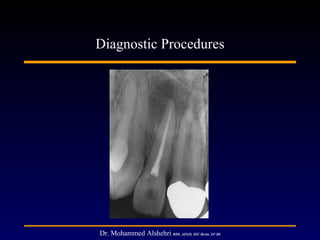
- 36. Radiographic Examination • Presence of caries that may involve or threat to involve the pulp. • May show the number, curve, length and width of root canals. • Presence of calcified materials in the pulp chamber or root canals. • Resorption of dentin • Thickening of PDL • Nature and extent of periapical and alveolar bone destruction
- 37. Periodontal Examination Consists use of a blunt calibrated probe to explore the integrity of gingival sulcus around each tooth. A significant pocket if present in the absence of periodontal disease it increases the probability of presence of vertical treatment. To distinguish disease of periodontal origin from pulp origin, thermal and EPT along with PDL probing are essential.
- 38. Periapical Lesions • Inflammation results in bone resorption, and creation of radiolucent lesion around apex.
- 39. Periapical lesion of endodontic origin • Usually have 4 characteristics : 1) Lamina dura of tooth socket is lost apically 2) The radiolucency remains at apex in radiographs made at different cone angles 3) Radiolucency tends to resemble a hanging drop 4) Tooth has a necrotic pulp
- 40. Pulpal lesion of endodontic origin • Usually have : 1) Altered pulp space enlargement- internal resorption 2) Diffuse calcification in chamber 3) Pulp stones in pulp chamber 4) Canal calcifications
- 41. Additional Diagnostic Tests Caries Removal • Determining the depth of caries penetration is important in pulp diagnosis. • Exposure by soft caries is irreversible pulpitis.
- 42. Anesthetic Test • Performed when usual tests have failed to enable one to identify the tooth. • Objective is to anaesthetize a single tooth at a time until the pain disappears and is localized to specific tooth.
- 43. Transillumination Test • Light from fibre optic is applied from buccal surface to illuminate the tooth to detect fractured lines when present.
- 44. Indicators for difficult diagnosis 1) Patient cannot localize the pain 2) No local dental cause for the pain 3) Pain is spontaneous or intermittent and not related to an initiating stimulus 4) Stimulation of suspected tooth does not produce the symptoms 5) Suspected tooth shows no clear etiology 6) When more than one tooth involved 7) Symptoms are bilateral 8) Selective anesthesia fails to localize the source of pain.