SKULL BASE IMAGING
•Download as PPT, PDF•
39 likes•13,563 views

IMAGING OF SKULL BASE

Recommended
Recommended
More Related Content
What's hot
What's hot (20)
Viewers also liked
Similar to SKULL BASE IMAGING
Similar to SKULL BASE IMAGING (20)
More from Stalinsurgeon Joseph Antonymuthu
More from Stalinsurgeon Joseph Antonymuthu (9)
Recently uploaded
Recently uploaded (20)
SKULL BASE IMAGING
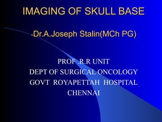
- 1. IMAGING OF SKULL BASEIMAGING OF SKULL BASE --Dr.A.Joseph Stalin(MCh PG)Dr.A.Joseph Stalin(MCh PG) PROF .R.R UNIT DEPT OF SURGICAL ONCOLOGY GOVT ROYAPETTAH HOSPITAL CHENNAI
- 2. CONTENTSCONTENTS 1.REVIEW OF ANATOMY 2.ROLE OF IMAGING 3.IMPORTANT PATHOLOGICAL LEISIONS IN IMAGING 4.CLINICAL EXAMPLES
- 4. Skull Base Anatomy Review Temporal Bone Temporal bone- petrous portion Sphenoid Bone Occipital Bone Key Fissures • Petrosphenoidal fissure • Petrooccipital fissure Key Sutures • Sphenosquamous Suture • Occipitomastoid Suture
- 5. Skull Base Anatomy Review Key Openings • Foramen spinosum • Foramen ovale • Foramen lacerum • Foramen rotundum • Foramen magnum • Foramen of vesalius • Jugular foramen • Superior orbital fissure • Inferior orbital fissure • Optic canal • Vidian canal • Hypoglossal canal • Pterygopalatine fossa
- 6. Skull Base Anatomy Review
- 7. Skull Base Anatomy Review Foramen spinosum Sphenoid spine- lower level Foramen rotundum- higher level Pterygopalatine fossa Foramen ovale Petro-occipital fissure Pterygoid canal f. lacerum
- 9. ROLE OF IMAGINGROLE OF IMAGING Diagnosis Deciding Resectability Planning of Treatment- Approach Specialist Help Reconstruction Follow up/Recurrance
- 10. DiagnosisDiagnosis Site Extend Consistency Vascularity Bony Involvement Perineural spread Vascular Involvement
- 11. Characterisation of the lesionCharacterisation of the lesion Morphology 1. tissue characterisation 2. pattern of bone involvment 3. vascularity Localisation 1. intrinsic to the skull base 2. arising from intracranial compartment 3. arising from extracranial head and neck Invasion of other structures 1. Direct extension • infiltrating bone, soft tissue, meninges, cerebrum • preformed channels and foramina 2. Hematogenous spread 3. Perineural spread
- 12. Agressive bone involvement patternAgressive bone involvement pattern Osteolysis Absent bone replaced by soft tissue Thinned bone with soft tissue mass on its both sides Abnormal signal of the bone marrow Calcifications within the soft tissue mass
- 13. Non-aggressive bone involvement patternNon-aggressive bone involvement pattern Bone remodeling with bowing, thin or demineralized walls Bone expansion with smooth contour or interrupted walls Enlarged intramedullary cavity Varying attenuation: ground-glass, radiolucent or sclerotic
- 14. INTRACRANIAL <> EXTRA CRANIALINTRACRANIAL <> EXTRA CRANIAL Pharygeal mucosal space PMS Sinus Morgagni Parapharyngeal space PPS Skull base Carotid space CS Carotid canal Jugular foramen Mandibular space MS Foramen ovale Retropharyngeal space RPS Basiocciput
- 15. Sinus frontalis Squamous CellSinus frontalis Squamous Cell Cancer with intracranialCancer with intracranial spreadspread Nodular dural enhancing have high specificity Dural thickness > 5 mm Coexistent leptomeningeal enhancement Hypointense leision Brain parenchymal changes
- 16. Perineural spreadPerineural spread Nerve enlargement and nerve enhancement Obliteration of the fat in the foramina, fosse or fissures Foraminal enlargement or destruction Enhancing soft tissue in the cavernous sinus and Meckel cave Neuropathic atrophy and fat replacement Tumor growthTumor growth Incresed permeability of endoneurial capillariesIncresed permeability of endoneurial capillaries Rupture of the blood-nerve barrierRupture of the blood-nerve barrier Contrast-enhancementContrast-enhancement
- 18. Ethmoidal Adenocarcinoma withEthmoidal Adenocarcinoma with perineural spread in pterigopalatine fossaperineural spread in pterigopalatine fossa
- 19. Cavernous sinus infiltrationCavernous sinus infiltration
- 20. Internal carotid arteryInternal carotid artery encasementencasement
- 21. ABOX CTABOX CT
- 22. ABOX-CTABOX-CT
- 23. Imaging ChecklistImaging Checklist Bony involvement- site/extension Scan all FORAMINA- content involvement SA plane/Dural/Brain involvement Carotid Sinus/other sinuses Internal Carotid Artery course/encasement Perineural spread
- 24. CRITERIA FOR NONCRITERIA FOR NON RESECTABILITYRESECTABILITY Cavernous sinus infiltration B/l optic nerve/optic chiasmal infiltration Sphenoid sinus infiltration (superior/lateral ) Extensive brain involvement- temporal lobe for anterior resection
- 25. Skull Base Pathology Chordoma Chondrosarcoma Dermoid tumors Epidermoid tumors Glomus tumors Meningioma Metastases Myeloma Neuroma Schwannoma Vascular Aneurysm
- 26. ANTERIOR SKULL BASEANTERIOR SKULL BASE MENINGIOMA SINONASAL MALIGNANCY OLFACTORY NEUROBLASTOMA
- 27. MIDDLE SKULL BASEMIDDLE SKULL BASE Pituitary adenoma Craniopharyngioma Sphenoid sinus malignancy Schwanoma
- 28. POSTERIOR SKULL BASEPOSTERIOR SKULL BASE Chordoma Acoustic neuroma Chondrosarcoma Paraganglioma
- 31. Bone marrow spaceBone marrow space
- 33. orbitorbit
- 35. Normal ethamoid sinusNormal ethamoid sinus
- 38. Anterior cranial fossa tumourAnterior cranial fossa tumour with dural involvementwith dural involvement
- 41. Cavernous sinus infiltrationCavernous sinus infiltration
- 43. Case 1
- 44. Chondrosarcoma CT Findings: • Irregular, destructive mass • Centered off midline • Petro-occipital fissure • Calcifications, 70%; “rings/arcs” MRI Findings: • Low T1 signal, high T2 signal • Enhance with contrast • Scalloped, well circumsribed margins
- 45. Chondrosarcoma Origin: • Preexisting cartilaginous lesion, synchondroses, cartilage endplates Location: • Paranasal sinuses, skull base, parasellar region • Long bones, pelvis, sternum, ribs Clinical: • 45 yo, median age • Classic, mesenchymal, or dedifferentiated
- 46. Case 2
- 47. CT/MRI Findings: • Expansile lytic lesion, midline • Well delineated mass arising from bone • Large soft tissue component • Variable calcification • Anteroposterior extension • Heterogeneous enhancement on T1, T2 • Dark on T1, bright on T2 Chordoma Differential Diagnosis: • Chondroma • Chondrosarcoma • Clivus meningioma
- 48. Chordoma Origin • Notochord remnants Location • Clivus 35% • Sacrum 50%, Vertebral bodies 15% Clinical • age 30-70 • Slow growing, locally aggressive • CN VI- CN deficits • Mets late • Tx: surgery, radiation
- 49. Case 3
- 50. Glomus Tumor Glomus jugulare CT/MRI Findings: • Center: jugular foramen • Limit: hyoid bone • Enhance w/ contrast • Salt and pepper appearance on MRI • Bone erosion
- 51. Glomus Tumor Origin: • Chemoreceptor cells Location: • 10% multiple • glomus jugulare: jugular bulb • glomus tympanicum: cochlear promontory Clinical: • Pulsatile tinnitus • Hearing loss • arrythmia, BP fluctuation
- 52. CONCLUSIONCONCLUSION Thorough anatomical knowledge essential. Both CT and MRI are needed. Histological diagnosis not needed for managing skull base tumours. Main role of imaging is to plan the recection . Treatment options for skull base tumours – resection +/_ radiotherapy
- 53. CONCLUSIONCONCLUSION Anatomy of skull base – complex – not the imaging Treatment options –simple- not the procedure
Editor's Notes
- Complex area Keep it simple
- Guidance for geography Don’t mistake for fx
- 7 foramina 2 fissures 3 canals All with fun stuff inside.
- Hypoglossal artery = not always present. Sometimes, can have a small emissary vein that runs through here that can protrude into the cerebellomedullary cistern and mimic a nerve sheath tumor.
- 2 parts: Pars nervosa = anteromedial Pars vascularis aka pars venosa = posterolateral Separated by the jugular spur. Pars nervosa: glossopharyngeal nerve, inferior petrosal sinus- runs along petrooccipital fissure Pars vascularis: vagus and spinal accessory nerve Jugular bulb: confluence b/w sigmoid sinus and internal jugular vein. Anterolateral to pars nervosa = petrous portion of the carotid artery.
- F.S.: V3 recurrent = mandibular branch. Emissary veins in FS and FO connect cavernous sinus with pterygoid plexus of veins = path for nasaopharyngeal tumors. Foramen of vesalius- inconstant. Anterior and medial to f. ovale. When it does occur, it contains (read slide)
- F.L.:read slide Vidian canal = aka pterygoid canal. Connects pterygopalatine fossa to foramen lacerum. Contains vidian artery (branch of maxillary artery) and nerve. Vidian nerve = formed by merger of greater superficial petrosal nerve (branch of facial nerve) and deep petrosal nerve.
- PPF = conduit for spread of tumor and infection Communicates with inf orbital fissure, orbital apex,… PPF - Sphenoplatine foramen = to nasal fossa PPF – pterygomaxillary fissure = to masticator space PPF- foramen rotundum = connection with Meckel’s cave, cavernous sinus PPF- Vidian canal = to foramen lacerum PPF- to greater/lesser palatine canals = to palate
- PPF – foramen rotundum = connection with meckel’s cave, cav sinus, since we’ve mentioned it a few times now and b/c contains a lot of key elements: Cav sinus- read slide. V2- lateral wall of CS- then to foramen rotundum V1- lateral wall of cav sinus- then to superior orbital fissure, along with CN III, IV, VI
- Speaking of the superior orbital fissure… Below SOF = IOF. Optic canal = more superior to SOF.
- Pitfalls calcified debris in fungal sinuitis and flow voids associated with aggressive rapidly growing tumors , malignant neoplasms or infectious process( osteocorsoma, osteosarcoma and metastases)
- High T2 signal and contrast enhancement meaning brain vasogen edema ; ddif linerar pattern of enhancemet rep reactiv dural changes of hyperemia, hypertrofia and inflammation
- Adenoid cystic carcinoma, squamos cell carcinoma but even lymfoma, melanoma desmoid type basal cell ca rhabdomyosarcoma neurogenic tum juv angifibroma Gd T1 and high resolusion T2 w ; Gd fs t1
- etmoidal sinus cancer
- Chordomas like the clivus Chondrosarcomas like the cartilaginous endplates- petrooccipital suture Glomus tumors- jugular bulb, middle ear, carotid body Meningiomas- most common (20% of all brain tumors). But most = cerebral convexity, along and lateral to the falx. 10% clivus 20% sphenoid ridge Schwannomas almost always develop from sensory nerves. Because the olfactory and optic nerves do not have a Schwann cell layer, they do not develop these tumors. The most common intracranial schwannoma = acoustic neuroma- it develops from the vestibular nerve. Bilateral acoustic neuromas = pathogomonic for NF2. (90% of the time, schwannomas are solitary). The second most common intracranial schwannomas develop from the trigeminal nerve. Trigeminal schwannomas usually arise from the root or ganglion and occupy the middle fossa and, sometimes, the posterior fossa. Schwannomas of the other cranial nerves are rare.
- T1 post contrast MR showing extraaxial lesion arising from the middle cranial fossa. Heterogeneous enhancement. Low signal areas = flow voids or calcs. Coronal = involvement with skull base. Mass effect on temporal lobe.
- Chondrosarcomas can occur anywhere in the skeletal system. Often = preexisting cartilaginous lesion like previously benign osteochondroma. In skull base = usually at cartilage endplates. E.g. petrooccipital suture. Location: read slide Clinical: Most commonly, patients are diagnosed with chondrosarcomas during the third or fourth decade of life. Males are affected more often than females. Chondrosarcomas can be divided into classic, mesenchymal, and dedifferentiated tumors. Mesenchymal, Dedifferentiated = high grade. Classic low grade = like chordoma. DDX: Chordoma- usually has marked bone destruction, midline (clivus) Chondrosarcoma = significant soft tissue component, little bone destruction, arcs/nodules of calcification, eccentric locations- often centered in framen lacerum.
- Sagittal T1-weighted MR image shows a large, hypointense soft-tissue mass that arises from the distal clivus with anterior extension into the nasopharynx (arrows) and extradural extension into the posterior fossa (arrowhead).
- CT to assess degree of bone involvement. MRI to evaluate extension of tumor. CT Findings: The most characteristic appearance of intracranial chordoma is of a centrally located homogeneous soft tissue mass arising from the clivus and causing adjacent bone destruction. Calcification is common but variable. Areas of low attenuation within the soft tissue mass occasionally are found on CT, representing the myxoid and gelatinous material found on pathologic examination. CT reliably demonstrates petrous apex involvement and lysis of the skull base foramina. MRI Findings: Mass originating from midline with extension primarily in the anteroposterior axis rather than laterally. Well delineated. Expands bone in the early stage = indicator that it arises from bone, not from adjacent structures. Post gad = lobulated area, heterogeneous on T1 and T2 b/c of mucinous and gelatinous contents. DDX: Chondroma- similar appearance but extend more laterally into sellar and cerebellopontine angles. Clivus meningioma – homegeneous signal, dural attachment
- Contrast enhanced T1 spin echo image. Chordoma of the upper part of the clivus with posterior extension into the pontine cistern. Chordomas = benign tumor but has significant problems b/c of location, local invasion, recurrence. Origin: Notochord = early fetal axial skeleton. Gets surrounded by cartilage. Cartilage ossifies and notochord = squeezed out into intervertebral regions = nucleus pulposus of intervertebral disks. Can get remnants then, along any position of the neural axis- turn into chordomas. Location: read slide. Rare. &lt;0.2% of all intracranial tumors. Clinical:read slide CN deficits: HA, dysphagia, facial pain, facial paresis, visual loss, hearing loss, and ataxia. CT to assess degree of bone involvement. MRI to evaluate extension of tumor. CT Findings: The most characteristic appearance of intracranial chordoma is of a centrally located homogeneous soft tissue mass arising from the clivus and causing adjacent bone destruction. Calcification is common but variable. Areas of low attenuation within the soft tissue mass occasionally are found on CT, representing the myxoid and gelatinous material found on pathologic examination. CT reliably demonstrates petrous apex involvement and lysis of the skull base foramina. MRI Findings: Mass originating from midline with extension primarily in the anteroposterior axis rather than laterally. Well delineated. Expands bone in the early stage = indicator that it arises from bone, not from adjacent structures. Post gad = lobulated area, heterogeneous on T1 and T2 b/c of mucinous and gelatinous contents.
- CT imaging demonstrates the extent of bony destruction (white and black arrows) by the tumor. The normal jugular foramen on the left (arrow head) is shown for comparison.
- Salt and pepper: multiple low signal intensity areas = flow voids in tumor. When large- erode bone.
- Glomus tumors arise from chemoreceptor cells. These tumors are slow-growing hypervascular tumors that usually occur in the temporal bone. Location: read slide- check other places for them b/c = multiple. E.g. Carotid body Patients usually present with gradual hearing loss, unilateral pulsatile tinnitus, and lower cranial nerve palsies. Approximately 1-3% of gangliogliomas produce catecholamines, so can get arrythmia, BP fluctuation. May be locally invasive but rarely metastasize.