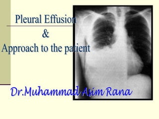
Approach to pleural effusion
•Download as PPSX, PDF•
327 likes•59,311 views

Apparently a lengthy presentation actually very good for junior physicians as it covers all aspects of assessment, diagnosis and treatment of pleural effusion

Recommended
More Related Content
What's hot
What's hot (20)
Viewers also liked
Similar to Approach to pleural effusion
Similar to Approach to pleural effusion (20)
More from Muhammad Asim Rana
More from Muhammad Asim Rana (20)
Recently uploaded
Recently uploaded (20)
Approach to pleural effusion
- 2. Definition Pleural effusion is the accumulation of fluid in the pleural space. The pleural space lies between the lung and chest wall and normally contains a very thin layer of fluid, which serves as a coupling system. A pleural effusion is present when there is an excess quantity of fluid in the pleural space.
- 3. Etiology Normally, fluid enters the pleural space from the capillaries in the parietal pleura and is removed via the lymphatics situated in the parietal pleura. Fluid can also enter the pleural space from the interstitial spaces of the lung via the visceral pleura or from the peritoneal cavity via small holes in the diaphragm.
- 4. Pleural fluid accumulates when pleural fluid formation exceeds pleural fluid absorption. The lymphatics have the capacity to absorb 20 times more fluid than is normally formed. Accordingly, a pleural effusion may develop when there is excess pleural fluid formation (from the interstitial spaces of the lung, the parietal pleura, or the peritoneal cavity) or when there is decreased fluid removal by the lymphatics.
- 5. Types of pleural effusion
- 6. Transudative pleural effusions result from alteration of hydrostatic and oncotic factors that increase the formation or decrease the absorption of pleural fluid (e.g., increased mean capillary pressure [heart failure] or decreased oncotic pressure [cirrhosis or nephrotic syndrome]).
- 7. Exudative pleural effusions occur when damage or disruption of the normal pleural membranes or vasculature (e.g., tumor involvement of the pleural space, infection, inflammatory conditions, or trauma) leads to increased capillary permeability or decreased lymphatic drainage.
- 9. Clinical Presentation The underlying cause of the effusion usually dictates the symptoms, although patients may be asymptomatic. Pleural inflammation, abnormal pulmonary mechanics, and worsened alveolar gas exchange produce symptoms and signs of disease.
- 10. symptoms and signs Inflammation of the parietal pleura leads to pain in local (intercostal) involved areas or referred (phrenic) distributions (shoulder). Dyspnea is frequent and may be present and out of proportion to the size of the effusion. Cough can occur.
- 11. Chest examination is notable for dullness to percussion, decreased or absent tactile fremitus, and decreased breath sounds. Tracheal shift to the contralateral side or an ipsilateral pleural rub may be present.
- 12. The clinical setting is crucial to establishing a proper diagnosis. A definitive diagnosis based solely upon pleural fluid analysis is possible in the minority of pleural effusions. History or physical examination findings suggestive of congestive heart failure, malignancy, pneumonia, pulmonary embolism, myocardial infarction, surgery, cirrhosis, or rheumatologic arthritis provide important clues to the underlying diagnosis.
- 14. Chest Roentginogram Pleural effusions are typically detected by chest radiography as blunting of the costophrenic angle or opacification of the base of the hemithorax without loss of volume of the hemithorax (which would suggest atelectasis), and may be accompanied by air bronchograms (which would suggest an alveolar filling process such as pneumonia).
- 15. Prior to invasive diagnostic or therapeutic procedures, the patient should undergo imaging to confirm the presence and size of the effusion. Preferred modalities include:
- 16. Decubitus chest radiography Showing layering fluid will confirm the presence of pleural effusion and demonstrates that at least a portion of the fluid is not loculated.
- 17. Thoracic ultrasonography Is one of the best modalities to assess for pleural fluid loculations. Ultrasonography can also provide real-time guidance for pleural procedures and can reduce both the complication and failure rate of thoracentesis.
- 18. Computed tomography of the chest With contrast helps differentiate pleural fluid from lung masses and atelectatic lung, and helps define the extent of pleural thickening, pleural nodularity, and other associated findings.
- 19. Pleural fluid analysis Thoracentesis can be performed safely at the bedside, in the absence of disorders of hemostasis, on effusions that extend >10 mm from the inner chest wall on a lateral decubitus film. Loculated effusions can be localized with ultrasonography or CT scan. Proper technique and sonographic guidance minimize the risk of pneumothorax and other complications.
- 20. The first step is to determine whether the effusion is a transudate or an exudate.
- 21. A transudative pleural effusion occurs when systemic factors that influence the formation and absorption of pleural fluid are altered. The leading causes of transudative pleural effusions are left ventricular failure and cirrhosis.
- 22. An exudative pleural effusion occurs when local factors that influence the formation and absorption of pleural fluid are altered. The leading causes of exudative pleural effusions are bacterial pneumonia, malignancy, viral infection, and pulmonary embolism.
- 23. The primary reason to make this differentiation is that additional diagnostic procedures are indicated with exudative effusions to define the cause of the local disease.
- 24. While pleural effusion occurs in a vast array of disease states, 90% of pleural effusions are the result of only five diseases. Congestive heart failure (36%) Pneumonia (22%) Malignancy (14%) Pulmonary embolism (11%) Viral disease (7%)
- 25. Check pleural fluid for Appearance, lactate dehydrogenase (LDH), protein, pH, glucose and albumin
- 26. Serum lactate dehydrogenase (LDH), protein, pH, glucose and albumin should be measured within hour of the thoracentesis to allow appropriate comparison.
- 27. Pleural fluid appearance Most transudates are clear, straw colored, nonviscid, and without odor Red-tinged pleural effusions indicate the presence of blood. In exudative pleural effusions, serosanguineous fluid is usually not helpful in narrowing the diagnosis.
- 28. Bloody pleural fluid If the blood is due to thoracentesis, the degree of discoloration should clear during the aspiration. Bloody pleural fluid usually indicates the presence of malignancy, pulmonary embolism (PE), or trauma.
- 29. Hemothorax The presence of gross blood should lead to the measurement of a pleural fluid hematocrit. Hemothorax is defined as a pleural fluid to blood hematocrit ratio of >0.5, and chest tube drainage should be considered.
- 30. Exudative pleural effusions meet at least one of the Light's criteria , whereas transudative pleural effusions meet none:
- 31. Light's criteria (a) a pleural fluid-to-serum protein ratio of >0.5, (b) a pleural fluid-to-serum LDH ratio of >0.6, (c) a pleural fluid LDH of more than two-thirds of the upper limit of normal for serum LDH
- 32. The above criteria misidentify ~25% of transudates as exudates. If one or more of the exudative criteria are met and the patient is clinically thought to have a condition producing a transudative effusion, like in whom clinical suspicion for heart, liver, or kidney disease is high what should be done?
- 33. The difference between the protein levels in the serum and the pleural fluid should be measured. If this gradient is greater than 31 g/L (3.1 g/dL), the exudative categorization by the above criteria can be ignored because almost all such patients have a transudative pleural effusion. In some texts a gradient of >1.2 g/dL suggests that the pleural fluid is transudate.
- 34. If a patient has an exudative pleural effusion Description of the fluid, Glucose level, Differential cell count, Microbiologic studies, Cytology. Cultures, Triglycerides, Amylase, and pH
- 35. WBC differential The WBC differential is often not diagnostic, although neutrophilia is suggestive of infection.
- 36. Eosinophilia (>10% of total nucleated cell count) is suggestive of air or blood in the pleural space. If air or blood is not present in the pleural space, consideration should be given to fungal and parasitic infection, druginduced disease, PE, asbestos-related disease, and Churg-Strauss syndrome.
- 37. Lymphocytosis (>50% of the total nucleated cell count) is suggestive of malignancy or tuberculosis. Mesothelial cells argues against the diagnosis of tuberculosis. Plasma cells suggest a diagnosis of multiple myeloma.
- 38. Exudative effusions with normal protein but high LDH are likely to be parapneumonic or secondary to malignancy. LDH is an indicator of the degree of pleural inflammation.
- 39. Glucose concentration A glucose concentration of <60 mg/dL is probably due to tuberculosis, malignancy, rheumatoid arthritis, or parapneumonic effusion. For parapneumonic pleural effusions with a glucose of <60 mg/dL, tube thoracostomy should be considered.
- 40. Pleural fluid with a low pH A pH of <7.3 is seen with empyema, tuberculosis, malignancy, collagen vascular disease, or esophageal rupture.
- 41. For parapneumonic pleural effusions with a pH of <7.20, tube thoracostomy should be considered. Pleural fluid for pH testing should be collected anaerobically in a heparinized syringe and placed on ice. Pleural fluid with a low pH usually has a low glucose and a high LDH; otherwise, the low pH may be due to poor sample collection technique.
- 42. Amylase An elevation of amylase suggests that the patient has pancreatic disease, malignancy, or esophageal rupture. Malignancy and esophageal rupture have salivary amylase elevations and not pancreatic amylase elevations.
- 43. Turbid or milky fluid should be centrifuged. If the supernatant clears, the cloudiness is likely due to cells and debris. If the supernatant remains turbid, pleural lipids should be measured. Elevation of triglycerides (>110 mg/dL) suggests that a chylothorax is present, usually due to disruption of the thoracic duct from trauma, surgery, or malignancy (i.e., lymphoma).
- 44. Cytology Cytology is positive in approximately 60% of malignant effusions. Priming the fluid collection bag with unfractionated heparin (UFH; e.g., 1,000 International Units) may increase the yield. The volume of pleural fluid analyzed does not impact the yield of cytologic diagnosis. Repeat thoracentesis increases the diagnostic yield.
- 46. Closed pleural biopsy Closed pleural biopsy adds little to the diagnostic yield of thoracentesis, except in the diagnosis of tuberculosis. For tuberculous effusions, pleural fluid cultures alone are positive in only 20% to 25% of cases. However, the combination of pleural fluid studies and pleural biopsy (demonstrating granulomas or organisms) is 90% sensitive in establishing tuberculosis as the etiology of the effusion.
- 47. Diagnostic thoracoscopy Diagnostic thoracoscopy has largely replaced closed pleural biopsy. Thoracoscopy allows directed biopsies that increase the diagnostic yield for malignancy while maintaining the high diagnostic yield of closed pleural biopsy for TB.
- 48. Indications for diagnostic thoracoscopy Pleural effusion of unknown etiology Mesothelioma Lung cancer Tuberculosis Other benign pleural disorders Pulmonary parenchymal disease
- 50. Other diagnostic procedures that are useful in establishing the etiology of a pleural effusion when the aforementioned tests are nondiagnostic include: Evaluation of liver function Renal function, Cardiac echo, Biopsy of other abnormal sites (e.g., a mediastinal or lung mass), and Evaluation of PE.
- 53. Differential Diagnoses of Pleural Effusions
- 54. Transudative Pleural Effusions 1. Congestive heart failure 2. Cirrhosis 3. Pulmonary embolization 4. Nephrotic syndrome 5. Peritoneal dialysis 6. Superior vena cava obstruction 7. Myxedema 8. Urinothorax
- 55. Exudative Pleural Effusions 1. Neoplastic diseases a. Metastatic disease b. Mesothelioma
- 56. 2. Infectious diseases a. Bacterial infections b. Tuberculosis c. Fungal infections d. Viral infections e. Parasitic infections
- 57. 3. Pulmonary embolization 4. Gastrointestinal disease a. Esophageal perforation b. Pancreatic disease c. Intraabdominal abscesses d. Diaphragmatic hernia e. After abdominal surgery f. Endoscopic variceal sclerotherapy g. After liver transplant
- 58. 5. Collagen-vascular diseases a. Rheumatoid pleuritis b. Systemic lupus erythematosus c. Drug-induced lupus d. Immunoblastic lymphadenopathy e. Sjögren's syndrome f. Wegener's granulomatosis g. Churg-Strauss syndrome
- 59. 6. Post-coronary artery bypass surgery 7. Asbestos exposure 8. Sarcoidosis 9. Uremia 10. Meigs' syndrome 11. Yellow nail syndrome
- 60. 12. Drug-induced pleural disease a. Nitrofurantoin b. Dantrolene c. Methysergide d. Bromocriptine e. Procarbazine f. Amiodarone
- 61. 13. Trapped lung 14. Radiation therapy 15. Post-cardiac injury syndrome 16. Hemothorax
- 62. 17. Iatrogenic injury 18. Ovarian hyperstimulation syndrome 19. Pericardial disease 20. Chylothorax
- 63. "Classic" exudates that can be transudates Malignancy: Due to early lymphatic obstruction, obstructive atelectasis, or concomitant disease (CHF). Pulmonary embolism: 23 percent incidence; due to atelectasis. Sarcoidosis: Stage II and III disease. Hypothyroid pleural effusion: From hypothyroid heart disease or hypothyroidism per se.
- 64. Treatment
- 65. Transudates resolve with treatment of the underlying heart, kidney, or liver disease. Uncommonly, more aggressive approaches including pleurodesis and shunts are required.
- 66. Parapneumonic effusions and empyema should be managed with tube drainage when indicated based on the size, gross appearance, or biochemical analysis of the pleural fluid or the presence of loculations Multiple tubes are sometimes required to adequately drain the pleural space.
- 67. Failure to adequately and quickly drain a complicated parapneumonic effusion can lead to organization of the pleural fluid and formation of a thick pleural adhesions which may necessitate surgical removal known as decortication.
- 68. Indications for Tube Thoracostomy in Parapneumonic Effusions
- 69. Radiographic criteria Pleural fluid loculations Effusion filling more than half the hemithorax Air fluid level
- 70. Microbiologic criteria Pus in the pleural space Positive stain for microorganisms Positive pleural fluid cultures
- 71. Chemical criteria Pleural fluid pH <7.2 Pleural fluid glucose <60 mg/dL
- 72. CONTRAINDICATIONS There are no absolute contraindications to tube thoracostomy, particularly if the patient is in respiratory distress. Anticoagulation or a bleeding diathesis is a relative contraindication in a patient undergoing elective chest tube placement for pleurodesis. Blind insertion of a chest tube is dangerous in a patient with adhesions from infection, previous pleurodesis, or a lung transplant; guidance by CT scan without contrast is preferred in these patients.
- 73. Type of tube Silastic® tubes are preferred because older rubber tubes have fewer drainage holes, are not well visualized on chest radiographs, and produce more pleural inflammation. Silastic chest tubes contain a radiopaque strip with a gap that serves to mark the most proximal drainage hole.
- 74. Size of tube A chest tube's internal diameter and length are the critical determinants of flow. Select the appropriate chest tube size to account for the viscosity and accumulation rate of the pleural material to be drained. As an example, drainage of viscous fluids requires a larger bore chest tube than that required for drainage of a similar volume of air.
- 75. Malignant effusion— A small-bore catheter (8 to 14 Fr) placed under ultrasound or CT guidance is usually adequate to drain a malignant pleural effusion and achieve pleurodesis. Empyema — For a complicated parapneumonic effusion or empyema that is amenable to drainage with a single catheter. Prefer initial image-guided placement of small-bore catheters (10 to 14 Fr), with or without intrapleural fibrinolytic agents.
- 76. It is preferred to use the smaller tube size as this is generally more comfortable for patients, particularly if more than one tube is needed. Alternatively, when the fluid appears viscous, a larger bore tube (16-24 Fr) may be used.
- 77. Unsuccessful drainage with a small-bore catheter either indicates the presence of multiple loculations or very viscous material. Multiple small-bore catheters may be used in multiloculated effusions or large bore catheters in case of very viscous material. Failure to drain with a single small-bore tube should also lead to thoracic surgery consultation to avoid delays in case video assisted thoracoscopy (VATS) becomes necessary.
- 78. Hemothorax — The goals of tube thoracostomy in acute hemothorax are drainage of fresh blood, quantification of the rate of bleeding, evacuation of any coexisting pneumothorax, and tamponade of the bleeding site. Large bore catheters (32 to 40 Fr) are required to reliably achieve these goals.
- 79. Once a hemothorax is defibrinated in situ, that is after the acute phase, success of drainage is less dependent on the size of the tube, than on the degree and mode of clot formation. Large amounts of clotted blood should be evacuated via video assisted thoracoscopy.
- 80. Occasionally, a hemothorax may result in a sonographically complex septate pattern and may be treated with small-bore catheters. Treatment of hemothorax should be individualized and done in consultation with thoracic surgery.
- 81. Malignant pleural effusions Observation without invasive interventions may be appropriate for some patients with malignant pleural effusions. Therapeutic thoracentesis may improve patient comfort and relieve dyspnea. The rapid removal of more than 1 L of pleural fluid may rarely result in re-expansion pulmonary edema, especially if the lung is unable to reexpand.
- 82. Repeated thoracenteses are reasonable if they achieve symptomatic relief and if fluid reaccumulation is slow. Unfortunately, 95% of malignant effusions will recur with a median time to recurrence of less than a week. When frequent or repeated thoracentesis is required for effusions that reaccumulate, early consideration should be given to tube drainage with pleurodesis or placement of a chronic indwelling pleural catheter.
- 83. Chemical pleurodesis Chemical pleurodesis is an effective therapy for recurrent effusions. This treatment is recommended in patients whose symptoms are relieved with initial drainage but who have rapid reaccumulation of fluid.
- 84. Talc pleurodesis Effective and inexpensive. Fever and hypoxia are common following instillation of talc into the pleural space, and respiratory failure has been described on occasion. Overall efficacy is similar for talc slurry delivered via chest tube versus dry talc insufflated during thoracoscopy.
- 85. Doxycycline or minocycline Doxycycline or minocycline can also be instilled into the pleural space via a chest tube. Pain is more prevalent and severe following doxycycline and minocycline than following talc.
- 86. Bleomycin Bleomycin appears to be less effective and more expensive than other drugs.
- 87. Systemic analgesics and the administration of lidocaine in the sclerosing agent solution help to decrease the appreciable discomfort associated with the procedure. If the chest tube drainage remains high (>100 mL/d) more than 2 days after the initial pleurodesis, a second dose of the sclerosing agent can be administered.
- 88. Chronic indwelling pleural catheters Provide good control of effusion-related symptoms via intermittent drainage. The Pleurx catheter is better at controlling symptoms than doxycycline administered via a chest tube. Furthermore, repeated drainage via a Pleurx catheter leads to pleurodesis in roughly 50% of patients, allowing the catheter to be removed.
- 89. Pleurectomy or pleural abrasion Requires thoracic surgery and should be reserved for patients with a good prognosis who have had ineffective pleurodesis or inadequate response to chronic pleural drainage.
- 90. Chemotherapy and mediastinal radiotherapy May control effusions in responsive tumors, such as lymphoma or small-cell bronchogenic carcinoma, although it has poor efficacy in metastatic carcinoma.
- 91. Thank you very much