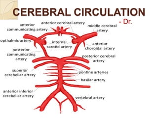
2. Cerebral Blood Flow
Normal blood flow through the brain of the adult person
averages 50 to 65 milliliters per 100 grams of brain tissue
per minute.
For the entire brain, this amounts to 750 to 900 ml/min, or
15 per cent of the resting cardiac output.
Three metabolic factors have potent effects in controlling
cerebral blood flow:
(1) carbon dioxide concentration,
(2) hydrogen ion concentration,
(3) oxygen concentration.
3. Cerebral Blood Flow
An increase in carbon dioxide concentration in the arterial blood
perfusing the brain greatly increases cerebral blood flow.
A 70 per cent increase in arterial PCO2 approximately doubles cerebral
blood flow.
Carbon dioxide is believed to increase cerebral blood flow by combining
first water in the body fluids to form carbonic acid, with subsequent
dissociation of this acid to form hydrogen ions.
The hydrogen ions then cause vasodilation of the cerebral vessels—the
dilation being almost directly proportional to the increase in hydrogen
ion concentration. Increased hydrogen ion concentration greatly
depresses neuronal activity.
Any other substance that increases the acidity of the brain tissue, and
therefore increases hydrogen ion concentration, will likewise increase
cerebral blood flow. Such substances include lactic acid, pyruvic acid,
and any other acidic material formed during the course of tissue
4.
5. Cerebral Blood Flow
Except during periods of intense brain activity, the rate of
utilization of oxygen by the brain tissue remains within
narrow limits—almost exactly 3.5 (± 0.2) ml of O2 /100 grams
of brain tissue / min.
If blood flow to the brain ever becomes insufficient to supply
this needed amount of oxygen, the oxygen deficiency
mechanism for causing vasodilation immediately causes
vasodilation, returning the brain blood flow and transport of
oxygen to the cerebral tissues to near normal.
Experiments have shown that a decrease in cerebral tissue
PO2 below about 30 mm Hg (normal value is 35 to 40 mm
Hg) immediately begins to increase cerebral blood flow.
6.
7. Autoregulation
Cerebral blood flow is ―autoregulated‖ extremely well
between arterial pressure limits of 60 and 140 mm Hg. That
is, mean arterial pressure can be decreased acutely to as
low as 60 mm Hg or increased to as high as 140 mm Hg
without significant change in cerebral blood flow.
And, in people who have hypertension, autoregulation of
cerebral blood flow occurs even when the mean arterial
pressure rises to as high as160 to 180 mm Hg.
If the arterial pressure falls below 60 mm Hg, cerebral blood
flow then becomes severely decreased.
8.
9. Role of Sympathetic
Transection of the sympathetic nerves or mild to moderate
stimulation of them usually causes very little change in
cerebral blood flow because the blood flow autoregulation
mechanism can override the nervous effects.
When mean arterial pressure rises acutely to an
exceptionally high level, such as during strenuous exercise
or during other states of excessive circulatory activity, the
sympathetic nervous system normally constricts the large
and intermediate sized brain arteries enough to prevent the
high pressure from reaching the smaller brain blood vessels.
This is important in preventing vascular hemorrhages into
the brain—that is, for preventing the occurrence of ―cerebral
stroke.‖
10. Cerebral Microcirculation
An important structural characteristic of the brain
capillaries is that they are much less ―leaky‖ than the
blood capillaries in almost any other tissue of the body.
Glial cells
The walls of the small arterioles leading to the brain
capillaries become greatly thickened in people who
develop high blood pressure, and these arterioles
remain significantly constricted all the time to prevent
transmission of the high pressure to the capillaries.
11. Cerebral “Stroke”
Most strokes are caused by arteriosclerotic plaques that
occur in one or more of the feeder arteries to the brain.
The plaques can activate the clotting mechanism of the
blood, causing a blood clot to occur and block blood flow in
the artery, thereby leading to acute loss of brain function in a
localized area.
In about one quarter of people who develop strokes, high
blood pressure makes one of the blood vessels burst;
hemorrhage then occurs, compressing the local brain tissue
and further compromising its functions.
The neurological effects of a stroke are determined by the
brain area affected.
12. Cerebral “Stroke”
One of the most common types of stroke is blockage of the
middle cerebral artery that supplies the midportion of one
brain hemisphere.
For instance, if the middle cerebral artery is blocked on the
left side of the brain, the person is likely to become almost
totally demented because of lost function in Wernicke’s
speech comprehension area in the left cerebral hemisphere.
He or she also becomes unable to speak words because of
loss of Broca’s motor area for word formation.
13. Cerebral “Stroke”
Loss of function of neural motor control areas of the left
hemisphere can create spastic paralysis of most muscles on
the opposite side of the body.
Blockage of a posterior cerebral artery will cause infarction of
the occipital pole of the hemisphere on the same side as the
blockage, which causes loss of vision in both eyes in the half
of the retina on the same side as the stroke lesion.
Strokes that involve the blood supply to the midbrain
because this can block nerve conduction in major pathways
between the brain and spinal cord, causing both sensory and
motor abnormalities.
14. Cerebro Spinal Fluid (CSF)
150 ml
Present in the ventricles of the brain, in the
cisterns around the outside of the brain, and in
the subarachnoid space around both the brain
and the spinal cord.
All these chambers are connected with one
another, and the pressure of the fluid is
maintained at a surprisingly constant level.
Monro kellie doctrine
15. Cerebro Spinal Fluid (CSF)
Cerebrospinal fluid is formed at a rate of about 500
milliliters each day.
About two thirds or more of this fluid originates as
secretion from the choroid plexuses in the four
ventricles, mainly in the two lateral ventricles.
Choroid plexus – lateral ventricles – foramen of
monro - 3rd ventricle - aqueduct of Sylvius – 4th
ventricle - two lateral foramina of Luschka and a
midline foramen of Magendie – cisterna magna –
subarachnoid space
Any extra fluid empties into the venous blood through
16.
17. Secretion by the Choroid Plexus
Secretion of fluid into the ventricles by the choroid plexus
depends mainly on active transport of sodium ions through the
epithelial cells lining the outside of the plexus.
The sodium ions in turn pull along large amounts of chloride ions
as well because the positive charge of the sodium ion attracts the
chloride ion’s negative charge.
The two of these together increase the quantity of osmotically
active sodium chloride in the CSF, which then causes almost
immediate osmosis of water through the membrane, thus
providing the fluid of the secretion.
Less important transport processes move small amounts of
glucose into the cerebrospinal fluid and both potassium and
bicarbonate ions out of the cerebrospinal fluid into the capillaries.
18. Secretion by the Choroid Plexus
Osmotic pressure, approximately equal to that of
plasma;
Sodium ion concentration, also approximately equal to
that of plasma;
Chloride ion, about 15 per cent greater than in plasma;
Potassium ion, approximately 40 per cent less;
Glucose, about 30 per cent less,
19. Absorption by the Arachnoidal Villi
The endothelial cells covering the arachnoid villi have
vesicular passages directly through the bodies of the cells
large enough to allow relatively free flow of
(1)
Cerebrospinal fluid,
(2)
Dissolved protein molecules,
(3)
Particles as large as red and white blood cells
into the venous blood.
20.
21. Cushioning Function
A major function of the cerebrospinal fluid is
to cushion the brain within its solid vault.
The brain simply floats in the fluid.
Therefore, a blow to the head, if it is not too
intense,
moves
the
entire
brain
simultaneously with the skull, causing no
one portion of the brain to be momentarily
distorted by the blow.
22. Countercoup
When a blow to the head is extremely severe, it may not
damage the brain on the side of the head where the blow is
struck but on the opposite side. This phenomenon is known
as ―contrecoup,‖
When the blow is struck, the fluid on the struck side is so
incompressible that as the skull moves, the fluid pushes the
brain at the same time in unison with the skull.
When the skull is no longer being accelerated by the blow,
the brain strikes the inner surface of the skull.
If the contusion occurs on the same side as the impact injury,
it is a coup injury; if it occurs on the opposite side, the
contusion is a countercoup injury.
23. Cerebrospinal Fluid Pressure
The normal pressure in the cerebrospinal fluid system when one
is lying in a horizontal position averages
130 millimeters of water (10 mm Hg),
although this may be as low as 65 millimeters of water or as high
as 195 millimeters of water even in the normal healthy person.
The normal rate of cerebrospinal fluid formation remains very
nearly constant.
The arachnoidal villi function like ―valves‖ that allow cerebrospinal
fluid and its contents to flow readily into the blood of the venous
sinuses while not allowing blood to flow backward in the opposite
24. Cerebrospinal Fluid Pressure
Normally, this valve action of the villi allows cerebrospinal fluid to
begin to flow into the blood when cerebrospinal fluid pressure is
about 1.5 mm Hg greater than the pressure of the blood in the
venous sinuses.
Then, if the cerebrospinal fluid pressure rises still higher, the
valves open
more widely, so that under normal conditions, the cerebrospinal
fluid pressure almost never rises more than a few millimeters of
mercury higher than the pressure in the cerebral venous sinuses.
In disease states, the villi sometimes become blocked by large
particulate matter, by fibrosis, or by excesses of blood cells that
have leaked into the cerebrospinal fluid in brain diseases.
25. High Cerebrospinal Fluid
Pressure
Often a large brain tumor elevates the cerebrospinal fluid
pressure by decreasing reabsorption of the cerebrospinal fluid
back into the blood. As a result, the cerebrospinal fluid pressure
can rise to as much as 500 millimeters of water (37 mm Hg) or
about four times normal.
The cerebrospinal fluid pressure also rises considerably when
hemorrhage or infection occurs in the cranial vault. In both these
conditions, large numbers of red and/or white blood cells
suddenly appear in the cerebrospinal fluid, and they can cause
serious blockage of the small absorption channels through the
arachnoidal villi.
Some babies are born with high cerebrospinal fluid pressure. This
is often caused by abnormally high resistance to fluid
26. Measurement
First, the person lies exactly horizontally on his or her side so
that the fluid pressure in the spinal canal is equal to the
pressure in the cranial vault.
A spinal needle is then inserted into the lumbar spinal canal
below the lower end of the cord, and the needle is connected
to a vertical glass tube that is open to the air at its top.
The spinal fluid is allowed to rise in the tube as high as it will.
If it rises to a level 136 millimeters above the level of the
needle, the pressure is said to be 136 millimeters of water
pressure or, about 10 mm Hg pressure.
Lumber puncture
27. Papilledema
Anatomically, the dura of the brain extends as a sheath
around the optic nerve and then connects with the sclera of
the eye.
When the pressure rises in the cerebrospinal fluid system, it
also rises inside the optic nerve sheath.
The retinal artery and vein pierce this sheath a few
millimeters behind the eye and then pass along with the
optic nerve fibers into the eye itself.
High cerebrospinal fluid pressure pushes fluid first into the
optic nerve sheath and then along the spaces between the
28. Papilledema
The high pressure decreases outward fluid flow in the optic
nerves, causing accumulation of excess fluid in the optic disc at
the center of the retina.
The pressure in the sheath also impedes flow of blood in the
retinal vein, thereby increasing the retinal capillary pressure
throughout the eye, which results in still more retinal edema.
The tissues of the optic disc are much more distensible than those
of the remainder of the retina, so that the disc becomes far more
edematous than the remainder of the retina and swells into the
cavity of the eye.
The swelling of the disc can be observed with an ophthalmoscope
and is called papilledema. Neurologists can estimate the
cerebrospinal fluid pressure by assessing the extent to which the
edematous optic disc protrudes into the eyeball.
29. Hydrocephalus
―Hydrocephalus‖ means excess water in the cranial vault.
This condition is frequently divided into communicating (external)
hydrocephalus and noncommunicating (internal) hydrocephalus.
In communicating hydrocephalus fluid flows readily from the ventricular
system into the subarachnoid space, whereas in noncommunicating
hydrocephalus fluid flow out of one or more of the ventricles is blocked.
Usually the noncommunicating type of hydrocephalus is caused by a
block in the aqueduct of Sylvius, resulting from atresia (closure) before
birth in many babies or from blockage by a brain tumor at any age.
As fluid is formed by the choroid plexuses in the two lateral and the third
ventricles, the volumes of these three ventricles increase greatly. In
neonates, the increased pressure also causes the whole head to swell
because the skull bones have not yet fused.
30. Hydrocephalus
The communicating type of hydrocephalus is usually caused
by blockage of the arachnoidal villi where the fluid is
normally absorbed into the venous sinuses.
Fluid therefore collects both on the outside of the brain and
to a lesser extent inside the ventricles.
This will also cause the head to swell tremendously if it
occurs in infancy when the skull is still pliable and can be
stretched, and it can damage the brain at any age.
A therapy for many types of hydrocephalus is surgical
placement of a silicone tube shunt all the way from one of
the brain ventricles to the peritoneal cavity where the excess
fluid can be absorbed into the blood.
31. Blood CSF Barrier, BBB
Barriers exist both at the choroid plexus and at the tissue capillary
membranes in essentially all areas of the brain parenchyma
except in some areas of the hypothalamus, PP, pineal gland,
OVLT, subfornical organ and area postrema (CVO), where
substances diffuse with greater ease into the tissue spaces.
The ease of diffusion in these areas is important because they
have sensory receptors that respond to specific changes in the
body fluids, such as changes in osmolality and in glucose
concentration, as well as receptors for peptide hormones that
regulate thirst, such as angiotensin II.
The blood-brain barrier also has specific carrier molecules that
facilitate transport of hormones, such as leptin, from the blood into
the hypothalamus where they bind to specific receptors that
control other functions such as appetite and sympathetic nervous
system activity.
32.
33. BBB
In general, the BBB are highly permeable to water, carbon dioxide,
oxygen, and most lipid-soluble substances such as alcohol and
anesthetics;
Slightly permeable to electrolytes such as sodium, chloride, and
potassium;
Almost totally impermeable to plasma proteins and most non–lipidsoluble large organic molecules.
Therefore, BBB often make it impossible to achieve effective
concentrations of therapeutic drugs, such as protein antibodies and
non–lipid-soluble drugs, in the cerebrospinal fluid or parenchyma of the
brain.
Endothelial cells are joined by so-called tight junctions. That is, the
membranes of the adjacent endothelial cells are tightly fused rather
34. Brain Edema
Because the brain is encased in a solid cranial vault,
accumulation of extra edema fluid compresses the blood
vessels, often causing seriously decreased blood flow and
destruction of brain tissue.
The usual cause of brain edema is either greatly increased
capillary pressure or damage to the capillary wall that makes
the wall leaky to fluid.
A very common cause is a serious blow to the head, leading
to brain concussion, in which the brain tissues and
capillaries are traumatized so that capillary fluid leaks into
the traumatized tissues.
35. Brain Edema
Once brain edema begins, it often initiates two vicious circles
because of the following positive feedbacks:
(1) Edema compresses the vasculature. This in turn
decreases blood flow and causes brain ischemia. The
ischemia in turn causes arteriolar dilation with still further
increase in capillary pressure. The increased capillary
pressure then causes more edema fluid, so that the edema
becomes progressively worse.
(2) The decreased cerebral blood flow also decreases
oxygen delivery. This increases the permeability of the
capillaries, allowing still more fluid leakage. It also turns off
the sodium pumps of the neuronal tissue cells, thus allowing
36. Brain Edema
Once these two vicious circles have begun, heroic
measures must be used to prevent total destruction of
the brain.
One such measure is to infuse intravenously a
concentrated osmotic substance, such as a very
concentrated mannitol solution. This pulls fluid by
osmosis from the brain tissue and breaks up the vicious
circles.
Another procedure is to remove fluid quickly from the
lateral ventricles of the brain by means of ventricular
needle puncture, thereby relieving the intracerebral
37. Brain Metabolism
Under resting but awake conditions, the metabolism of the
brain accounts for about 15 per cent of the total metabolism
in the body, even though the mass of the brain is only 2 per
cent of the total body mass.
Special Requirement of the Brain for Oxygen—Lack of
Significant Anaerobic Metabolism.
Under Normal Conditions Most Brain Energy Is Supplied by
Glucose.