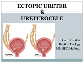
2. INTRODUCTION
Ureteral duplication:
Most common congenital renal abnormality.
Found in 1% population & 10% of children diagnosed
with UTIs.
Incomplete ureteral duplication- one common
ureter enters bladder, rarely clinically significant.
Complete ureteral duplication- two ureters
ipsilaterally enter the bladder.
3. Propensity for VUR into lower pole and
obstruction of upper pole.
Upper-pole ureter may be ectopic in its insertion into
bladder or may end in a ureterocele.
Both conditions are more common in duplicated
collecting systems but may also be seen in single
systems.
4. Ectopic ureter & Ureterocele:
Distinct entities, but share many common features.
Same underlying developmental mechanisms.
A continuum of embryologic development.
Similar clinical presentations.
Approached in a similar manner.
Slight variation in management.
5. DEFINITION
Ectopic ureter:
Any ureter, single or duplex, that does not enter
trigonal area of bladder.
In a duplex system, inevitably upper pole ureter,
because of its budding from mesonephric duct later
than lower pole with later incorporation into the
developing urogenital sinus.
6. In females, entry anywhere from bladder neck to
perineum and into vagina, uterus, and even rectum.
May be a/w dilated Gartner duct cyst (Wolffian duct
remnant from which ureter buds)→ Rupture →
vaginal communication→ incontinence.
In males, entry always above external sphincter or
pelvic floor, usually into wolffian structures,
including vas deferens, seminal vesicles, or
ejaculatory duct.
No incontinence, but infection and pain of affected
organs (testicles and epididymis).
8. Retrograde injection study
of boy with abdominal
pain and a ureterocele
associated with a
hypoplastic right
kidney. The intravesical
ureterocele (UC) is being
injected and demonstrates
communication with
the right seminal vesicle
(arrowhead) and vas
deferens (arrows), with
the ureter (UR) leading to
the dysplastic kidney. At
surgical resection, the
ureter and vas joined just
above the seminal vesicles
9. Single-system ectopic ureters & ureteroceles →
apparently absent kidney on USG → small, poorly-
functioning renal unit on CT Urogram.
Rare B/L single-system ectopic ureters may be a/w
hypoplastic bladder & B/L renal
abnormalities/dysplasia (apparent bladder agenesis).
10. Ureterocele:
Cystic dilatation of terminal intravesical ureter.
Intravesical ureterocele- entirely contained within
the bladder; may prolapse into urethra during
voiding.
Ectopic ureterocele- if any portion is permanently
situated at bladder neck or urethra, regardless of the
position of orifice(bladder, bladder neck or urethra).
Do not form entirely within urethra, nor do they
attach to wolffian ductal structures.
Single or duplex system, and in duplex systems
invariably affects upper pole.
13. A, Sphincterostenotic ectopic ureterocele.
B, Cecoureterocele lumen extends distal to the orifice as a long tongue
beneath the ureteral submucosa. The orifice communicates with the
lumen of the bladder and is large and incompetent.
14. Churchill’s Functional classification system:
based on impact of ureterocele on upper urinary
tract, including all renal units.
1. Only upper pole affected, 2. Entire ipsilateral
kidney involved, & 3. Contralateral system also at
risk d/t reflux or B.O.O.
15. Non-obstructive ureterocele with duplication or
“Ureterocele disproportion”:
A/w a duplex kidney, but affected upper pole &
ureter non-dilated and dysplastic→ not readily
detected on most imaging.
Typical ureterocele seen in bladder, but ipsilateral
kidney completely normal.
17. PATHOPHYSIOLOGY & EMBRYOLOGY
Ureteral bud branch from mesonephric/Wolffian duct
Extends into nephrogenic blastema(undifferentiated
mesenchyma)
Formation of the entire renal collecting system
Distal to ureteric bud, mesonephric duct incorporates into
UGS
U.O. superolaterally moves to its normal position on
trigone
18. Distal segment of mesonephric duct is carried
inferomedially→incorporated into bladder neck.
In male fetus, it also develops into seminal vesicle, vas
deferens, and epididymis.
In females, it becomes Gartner duct, located between
vagina and urethra.
19. In ureteral duplication, two ureteric buds arise from
mesonephric duct.
Lower - earlier insertion into UGS & superolateral
location of orifice; poor trigonal support & short
intramural tunnel → predisposed to VUR.
Upper- inserts later & low on trigone inferomedially
→ inserts ectopically at bladder neck, ejaculatory
duct, seminal vesicle, or vas deferens in males & in
Gartner duct in females.
20. Ureteral ectopia without duplication result from
delayed incorporation of distal ureter into
developing bladder.
Ureterocele development- two theories: 1. failure of
Chwalle membrane to break down at the distal
ureter during development -results in obstruction
and saccular dilation. 2. Aberrant signaling from
expanding urogenital sinus results in dilation of
distal ureter.
21. EPIDEMIOLOGY
Incidence of ureteral duplication- 1% (autopsy series).
Ureteroceles- 1 per 5000-12000 population; 10%
bilateral, 60-80% ectopic, 80% a/w upper-pole ureter
of a duplex system. Single system ureteroceles a/w
cardiac & genital anomalies.
More common in females.
More common in whites.
22. PRENATAL IMAGING DETECTION
Majority of ectopic ureters & ureteroceles detected
on prenatal USG, even if no specific diagnosis.
Duplex system prenatal Dx difficult, except in dilated
upper moiety.
Upper pole “cyst” in a fetus – upper pole
hydronephrosis until proven otherwise.
Bladder inspection mandatory to identify ureterocele
in all cases- wait for bladder filling.
Character of upper pole parenchyma- thickness &
echogenicity.
23. A large ectopic ureter may impinge on the bladder appear
as intravesical structure, “Pseudoureterocele.”
Careful evaluation of other renal units & bladder.
Ipsilateral lower pole or contralateral dilation suggests
reflux or less commonly obstruction from ureterocele or
dilated ectopic ureter.
B.O.O. by a ureterocele can manifest as hydronephrosis of
all renal units.
Oligohydramnios, contralateral renal dysplasia- rare.
Prenatal intervention or early delivery- no benefit.
24. CLINICAL PRESENTATION
INCIDENTAL:
Significant HN with an ectopic ureter or ureterocele.
During evaluation for cause of general abdominal
pain.
Cases of presumed ovarian cysts may be markedly
dilated ureters.
25. INFECTION:
UTI in first few months of life- MC presentation.
Generalised urosepsis d/t infected obstructed system.
Ongoing low-grade fever with periodic spikes.
Purulent discharge from the perineum
Bacterial epididymitis/orchitis- recurrent episodes.
26. INCONTINENCE:
Caused by an ectopic ureter in a girl, but never in boys.
Persistent low-volume dampness at all time; Child
can’t remain dry for even 30-60 min.
Diagnosis difficult before toilet training.
Rare pts.- intermittent leakage through a Gartner duct
membrane.
Untreated ureteroceles not a/w incontinence.
27. PAIN: Uncommonly a/w acute infection, episodic
obstruction of ectopic ureter or bladder pain caused
by an obstructing ureterocele.
PROLAPSE: Ureterocele prolapse unusual; smooth,
congested, mucosa covered interlabial masses
protruding from urethra; non-circumferential, non-
lobulated.
LATE PRESENTATION: Infection,
abdominal/flank pain, Incontinence, Stone in
ureterocele.
31. EVALUATION
PHYSICAL EXAMINATION:
May facilitate diagnosis.
Prolapsed ureterocele, ectopic perineal ureteral
orifice in a child with H/O continuous dampness
Dilated Gartner duct cyst- rare.
Palpable dilated upper pole of ectopic ureter or
ureterocele in a relaxed infant.
32. Perineal ectopic ureteral orifice (bottom arrow) cannulated with an angiocatheter,
situated
between the urethral orifice (top arrow) and the vagina, just to the left of midline
33. A, Gartner duct cyst (bottom right arrow) in newborn with a left multicystic dysplastic
kidney. B, Injection of the cyst communicated with the ureter and dysplastic kidney.
34. ULTRASOUND:
Typical findings-dilated upper pole with ureteral
dilation or dilated single system.
Bladder images differentiate ureterocele from ectopic
ureter- thin-walled cystic dilation within the bladder,
not extending beyond its walls.
36. Ultrasound demonstrating dilated upper pole (UP) and lower pole (LP) associated with a
ureterocele. The upper pole has evident renal parenchyma. The lower pole is dilated because of
compression of the dilated upper pole ureter on the lower pole system, creating a partial obstruction.
37. Ultrasound image of dilated upper pole (UP) associated with a
ureterocele, demonstrating limited renal parenchyma
39. MRI:
Provides most detailed imaging.
Currently reseved for patients with distorted,
complex anatomy.
Added advantage- functional information.
40. RENAL FUNCTION- NUCLEAR IMAGING:
Gold standard for renal functional assessment- DMSA.
Prime role- function of affected upper pole, also status of
other renal moieties, if lower pole reflux of HN of any unit.
To assess drainage function in ureteroceles in which
Observation is planned- Diuretic renal scan replaces DMSA-
provides both funvtional & drainage information.
IVU:
Less useful baseline study.
Functional assessment only qualitative.
Ureterocele- a "cobra head" or "spring onion"
configuration at bladder level.
43. VOIDING CYSTOURETHROGRAM (VCUG):
Most definitive test for bladder,distal ureters &urethra.
Obligatory to define baseline situation before any
intervention.
Omitted in emergency TUI for ureterocele producing
BOO, urosepsis or B/L upper tract obstruction.
Duplicated collecting systems with lower-pole reflux &
nonrefluxing upper pole, give appearance of a
"drooping lily“.
45. VCUG of duplex system ureterocele with reflux into
lower moiety
ureterocele
Reflux into lower moiety
Everting
ureterocele
46. Voiding cystourethrogram image of a cecoureterocele where the ureterocele
(black arrow) is attached to the urethra (white arrow) and the lumen extends
into the urethra
48. REFLUX:
Reflux of ipsilateral lower pole – 50%.
Contralateral reflux in 25% of cases, and
Reflux into ureterocele in 10% of cases.
In an ectopic ureter, ipsilateral lower pole reflux is
unlikely to resolve spontaneously.
49. ENDOSCOPIC EVALUATION:
Assess character of urethra, bladder neck and trigone
relative to ureterocele or ectopic ureter.
Location of other ureteral orifices should be documented.
Orifice of affected ureter should be sought but may not be
identified.
Urethra is examined carefully for orifice if not seen in
bladder.
Appearance of ureterocele will vary with bladder filling;
start with little filling and slowly increase bladder volume.
Lowest portion – best site for incision.
Retrograde contrast can confirm ureterocele disproportion
& unusual connections with genital ducts.
50.
51. CLINICAL MANAGEMENT
Before intervention, obtain maximum information
about pts’ altered anatomy & physiology.
No criteria to decide how much upper pole renal
function in worth preseving.
MANAGEMENT GOALS:
1. Preservation of renal function;
2. Elimination of infection, obstruction, and reflux;
3. Maintenance of urinary continence; and
4. Minimizing surgical morbidity.
52. ACUTE DECOMPRESSION:
Indications:
Ureterocele producing BOO or severe B/L upper tract
obstruction.
Severe urosepsis.
Sepsis not responding to appropriate therapy.
Methods:
For ureteroceles- Transurethral Incision (TUI).
For ectopic ureters- end ureterostomy near bladder.
53. DEFINITIVE SURGICAL OPTIONS:
For Ectopic ureter- common sheath
reimplantation or ureteroureterostomy, either
low/distal or high proximal near the renal pelvis.
For Ureterocele- TUI, ureterocele excision and
common sheath reimplantation or
ureteroureterostomy.
54. OBSERVATIONAL MANAGEMENT:
Non-operative management of ureteroceles meeting
certain criteria, in carefully selected pt.& parental
education-
1. no obstruction of ipsilateral lower pole or
contralateral kidney,
2. limited reflux to lower pole (grade III or less),
3. no function of upper pole, or
4. no obstruction on diuretic renography.
Potential for later unpredictable acute presentation.
55. TOTAL RECONSTRUCTION:
Upper pole nephrectomy with ureterocele excision
and reimplantation of lower pole ureter is definitive
but extensive operation performed with two
incisions.
Ideal candidate- older child with a massive
ureterocele and no function of an upper pole with
significant lower pole reflux.
56. UPPER POLE PARTIAL OR HEMI-
NEPHRECTOMY:
Preferred treatment when no function in the upper pole.
Open surgery conventional laparoscopy, Robotic
laparoscopy, Laparoendoscopic single-site
surgery(LESS) nephrectomy.
Results in ureteroceles with/without lower pole reflux:
resolution- 20%, New reflux- 15-50%, secondary surgery
rate- 40-50%.
58. Surgical management of the
refluxing ureteral stump.
A It is difficult to completely
separate the distal 2 to 3 cm
of upper pole ureter from
lower pole ureter. The ectopic
ureter is excised to this point.
B The outer wall of ectopic
ureter is excised to the
bladder level.
C A transfixing suture
obliterates its lumen, with
care being taken not to injure
the orthotopic ureter.
59. COMPLICATIONS OF UPPER POLAR
NEPHRECTOMY:
1. Loss of lower pole function,
2. Postoperative upper pole urinoma
3. IVC laceration,
4. Duodenal perforation,
5. Total nephrectomy,
6. Peritoneal tears.
60. LOWER TRACT RECONSTRUCTION:
A definitive reconstruction at bladder is suitable for
both ectopic ureter and ureterocele.
Advantage: relieving obstruction as well as
correcting reflux.
Disadvantages: potential for injury to bladder neck
and vagina, complexity of the procedure.
If clinically significant reflux persists after other
procedures, lower tract reconstruction may be
necessary.
61. Results:Very good.
Persisting reflux-
5-10%, more
common when
ureteral tapering
done.
62. PYELOURETEROSTOMY &
URETEROURETEROSTOMY:
When upper pole of an ectopic ureter or ureterocele is preserved
owing to function or surgeon preference.
Anastomosis between upper pole ureter & lower pole ureter in
an end-to-side fashion. Proximal & distal approaches used.
Proximal anastomoses preferable to a distal
ureteroureterostomy with a dilated upper pole, because the
latter may result in more urinary stasis .
63. TRANSURETHRAL INCISION (TUI):
Transverse incision through full thickness of
ureterocele wall using cutting current, as distally &
close to the bladder floor as possible.
Bugbee electrode, angled-tip wire, Cold knife,
resectoscope with Collins hot knife, Laser incision.
Deep incision to incise thick wall, see for urine-jet or
inner urothelium.
Ectopic ureterocele:Longitudinal incision from
intravesical into urethral portion, or two incisions.
64. No catheter required.
Follow-up USG after 4-6 weeks to assess degree of
decompression.
VCUG at 2-3 months to determine status of lower
pole reflux.
Risk of reoperation high with extravesical
ureteroceles & lower pole reflux (persisting or new).
65. TEMPORARY END URETEROSTOMY FOR
ECTOPIC URETER:
Ectopic ureter in infant with sepsis or massive
dilation.
Advantage- Acute decompression to manage sepsis
and permit later assessment(in 4 mths or 6 mths
age) of any function in affected renal unit before
definitive management.
66. CLINICAL DECISION MAKING
ECTOPIC URETERS:
Duplex System
Single System
Preservation or
Excision (based
on function &
Surgeon
preference)
Lower pole
reflux
No reflux
Proximal or
distal
uretero-
ureterostomy
Reflux
Common sheath
reimplantation or
lower pole
reimplantation with
distal upper to lower
ureteroureterostomy
Massively dilated ureter
Temporary end
ureterostomy
68. URETEROCELE:
TUI reasonable to offer before more complex
reconstructions, specially young infants.
May make a subsequent surgical procedure less
complex by decompressing a dilated upper pole
ureter. Reimplantation may be much more effective
and not require excisional tapering.
Older child with a massive upper pole, removal &
definitive surgery perform at diagnosis.