Renal cell carcinoma for students
•Download as PPT, PDF•
31 likes•9,247 views

Causes, pathogenesis, Morphology, stages and prognosis

Recommended
More Related Content
What's hot
What's hot (20)
Viewers also liked
Viewers also liked (20)
Similar to Renal cell carcinoma for students
Similar to Renal cell carcinoma for students (20)
More from Mohammad Manzoor
More from Mohammad Manzoor (20)
Recently uploaded
Recently uploaded (20)
Renal cell carcinoma for students
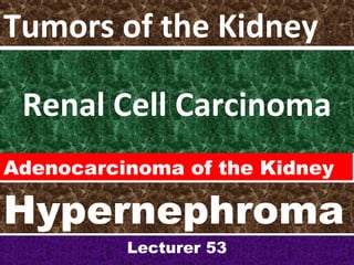
- 1. Renal Cell Carcinoma Lecturer 53 Tumors of the Kidney Adenocarcinoma of the Kidney Hypernephroma
- 2. HYPERNEPHROMA- RCC • Because of their gross yellow color and the resemblance of the tumor cells to clear cells of the adrenal cortex, they were at one time called Hypernephroma.
- 3. Renal Adenocarcinoma • It is now clear that all these tumors arise from tubular epithelium and are therefore RENAL ADENOCARCINOMAS.
- 4. Epidemiology • Male predominance (1.6:1.0 M:F) • Highest incidence between age 60-80 -Median age of diagnosis is 66 years -Median age of death 70 years • Highest incidence in Scandinavia and North America, lowest in Africa
- 5. Risk Factors
- 6. Causes/ Risk Factors • There is also an increased incidence in patients with chronic renal failure and acquired cystic disease and in tuberous sclerosis.
- 7. RCC • Most renal cancer is sporadic, but unusual forms of autosomal dominant familial cancers occur, usually in youngerindividuals.
- 8. Classification of RCC The major types of RCC are as follows: • 1. Clear cell carcinoma -70-80%, 95% Sporadic • 2. Papillary carcinoma- 10-15% • 3. Chromophobe renal carcinoma- 5% • 4. Collecting duct (Bellini duct) carcinoma- 1% 0r less
- 11. Pathogenesis of VHL • Von Hippel-Lindau protein, product of VHL gene, is a tumor suppressor • VHL inhibits hypoxia-inducible genes involved in angiogenesis such as VEGF, TGF-a, GLUT-1 • VHL destabilizes and promotes ubiquination of HIF-a (hypoxia-inducible factor) • Loss of VHL results in tumor angiogenesis, tumor-cell proliferation, epithelial cell proliferation Glucose transporter 1 Ubiquination: a small polypeptide, found in most eukaryotic cells, that combines with other proteins to make them susceptible to degradation
- 12. Morphology •Renal cell carcinomas may arise in any portion of the kidney, but more commonly affects the poles.
- 14. RCC Morphology • One of the striking characteristics of renal cell carcinoma is its tendency to invade the renal vein and grow as a solid column of cells within this vessel.
- 20. Morphology- Papillary Carcinomas • Papillary carcinomas are the most common type of renal cancer in patients who develop dialysis-associated cystic disease.
- 21. Microscopy- Papillary Carcinoma • Papillary carcinoma is composed of cuboidal or low columnar cells arranged in papillary formations. • Interstitial foam cells are common in the papillary cores. • Psammoma bodies may be present. • The stroma is usually scanty but highly vascularized.
- 22. Chromophobe renal carcinoma • Chromophobe renal carcinoma is made up of pale eosinophilic cells, often with a perinuclear halo, arranged in solid sheets with a concentration of the largest cells around blood vessels.
- 23. Collecting duct carcinoma • Collecting duct carcinoma is a rare variant showing irregular channels lined by highly atypical epithelium with a hobnail pattern.
- 24. RCC •Sarcomatoid changes arise infrequently in all types of renal cell carcinoma and are a decidedly ominous feature.
- 25. Renal cell carcinoma. Typical cross-section of yellowish, spherical neoplasm in one pole of the kidney. Note the tumor in the dilated thrombosed renal vein.
- 27. Paraneoplastic Syndromes 1. Polycythemia, 2. Hypercalcemia, 3. Hypertension, 4. Hepatic dysfunction, 5. Feminization or masculinization, 6. Cushing syndrome, 7. Eosinophilia, 8. Leukemoid reactions, and 9. Amyloidosis.
- 28. RCC
- 29. Clinical Presentation • Most asymptomatic • Hematuria present 40% of patients • Classic triad: flank pain, hematuria, palpable abdominal mass occur in 9% of patients • 45% present with localized disease, • 25% with locally advanced disease, • 30% with metastatic disease
- 32. Treatment •Nephrectomyhas been the treatment of choice, but •Partial nephrectomy to preserve renal function is being done with increasing frequency and similar outcome.