SlideShare a Scribd company logo
1 of 73
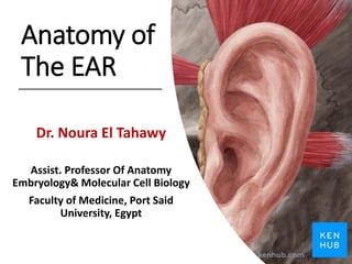
Anatomy of
The EAR
Dr. Noura El Tahawy
Assist. Professor Of Anatomy
Embryology& Molecular Cell Biology
Faculty of Medicine, Port Said
University, Egypt
1. Describe the external ear. (auricle& ext. acoustic meatus,
tympanic membrane.)
2. Describe blood &nerve supply& lymphatic drainage of the
external ear.
3. Describe the middle ear. (walls, special features in the cavity of
middle ear, auditory ossicles& muscles)
4. Describe the course& branches of the facial nerve in the
middle ear.
5. Describe divisions and boundaries of mouth cavity.
objectives
Auricle
Auricle
Auricle
Auricle: is composed mainly of
cartilage, except for the lobule.
The auricle collects the sound
waves and directs vibrations into
the external auditory canal.
Has the following features:
1. Helix
■ The slightly curved rim of the
auricle.
2. Antihelix
■ A broader curved eminence
internal to the helix, which
divides the auricle into an outer
scaphoid fossa and the deeper
concha.
3. Concha
■ The deep cavity in front of the
antihelix.
4. Tragus
■ A small projection from the
anterior portion of the external
ear anterior to the concha
5. Lobule
■ A structure made up of areolar
tissue and fat but no cartilage.
The Auricle:
External acoustic meatus
• External Acoustic (Auditory)
Meatus
• ■ Is approximately 2.5 cm
long, extending from the
concha to the tympanic
membrane.
• ■ Its external one-third is
formed by cartilage, and the
internal two-thirds is formed
by bone. produces earwax.
• ■ Is innervated by the
trigeminal nerve and vagus
nerve, which is joined by a
branch of the facial nerve and
the glossopharyngeal nerve.
External acoustic meatus
External acoustic
meatus (bony
part)
External acoustic meatus
Tympanic membrane
(Ear drum)
■ Conducts sound waves to the middle ear.
■ Its external (lateral) concave surface is covered by skin and is innervated (sensory) by the
trigeminal nerve and the vagus nerve.
Eardrum
Tympanic membrane
Consists of three layers: an outer (cutaneous), an intermediate (fibrous), and an inner (mucous) layer. ■ Has a thickened
fibrocartilaginous ring at the greater part of its circumference, which is fixed in the tympanic sulcus at the inner end of the
meatus. ■ Has a small triangular portion between the anterior and posterior malleolar folds called the pars flaccida
(deficient ring and lack of fibrous layer). The remainder of the membrane is called the pars tensa. ■ Contains the cone of
light, which is a triangular reflection of light seen in the anterior–inferior quadrant. ■ Contains the most depressed canter
point of the concavity, called the umbo (Latin for “knob”).
Otoscope ‫االذن‬ ‫منظار‬Tympanic membrane
Middle Ear Walls and boundaries
Dr. Noura El Tahawy
Pterous temporal Bone
Cranial Cavity
erous temporal Bone
Temporal bone
Temporal bone
Tympanic cavity (Middle Ear Cavity)
Tympanic cavity
Eardrum
Tympanic membrane
The lateral wall of the Middle Ear
Medial wall of the Middle Ear
Promontory
Tegmen tympani
Roof of Middle ear
Floor of the Middle Ear
Bulb of Internal Jugular vein
Eustachian tube
(Pharyngo-tympanic tube)
Anterior wall of Middle Ear
nasoPharynx
NasoPharynx
Walls of the Middle ear
1. Boundaries of the Tympanic Cavity ■ Roof: tegmen tympani (temporal bone).■ Floor:
jugular fossa. Anterior: carotid canal and Eustachian tube. ■ Posterior: mastoid air cells and
mastoid antrum through the aditus ad antrum. ■ Lateral: tympanic membrane. ■ Medial: lateral
wall of the inner ear, presenting the promontory formed by the basal turn of the cochlea, the
fenestra vestibuli (oval window), the fenestra cochlea (round window), and the prominence of
the facial canal.
2. Oval Window (Fenestra Vestibuli)
■ Is pushed back and forth by the footplate of the stapes and transmits the sonic vibrations
of the ossicles into the perilymph of the scala vestibuli in the inner ear.
3. Round Window (Fenestra Cochlea or Tympani)
■ Is closed by the secondary tympanic (mucous) membrane of the middle ear and
accommodates the pressure waves transmitted to the perilymph of the scala tympani.
Consists of the tympanic cavity with its ossicles and is located within the petrous portion of
the temporal bone.
■ Transmits the sound waves from air to auditory ossicles and then to the inner ear. A.
Tympanic (Middle Ear) Cavity
■ Includes the tympanic cavity proper (the space internal to the tympanic membrane) and the
epitympanic recess (the space superior to the tympanic membrane that contains the head of
the malleus and the body of the incus).
■ Communicates anteriorly with the nasopharynx via the auditory (eustachian) tube and
posteriorly with the mastoid air cells and the mastoid antrum through the aditus ad antrum.
■ Is traversed by the chorda tympani and lesser petrosal nerve.
Tympanic cavity (Middle Ear cavity)
Auditory (Pharyngotympanic or Eustachian) Tube
■ Connects the middle ear to the nasopharynx.
■ Allows air to enter or leave the middle ear cavity and thus balances the pressure
in the middle ear with atmospheric pressure, allowing free movement of the
tympanic membrane.
■ Has cartilaginous portion that remains closed except during swallowing or
yawning.
■ Is opened by the simultaneous contraction of the tensor veli palatini and
salpingopharyngeus muscles.
Contents of the Middle Ear
(tympanic cavity)
Ear Ossicles
Malleus
Malleus
Incus
Stapes
Transmission of Vioce
Muscles inside the Middle ear
Inner Ear
Vestibulocochlear nerve
Cochlear
part
Vestibular part
Facial nerve
Vestibulocochlear and Facial nerves
Facial Nerve
Facial nerve
Cochlear
part
Vestibular part
Facial nerve
Vestibulocochlear and Facial nerves
Facial nerve
Branches of Facial nerve
1. The motor nucleus supplies the muscles of facial expression, the auricular muscles,
the stapedius, the posterior belly of the digastric, and the stylohyoid muscles
2. Parasympathetic (secretomotor) to submandibular and sublingual salivary glands
and the nasal and palatine glands.
3. Parasympathetic (secretomotor) supplies the lacrimal gland.
4. The sensory part receives taste fibbers from the anterior two-thirds of the tongue,
the floor of the mouth, and the palate.
Distribution
Facial nerve
Course:
1. The two roots (sensory and motor) of the facial nerve emerge between the pons and
the medulla oblongata. They pass laterally in the posterior cranial fossa with the
vestibulocochlear nerve (CVIII) and enter the internal acoustic meatus (internal
auditory canal) in the petrous part of the temporal bone.
2. At the bottom of the meatus, the nerve enters the facial bony canal and runs laterally
through the inner ear.
3. On reaching the medial wall of the middle ear (tympanic cavity) the nerve expands to
form the sensory geniculate ganglion and turns sharply backward at the roof of
middle ear (above the promontory).
4. Reaching the posterior wall of the tympanic cavity, the facial nerve turns downward,
descends to emerge from the stylomastoid foramen to exit the skull and enters the
parotid gland. Inside the parotid g. it terminates by dividing into five terminal
branches which supply muscles of facial expression.
Atlas of The Ear
Gross Anatomy of the Middle Ear
End

More Related Content

What's hot

Anatomy of the external and middle ear
Anatomy of the external and middle earAnatomy of the external and middle ear
Anatomy of the external and middle earShweta Sharma
 
Larynx anatomy of larynx (1)
Larynx anatomy of larynx (1)Larynx anatomy of larynx (1)
Larynx anatomy of larynx (1)KGMU, Lucknow
 
Anatomy of temporal bone
Anatomy of temporal boneAnatomy of temporal bone
Anatomy of temporal bonepraneeth koduru
 
Inner ear anatomy
Inner ear anatomy Inner ear anatomy
Inner ear anatomy yatish88
 
TRIANGLES OF NECK - BY DR. JUVERIA MAJEED MS ENT
TRIANGLES OF NECK - BY DR. JUVERIA MAJEED MS ENTTRIANGLES OF NECK - BY DR. JUVERIA MAJEED MS ENT
TRIANGLES OF NECK - BY DR. JUVERIA MAJEED MS ENTDr.Juveria Majeed
 
Middle ear anatomy
Middle ear anatomyMiddle ear anatomy
Middle ear anatomykbn2914
 
Anatomy temporal bone
Anatomy temporal bone Anatomy temporal bone
Anatomy temporal bone KevinMungasia
 
Pterygopalatine fossa and approaches by Dr.Ashwin Menon
Pterygopalatine fossa and approaches by Dr.Ashwin MenonPterygopalatine fossa and approaches by Dr.Ashwin Menon
Pterygopalatine fossa and approaches by Dr.Ashwin MenonDr.Ashwin Menon
 
anatomy of larynx by ravindra daggupati
anatomy of larynx by ravindra daggupatianatomy of larynx by ravindra daggupati
anatomy of larynx by ravindra daggupatiRavindra Daggupati
 
Middle Ear Anatomy - Arjun Antony Graison
Middle Ear Anatomy - Arjun Antony GraisonMiddle Ear Anatomy - Arjun Antony Graison
Middle Ear Anatomy - Arjun Antony GraisonArjun Graison
 
Embryology & anatomy of external ear
Embryology &  anatomy of external earEmbryology &  anatomy of external ear
Embryology & anatomy of external earDr. Pruthvi Raj S
 
Surgical anatomy of deep neck spaces
Surgical anatomy of deep neck spacesSurgical anatomy of deep neck spaces
Surgical anatomy of deep neck spacesgoogle
 
External ear anatomy
External ear anatomyExternal ear anatomy
External ear anatomySupreet Sn
 
Skull base anatomy by Dr. Aditya Tiwari
Skull base anatomy by Dr. Aditya TiwariSkull base anatomy by Dr. Aditya Tiwari
Skull base anatomy by Dr. Aditya TiwariAditya Tiwari
 

What's hot (20)

Anatomy of the external and middle ear
Anatomy of the external and middle earAnatomy of the external and middle ear
Anatomy of the external and middle ear
 
Larynx anatomy of larynx (1)
Larynx anatomy of larynx (1)Larynx anatomy of larynx (1)
Larynx anatomy of larynx (1)
 
Anatomy of temporal bone
Anatomy of temporal boneAnatomy of temporal bone
Anatomy of temporal bone
 
Anatomy of ear
Anatomy of earAnatomy of ear
Anatomy of ear
 
Anatomy of Larynx
Anatomy of LarynxAnatomy of Larynx
Anatomy of Larynx
 
Inner ear anatomy
Inner ear anatomy Inner ear anatomy
Inner ear anatomy
 
Anatomy of Pharynx
Anatomy of PharynxAnatomy of Pharynx
Anatomy of Pharynx
 
TRIANGLES OF NECK - BY DR. JUVERIA MAJEED MS ENT
TRIANGLES OF NECK - BY DR. JUVERIA MAJEED MS ENTTRIANGLES OF NECK - BY DR. JUVERIA MAJEED MS ENT
TRIANGLES OF NECK - BY DR. JUVERIA MAJEED MS ENT
 
Tympanic membrane
Tympanic membraneTympanic membrane
Tympanic membrane
 
Middle ear anatomy
Middle ear anatomyMiddle ear anatomy
Middle ear anatomy
 
Anatomy temporal bone
Anatomy temporal bone Anatomy temporal bone
Anatomy temporal bone
 
Pterygopalatine fossa and approaches by Dr.Ashwin Menon
Pterygopalatine fossa and approaches by Dr.Ashwin MenonPterygopalatine fossa and approaches by Dr.Ashwin Menon
Pterygopalatine fossa and approaches by Dr.Ashwin Menon
 
anatomy of larynx by ravindra daggupati
anatomy of larynx by ravindra daggupatianatomy of larynx by ravindra daggupati
anatomy of larynx by ravindra daggupati
 
Middle Ear Anatomy - Arjun Antony Graison
Middle Ear Anatomy - Arjun Antony GraisonMiddle Ear Anatomy - Arjun Antony Graison
Middle Ear Anatomy - Arjun Antony Graison
 
Anatomy of ear
Anatomy of earAnatomy of ear
Anatomy of ear
 
Anatomy of Facial Nerve
Anatomy of Facial NerveAnatomy of Facial Nerve
Anatomy of Facial Nerve
 
Embryology & anatomy of external ear
Embryology &  anatomy of external earEmbryology &  anatomy of external ear
Embryology & anatomy of external ear
 
Surgical anatomy of deep neck spaces
Surgical anatomy of deep neck spacesSurgical anatomy of deep neck spaces
Surgical anatomy of deep neck spaces
 
External ear anatomy
External ear anatomyExternal ear anatomy
External ear anatomy
 
Skull base anatomy by Dr. Aditya Tiwari
Skull base anatomy by Dr. Aditya TiwariSkull base anatomy by Dr. Aditya Tiwari
Skull base anatomy by Dr. Aditya Tiwari
 

Similar to Anatomy of the ear (ecture 2) by dr, noura 2018

Dr Hoda _Hassan_Anatomy of EAR 20-21.pptx
Dr Hoda _Hassan_Anatomy of EAR 20-21.pptxDr Hoda _Hassan_Anatomy of EAR 20-21.pptx
Dr Hoda _Hassan_Anatomy of EAR 20-21.pptxMohamedAli961533
 
Anatomy of the ear
Anatomy of the earAnatomy of the ear
Anatomy of the earRafid Rashid
 
Presentation Ear.pptx
Presentation Ear.pptxPresentation Ear.pptx
Presentation Ear.pptxssuser1d41c52
 
Ear anatomy (1)-1.pptx
Ear anatomy (1)-1.pptxEar anatomy (1)-1.pptx
Ear anatomy (1)-1.pptxsafatullah50
 
The Ear, Anatomy, Physiology, Clinical diseases, and pathology, hearing tests
The Ear, Anatomy, Physiology, Clinical diseases, and pathology, hearing testsThe Ear, Anatomy, Physiology, Clinical diseases, and pathology, hearing tests
The Ear, Anatomy, Physiology, Clinical diseases, and pathology, hearing testsHamzehKYacoub
 
functional anatomy of the ear.ppt
functional anatomy of the ear.pptfunctional anatomy of the ear.ppt
functional anatomy of the ear.pptBaksantino123
 
Anatomy of Ear | SurgicoMed.com
Anatomy of Ear | SurgicoMed.comAnatomy of Ear | SurgicoMed.com
Anatomy of Ear | SurgicoMed.comMukhdoom BaharAli
 
Vestibular and auditory apparatus-Dr.B.B.Gosai
Vestibular and auditory apparatus-Dr.B.B.GosaiVestibular and auditory apparatus-Dr.B.B.Gosai
Vestibular and auditory apparatus-Dr.B.B.GosaiDr.B.B. Gosai
 
earanatomy-190219150351.pdf
earanatomy-190219150351.pdfearanatomy-190219150351.pdf
earanatomy-190219150351.pdfAliyahJohanis1
 
anatomy of ear nn.pptx
anatomy of ear nn.pptxanatomy of ear nn.pptx
anatomy of ear nn.pptxAzeemHamdani
 
Group 2 Medsurg ppt on Anatomy and physiology of ear-1.pptx
Group 2 Medsurg ppt on Anatomy and physiology of ear-1.pptxGroup 2 Medsurg ppt on Anatomy and physiology of ear-1.pptx
Group 2 Medsurg ppt on Anatomy and physiology of ear-1.pptxIjeomaNwokoro
 

Similar to Anatomy of the ear (ecture 2) by dr, noura 2018 (20)

ear anatomy.pptx
ear anatomy.pptxear anatomy.pptx
ear anatomy.pptx
 
Dr Hoda _Hassan_Anatomy of EAR 20-21.pptx
Dr Hoda _Hassan_Anatomy of EAR 20-21.pptxDr Hoda _Hassan_Anatomy of EAR 20-21.pptx
Dr Hoda _Hassan_Anatomy of EAR 20-21.pptx
 
EAR AND BALANCE.pptx
EAR AND BALANCE.pptxEAR AND BALANCE.pptx
EAR AND BALANCE.pptx
 
Ear ppt
Ear pptEar ppt
Ear ppt
 
Anatomy of the ear
Anatomy of the earAnatomy of the ear
Anatomy of the ear
 
Presentation Ear.pptx
Presentation Ear.pptxPresentation Ear.pptx
Presentation Ear.pptx
 
Ear anatomy (1)-1.pptx
Ear anatomy (1)-1.pptxEar anatomy (1)-1.pptx
Ear anatomy (1)-1.pptx
 
The Ear, Anatomy, Physiology, Clinical diseases, and pathology, hearing tests
The Ear, Anatomy, Physiology, Clinical diseases, and pathology, hearing testsThe Ear, Anatomy, Physiology, Clinical diseases, and pathology, hearing tests
The Ear, Anatomy, Physiology, Clinical diseases, and pathology, hearing tests
 
Anatomy of Ear
Anatomy of EarAnatomy of Ear
Anatomy of Ear
 
functional anatomy of the ear.ppt
functional anatomy of the ear.pptfunctional anatomy of the ear.ppt
functional anatomy of the ear.ppt
 
Anatomy of Ear | SurgicoMed.com
Anatomy of Ear | SurgicoMed.comAnatomy of Ear | SurgicoMed.com
Anatomy of Ear | SurgicoMed.com
 
Anatomy of ear and mastoid
Anatomy of ear and mastoidAnatomy of ear and mastoid
Anatomy of ear and mastoid
 
Nose, paranasal sinuses and ear
Nose, paranasal sinuses and earNose, paranasal sinuses and ear
Nose, paranasal sinuses and ear
 
Vestibular and auditory apparatus-Dr.B.B.Gosai
Vestibular and auditory apparatus-Dr.B.B.GosaiVestibular and auditory apparatus-Dr.B.B.Gosai
Vestibular and auditory apparatus-Dr.B.B.Gosai
 
Ears
EarsEars
Ears
 
Ear functions
Ear functionsEar functions
Ear functions
 
earanatomy-190219150351.pdf
earanatomy-190219150351.pdfearanatomy-190219150351.pdf
earanatomy-190219150351.pdf
 
Ear Anatomy
Ear AnatomyEar Anatomy
Ear Anatomy
 
anatomy of ear nn.pptx
anatomy of ear nn.pptxanatomy of ear nn.pptx
anatomy of ear nn.pptx
 
Group 2 Medsurg ppt on Anatomy and physiology of ear-1.pptx
Group 2 Medsurg ppt on Anatomy and physiology of ear-1.pptxGroup 2 Medsurg ppt on Anatomy and physiology of ear-1.pptx
Group 2 Medsurg ppt on Anatomy and physiology of ear-1.pptx
 

More from Dr. Noura El Tahawy

Development of togue& glands by Dr. Noura 2014
Development of togue& glands by Dr. Noura 2014Development of togue& glands by Dr. Noura 2014
Development of togue& glands by Dr. Noura 2014Dr. Noura El Tahawy
 
Development of face, nose, palate by dr. noura 2014
Development of face, nose, palate by dr. noura 2014Development of face, nose, palate by dr. noura 2014
Development of face, nose, palate by dr. noura 2014Dr. Noura El Tahawy
 
Introduction of anatomy lecture1 dr. noura
Introduction of  anatomy lecture1 dr. nouraIntroduction of  anatomy lecture1 dr. noura
Introduction of anatomy lecture1 dr. nouraDr. Noura El Tahawy
 
questions for peroidic examination (thorax) december 2011
questions for peroidic examination (thorax)  december 2011questions for peroidic examination (thorax)  december 2011
questions for peroidic examination (thorax) december 2011Dr. Noura El Tahawy
 
Lecture 2 thoracic wall & Diaphragm
Lecture 2 thoracic wall & DiaphragmLecture 2 thoracic wall & Diaphragm
Lecture 2 thoracic wall & DiaphragmDr. Noura El Tahawy
 
Questions on the lecture 2 thorax
Questions on the lecture 2 thoraxQuestions on the lecture 2 thorax
Questions on the lecture 2 thoraxDr. Noura El Tahawy
 
Peritoneum and peritoneal cavity
Peritoneum and peritoneal cavityPeritoneum and peritoneal cavity
Peritoneum and peritoneal cavityDr. Noura El Tahawy
 

More from Dr. Noura El Tahawy (20)

Lecture 1 orbit dr. noura
Lecture 1 orbit  dr. nouraLecture 1 orbit  dr. noura
Lecture 1 orbit dr. noura
 
Development of togue& glands by Dr. Noura 2014
Development of togue& glands by Dr. Noura 2014Development of togue& glands by Dr. Noura 2014
Development of togue& glands by Dr. Noura 2014
 
Development of face, nose, palate by dr. noura 2014
Development of face, nose, palate by dr. noura 2014Development of face, nose, palate by dr. noura 2014
Development of face, nose, palate by dr. noura 2014
 
Introduction of anatomy lecture1 dr. noura
Introduction of  anatomy lecture1 dr. nouraIntroduction of  anatomy lecture1 dr. noura
Introduction of anatomy lecture1 dr. noura
 
questions for peroidic examination (thorax) december 2011
questions for peroidic examination (thorax)  december 2011questions for peroidic examination (thorax)  december 2011
questions for peroidic examination (thorax) december 2011
 
Questions on the thorax
Questions on the thoraxQuestions on the thorax
Questions on the thorax
 
Lecture 4 Heart Anatomy
Lecture 4 Heart AnatomyLecture 4 Heart Anatomy
Lecture 4 Heart Anatomy
 
Questions on the thorax
Questions on the thoraxQuestions on the thorax
Questions on the thorax
 
Lecture 3 lungs & pleura
Lecture 3 lungs & pleuraLecture 3 lungs & pleura
Lecture 3 lungs & pleura
 
Lecture 2 thoracic wall & Diaphragm
Lecture 2 thoracic wall & DiaphragmLecture 2 thoracic wall & Diaphragm
Lecture 2 thoracic wall & Diaphragm
 
Questions on the lecture 2 thorax
Questions on the lecture 2 thoraxQuestions on the lecture 2 thorax
Questions on the lecture 2 thorax
 
Lecture 1 thoracic wall
Lecture 1 thoracic wall  Lecture 1 thoracic wall
Lecture 1 thoracic wall
 
Peritoneum and peritoneal cavity
Peritoneum and peritoneal cavityPeritoneum and peritoneal cavity
Peritoneum and peritoneal cavity
 
Skull, dura & sinuses
Skull, dura & sinusesSkull, dura & sinuses
Skull, dura & sinuses
 
Pituitary gland anatomy
Pituitary gland anatomyPituitary gland anatomy
Pituitary gland anatomy
 
Cavernous sinus anatomy
Cavernous sinus anatomy Cavernous sinus anatomy
Cavernous sinus anatomy
 
1. skull & mandible
1. skull & mandible 1. skull & mandible
1. skull & mandible
 
Blood supply of the brain
Blood supply of the brainBlood supply of the brain
Blood supply of the brain
 
Cerebellum
CerebellumCerebellum
Cerebellum
 
Fourth ventricle
Fourth ventricleFourth ventricle
Fourth ventricle
 

Recently uploaded

POST NATAL EXERCISES AND ITS IMPACT.pptx
POST NATAL EXERCISES AND ITS IMPACT.pptxPOST NATAL EXERCISES AND ITS IMPACT.pptx
POST NATAL EXERCISES AND ITS IMPACT.pptxvirengeeta
 
History and Development of Pharmacovigilence.pdf
History and Development of Pharmacovigilence.pdfHistory and Development of Pharmacovigilence.pdf
History and Development of Pharmacovigilence.pdfSasikiranMarri
 
Culture and Health Disorders Social change.pptx
Culture and Health Disorders Social change.pptxCulture and Health Disorders Social change.pptx
Culture and Health Disorders Social change.pptxDr. Dheeraj Kumar
 
Biomechanics- Shoulder Joint!!!!!!!!!!!!
Biomechanics- Shoulder Joint!!!!!!!!!!!!Biomechanics- Shoulder Joint!!!!!!!!!!!!
Biomechanics- Shoulder Joint!!!!!!!!!!!!ibtesaam huma
 
Pharmaceutical Marketting: Unit-5, Pricing
Pharmaceutical Marketting: Unit-5, PricingPharmaceutical Marketting: Unit-5, Pricing
Pharmaceutical Marketting: Unit-5, PricingArunagarwal328757
 
Introduction to Sports Injuries by- Dr. Anjali Rai
Introduction to Sports Injuries by- Dr. Anjali RaiIntroduction to Sports Injuries by- Dr. Anjali Rai
Introduction to Sports Injuries by- Dr. Anjali RaiGoogle
 
Report Back from SGO: What’s New in Uterine Cancer?.pptx
Report Back from SGO: What’s New in Uterine Cancer?.pptxReport Back from SGO: What’s New in Uterine Cancer?.pptx
Report Back from SGO: What’s New in Uterine Cancer?.pptxbkling
 
April 2024 ONCOLOGY CARTOON by DR KANHU CHARAN PATRO
April 2024 ONCOLOGY CARTOON by  DR KANHU CHARAN PATROApril 2024 ONCOLOGY CARTOON by  DR KANHU CHARAN PATRO
April 2024 ONCOLOGY CARTOON by DR KANHU CHARAN PATROKanhu Charan
 
Glomerular Filtration and determinants of glomerular filtration .pptx
Glomerular Filtration and  determinants of glomerular filtration .pptxGlomerular Filtration and  determinants of glomerular filtration .pptx
Glomerular Filtration and determinants of glomerular filtration .pptxDr.Nusrat Tariq
 
Lippincott Microcards_ Microbiology Flash Cards-LWW (2015).pdf
Lippincott Microcards_ Microbiology Flash Cards-LWW (2015).pdfLippincott Microcards_ Microbiology Flash Cards-LWW (2015).pdf
Lippincott Microcards_ Microbiology Flash Cards-LWW (2015).pdfSreeja Cherukuru
 
call girls in aerocity DELHI 🔝 >༒9540349809 🔝 genuine Escort Service 🔝✔️✔️
call girls in aerocity DELHI 🔝 >༒9540349809 🔝 genuine Escort Service 🔝✔️✔️call girls in aerocity DELHI 🔝 >༒9540349809 🔝 genuine Escort Service 🔝✔️✔️
call girls in aerocity DELHI 🔝 >༒9540349809 🔝 genuine Escort Service 🔝✔️✔️saminamagar
 
epilepsy and status epilepticus for undergraduate.pptx
epilepsy and status epilepticus  for undergraduate.pptxepilepsy and status epilepticus  for undergraduate.pptx
epilepsy and status epilepticus for undergraduate.pptxMohamed Rizk Khodair
 
See the 2,456 pharmacies on the National E-Pharmacy Platform
See the 2,456 pharmacies on the National E-Pharmacy PlatformSee the 2,456 pharmacies on the National E-Pharmacy Platform
See the 2,456 pharmacies on the National E-Pharmacy PlatformKweku Zurek
 
The next social challenge to public health: the information environment.pptx
The next social challenge to public health:  the information environment.pptxThe next social challenge to public health:  the information environment.pptx
The next social challenge to public health: the information environment.pptxTina Purnat
 
PULMONARY EDEMA AND ITS MANAGEMENT.pdf
PULMONARY EDEMA AND  ITS  MANAGEMENT.pdfPULMONARY EDEMA AND  ITS  MANAGEMENT.pdf
PULMONARY EDEMA AND ITS MANAGEMENT.pdfDolisha Warbi
 
VarSeq 2.6.0: Advancing Pharmacogenomics and Genomic Analysis
VarSeq 2.6.0: Advancing Pharmacogenomics and Genomic AnalysisVarSeq 2.6.0: Advancing Pharmacogenomics and Genomic Analysis
VarSeq 2.6.0: Advancing Pharmacogenomics and Genomic AnalysisGolden Helix
 
call girls in Connaught Place DELHI 🔝 >༒9540349809 🔝 genuine Escort Service ...
call girls in Connaught Place  DELHI 🔝 >༒9540349809 🔝 genuine Escort Service ...call girls in Connaught Place  DELHI 🔝 >༒9540349809 🔝 genuine Escort Service ...
call girls in Connaught Place DELHI 🔝 >༒9540349809 🔝 genuine Escort Service ...saminamagar
 
LUNG TUMORS AND ITS CLASSIFICATIONS.pdf
LUNG TUMORS AND ITS  CLASSIFICATIONS.pdfLUNG TUMORS AND ITS  CLASSIFICATIONS.pdf
LUNG TUMORS AND ITS CLASSIFICATIONS.pdfDolisha Warbi
 
SYNDESMOTIC INJURY- ANATOMICAL REPAIR.pptx
SYNDESMOTIC INJURY- ANATOMICAL REPAIR.pptxSYNDESMOTIC INJURY- ANATOMICAL REPAIR.pptx
SYNDESMOTIC INJURY- ANATOMICAL REPAIR.pptxdrashraf369
 
Statistical modeling in pharmaceutical research and development.
Statistical modeling in pharmaceutical research and development.Statistical modeling in pharmaceutical research and development.
Statistical modeling in pharmaceutical research and development.ANJALI
 

Recently uploaded (20)

POST NATAL EXERCISES AND ITS IMPACT.pptx
POST NATAL EXERCISES AND ITS IMPACT.pptxPOST NATAL EXERCISES AND ITS IMPACT.pptx
POST NATAL EXERCISES AND ITS IMPACT.pptx
 
History and Development of Pharmacovigilence.pdf
History and Development of Pharmacovigilence.pdfHistory and Development of Pharmacovigilence.pdf
History and Development of Pharmacovigilence.pdf
 
Culture and Health Disorders Social change.pptx
Culture and Health Disorders Social change.pptxCulture and Health Disorders Social change.pptx
Culture and Health Disorders Social change.pptx
 
Biomechanics- Shoulder Joint!!!!!!!!!!!!
Biomechanics- Shoulder Joint!!!!!!!!!!!!Biomechanics- Shoulder Joint!!!!!!!!!!!!
Biomechanics- Shoulder Joint!!!!!!!!!!!!
 
Pharmaceutical Marketting: Unit-5, Pricing
Pharmaceutical Marketting: Unit-5, PricingPharmaceutical Marketting: Unit-5, Pricing
Pharmaceutical Marketting: Unit-5, Pricing
 
Introduction to Sports Injuries by- Dr. Anjali Rai
Introduction to Sports Injuries by- Dr. Anjali RaiIntroduction to Sports Injuries by- Dr. Anjali Rai
Introduction to Sports Injuries by- Dr. Anjali Rai
 
Report Back from SGO: What’s New in Uterine Cancer?.pptx
Report Back from SGO: What’s New in Uterine Cancer?.pptxReport Back from SGO: What’s New in Uterine Cancer?.pptx
Report Back from SGO: What’s New in Uterine Cancer?.pptx
 
April 2024 ONCOLOGY CARTOON by DR KANHU CHARAN PATRO
April 2024 ONCOLOGY CARTOON by  DR KANHU CHARAN PATROApril 2024 ONCOLOGY CARTOON by  DR KANHU CHARAN PATRO
April 2024 ONCOLOGY CARTOON by DR KANHU CHARAN PATRO
 
Glomerular Filtration and determinants of glomerular filtration .pptx
Glomerular Filtration and  determinants of glomerular filtration .pptxGlomerular Filtration and  determinants of glomerular filtration .pptx
Glomerular Filtration and determinants of glomerular filtration .pptx
 
Lippincott Microcards_ Microbiology Flash Cards-LWW (2015).pdf
Lippincott Microcards_ Microbiology Flash Cards-LWW (2015).pdfLippincott Microcards_ Microbiology Flash Cards-LWW (2015).pdf
Lippincott Microcards_ Microbiology Flash Cards-LWW (2015).pdf
 
call girls in aerocity DELHI 🔝 >༒9540349809 🔝 genuine Escort Service 🔝✔️✔️
call girls in aerocity DELHI 🔝 >༒9540349809 🔝 genuine Escort Service 🔝✔️✔️call girls in aerocity DELHI 🔝 >༒9540349809 🔝 genuine Escort Service 🔝✔️✔️
call girls in aerocity DELHI 🔝 >༒9540349809 🔝 genuine Escort Service 🔝✔️✔️
 
epilepsy and status epilepticus for undergraduate.pptx
epilepsy and status epilepticus  for undergraduate.pptxepilepsy and status epilepticus  for undergraduate.pptx
epilepsy and status epilepticus for undergraduate.pptx
 
See the 2,456 pharmacies on the National E-Pharmacy Platform
See the 2,456 pharmacies on the National E-Pharmacy PlatformSee the 2,456 pharmacies on the National E-Pharmacy Platform
See the 2,456 pharmacies on the National E-Pharmacy Platform
 
The next social challenge to public health: the information environment.pptx
The next social challenge to public health:  the information environment.pptxThe next social challenge to public health:  the information environment.pptx
The next social challenge to public health: the information environment.pptx
 
PULMONARY EDEMA AND ITS MANAGEMENT.pdf
PULMONARY EDEMA AND  ITS  MANAGEMENT.pdfPULMONARY EDEMA AND  ITS  MANAGEMENT.pdf
PULMONARY EDEMA AND ITS MANAGEMENT.pdf
 
VarSeq 2.6.0: Advancing Pharmacogenomics and Genomic Analysis
VarSeq 2.6.0: Advancing Pharmacogenomics and Genomic AnalysisVarSeq 2.6.0: Advancing Pharmacogenomics and Genomic Analysis
VarSeq 2.6.0: Advancing Pharmacogenomics and Genomic Analysis
 
call girls in Connaught Place DELHI 🔝 >༒9540349809 🔝 genuine Escort Service ...
call girls in Connaught Place  DELHI 🔝 >༒9540349809 🔝 genuine Escort Service ...call girls in Connaught Place  DELHI 🔝 >༒9540349809 🔝 genuine Escort Service ...
call girls in Connaught Place DELHI 🔝 >༒9540349809 🔝 genuine Escort Service ...
 
LUNG TUMORS AND ITS CLASSIFICATIONS.pdf
LUNG TUMORS AND ITS  CLASSIFICATIONS.pdfLUNG TUMORS AND ITS  CLASSIFICATIONS.pdf
LUNG TUMORS AND ITS CLASSIFICATIONS.pdf
 
SYNDESMOTIC INJURY- ANATOMICAL REPAIR.pptx
SYNDESMOTIC INJURY- ANATOMICAL REPAIR.pptxSYNDESMOTIC INJURY- ANATOMICAL REPAIR.pptx
SYNDESMOTIC INJURY- ANATOMICAL REPAIR.pptx
 
Statistical modeling in pharmaceutical research and development.
Statistical modeling in pharmaceutical research and development.Statistical modeling in pharmaceutical research and development.
Statistical modeling in pharmaceutical research and development.
 

Anatomy of the ear (ecture 2) by dr, noura 2018

  • 1. Anatomy of The EAR Dr. Noura El Tahawy Assist. Professor Of Anatomy Embryology& Molecular Cell Biology Faculty of Medicine, Port Said University, Egypt
  • 2. 1. Describe the external ear. (auricle& ext. acoustic meatus, tympanic membrane.) 2. Describe blood &nerve supply& lymphatic drainage of the external ear. 3. Describe the middle ear. (walls, special features in the cavity of middle ear, auditory ossicles& muscles) 4. Describe the course& branches of the facial nerve in the middle ear. 5. Describe divisions and boundaries of mouth cavity. objectives
  • 6. Auricle: is composed mainly of cartilage, except for the lobule. The auricle collects the sound waves and directs vibrations into the external auditory canal. Has the following features: 1. Helix ■ The slightly curved rim of the auricle. 2. Antihelix ■ A broader curved eminence internal to the helix, which divides the auricle into an outer scaphoid fossa and the deeper concha. 3. Concha ■ The deep cavity in front of the antihelix. 4. Tragus ■ A small projection from the anterior portion of the external ear anterior to the concha 5. Lobule ■ A structure made up of areolar tissue and fat but no cartilage. The Auricle:
  • 7. External acoustic meatus • External Acoustic (Auditory) Meatus • ■ Is approximately 2.5 cm long, extending from the concha to the tympanic membrane. • ■ Its external one-third is formed by cartilage, and the internal two-thirds is formed by bone. produces earwax. • ■ Is innervated by the trigeminal nerve and vagus nerve, which is joined by a branch of the facial nerve and the glossopharyngeal nerve.
  • 11. Tympanic membrane (Ear drum) ■ Conducts sound waves to the middle ear. ■ Its external (lateral) concave surface is covered by skin and is innervated (sensory) by the trigeminal nerve and the vagus nerve.
  • 13. Consists of three layers: an outer (cutaneous), an intermediate (fibrous), and an inner (mucous) layer. ■ Has a thickened fibrocartilaginous ring at the greater part of its circumference, which is fixed in the tympanic sulcus at the inner end of the meatus. ■ Has a small triangular portion between the anterior and posterior malleolar folds called the pars flaccida (deficient ring and lack of fibrous layer). The remainder of the membrane is called the pars tensa. ■ Contains the cone of light, which is a triangular reflection of light seen in the anterior–inferior quadrant. ■ Contains the most depressed canter point of the concavity, called the umbo (Latin for “knob”). Otoscope ‫االذن‬ ‫منظار‬Tympanic membrane
  • 14. Middle Ear Walls and boundaries Dr. Noura El Tahawy
  • 19.
  • 20. Tympanic cavity (Middle Ear Cavity)
  • 22. Eardrum Tympanic membrane The lateral wall of the Middle Ear
  • 23. Medial wall of the Middle Ear Promontory
  • 25. Floor of the Middle Ear Bulb of Internal Jugular vein
  • 26.
  • 27. Eustachian tube (Pharyngo-tympanic tube) Anterior wall of Middle Ear nasoPharynx
  • 29.
  • 30. Walls of the Middle ear
  • 31. 1. Boundaries of the Tympanic Cavity ■ Roof: tegmen tympani (temporal bone).■ Floor: jugular fossa. Anterior: carotid canal and Eustachian tube. ■ Posterior: mastoid air cells and mastoid antrum through the aditus ad antrum. ■ Lateral: tympanic membrane. ■ Medial: lateral wall of the inner ear, presenting the promontory formed by the basal turn of the cochlea, the fenestra vestibuli (oval window), the fenestra cochlea (round window), and the prominence of the facial canal. 2. Oval Window (Fenestra Vestibuli) ■ Is pushed back and forth by the footplate of the stapes and transmits the sonic vibrations of the ossicles into the perilymph of the scala vestibuli in the inner ear. 3. Round Window (Fenestra Cochlea or Tympani) ■ Is closed by the secondary tympanic (mucous) membrane of the middle ear and accommodates the pressure waves transmitted to the perilymph of the scala tympani. Consists of the tympanic cavity with its ossicles and is located within the petrous portion of the temporal bone. ■ Transmits the sound waves from air to auditory ossicles and then to the inner ear. A. Tympanic (Middle Ear) Cavity ■ Includes the tympanic cavity proper (the space internal to the tympanic membrane) and the epitympanic recess (the space superior to the tympanic membrane that contains the head of the malleus and the body of the incus). ■ Communicates anteriorly with the nasopharynx via the auditory (eustachian) tube and posteriorly with the mastoid air cells and the mastoid antrum through the aditus ad antrum. ■ Is traversed by the chorda tympani and lesser petrosal nerve. Tympanic cavity (Middle Ear cavity)
  • 32. Auditory (Pharyngotympanic or Eustachian) Tube ■ Connects the middle ear to the nasopharynx. ■ Allows air to enter or leave the middle ear cavity and thus balances the pressure in the middle ear with atmospheric pressure, allowing free movement of the tympanic membrane. ■ Has cartilaginous portion that remains closed except during swallowing or yawning. ■ Is opened by the simultaneous contraction of the tensor veli palatini and salpingopharyngeus muscles.
  • 33. Contents of the Middle Ear (tympanic cavity)
  • 35.
  • 38. Incus
  • 41. Muscles inside the Middle ear
  • 43.
  • 48.
  • 51.
  • 52.
  • 53.
  • 54.
  • 55.
  • 56.
  • 58.
  • 59.
  • 60.
  • 61. 1. The motor nucleus supplies the muscles of facial expression, the auricular muscles, the stapedius, the posterior belly of the digastric, and the stylohyoid muscles 2. Parasympathetic (secretomotor) to submandibular and sublingual salivary glands and the nasal and palatine glands. 3. Parasympathetic (secretomotor) supplies the lacrimal gland. 4. The sensory part receives taste fibbers from the anterior two-thirds of the tongue, the floor of the mouth, and the palate. Distribution Facial nerve Course: 1. The two roots (sensory and motor) of the facial nerve emerge between the pons and the medulla oblongata. They pass laterally in the posterior cranial fossa with the vestibulocochlear nerve (CVIII) and enter the internal acoustic meatus (internal auditory canal) in the petrous part of the temporal bone. 2. At the bottom of the meatus, the nerve enters the facial bony canal and runs laterally through the inner ear. 3. On reaching the medial wall of the middle ear (tympanic cavity) the nerve expands to form the sensory geniculate ganglion and turns sharply backward at the roof of middle ear (above the promontory). 4. Reaching the posterior wall of the tympanic cavity, the facial nerve turns downward, descends to emerge from the stylomastoid foramen to exit the skull and enters the parotid gland. Inside the parotid g. it terminates by dividing into five terminal branches which supply muscles of facial expression.
  • 62.
  • 64.
  • 65.
  • 66.
  • 67. Gross Anatomy of the Middle Ear
  • 68.
  • 69.
  • 70.
  • 71.
  • 72.
  • 73. End