Arboviruses in indonesia
•
3 likes•863 views

1) Arthropod-borne viruses (arboviruses) are transmitted between hosts by mosquitoes, ticks, and sandflies. Some important arboviruses in Indonesia include dengue virus, chikungunya virus, Japanese encephalitis virus, and Zika virus. 2) Dengue virus is the most widespread arbovirus globally, with 50 million annual infections and 2.5 billion people at risk. It causes dengue fever and the potentially lethal dengue hemorrhagic fever/dengue shock syndrome. 3) Japanese encephalitis virus is a mosquito-borne virus that causes Japanese encephalitis, a severe disease with a 20-30% case fatality

Recommended
Recommended
More Related Content
What's hot
What's hot (20)
Similar to Arboviruses in indonesia
Similar to Arboviruses in indonesia (20)
More from Fadel Muhammad Garishah
More from Fadel Muhammad Garishah (18)
Recently uploaded
Recently uploaded (20)
Arboviruses in indonesia
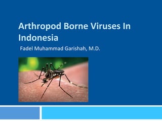
- 2. Arthropod-Borne Viruses ¨ Comprises 500 different viruses ¨ Vectors ¤ Mosquitoes: Aedes aegyp*; A. albopictus ¤ Ticks ¤ Sandflies ¨ Life cycle involving host blood and vector’s gut ¨ Transmission from infected person to healthy person via vector ¨ Trans-ovarial transmission occurs in vectors life cycle ¨ Diseases: Fever, arthriJs, arthralgia, rash, hemorrhages, plasma leakage, meningiJs, encephaliJs, meningoencephaliJs,
- 3. Iden3fied ARBOviruses in Indonesia ¨ Dengue Virus ¨ Chikungunya Virus ¨ Japanese EncephaliJs Virus ¨ Zika Virus
- 4. 1. Dengue Virus Infec3on ¨ The most spreading Arbovirus all over the world ¨ 50 millions infecJons annually with 2.5 billion people living in endemic area which includes dengue as an example of a disease that may constitute a public health emergency of international concern with implications for health security due to disruption and rapid epidemic spread beyond national borders. Figure 1.1 Countries/areas at risk of dengue transmission, 2008 Data Source: World Health Organization Map Production: Public Health Infrmation and Geographic Information Systems (GIS) World Health Organization The boundaries and names shown and the designations used on this map do not imply the expression of any opinion whatsoever on the part of the World Health Organization concerning the legal status of any country, territory, city or area or of its authorities, or concerning the delimitation of its frontiers or boundaries. Dotted lines or maps represent approximate border lines for which there may not yest be fill agreement. countries or areas at risk (As of 1 November 2008) The contour lines of the January and July isotherms indicate the potential geographical limits of the northern and southern hemispheres for year-round survival of Adeas aegypti, the principal mosquito vector of dengue viruses. July isotherm 10.C January isotherm 10.C © World Health Organization 2008
- 5. Clinical Manifesta3ons the clinicians’ decision as to where and how intensively the patient should be observed and treated (i.e. triage, which is particularly useful in outbreaks), in more consistent reporting in the national and international surveillance system, and as an end-point measure in dengue vaccine and drug trials. Figure 1.4 Suggested dengue case classification and levels of severity Probable dengue live in /travel to dengue endemic area. Fever and 2 of the following criteria: Laboratory-confirmed dengue (important when no sign of plasma leakage) Warning signs* concurrent with rapid decrease in platelet count *(requiring strict observation and medical intervention) Severe plasma leakage leading to: distress Severe bleeding as evaluated by clinician Severe organ involvement CRITERIA FOR DENGUE ± WARNING SIGNS CRITERIA FOR SEVERE DENGUE DENGUE ± WARNING SIGNS SEVERE DENGUE 1. Severe plasma leakage 2. Severe haemorrhage 3.Severe organ impairmentwithout with warning signs
- 6. Virology ¨ Single-stranded RNA virus ¨ Genus: Flavivirus ¨ Family: Flaviviridae ¨ Four serotypes DENV 1-4 possibly 5th serotype ¨ DENV-2 and -3 are associated with severe outcome ¨ AnJgen NS1 is useful for detecJon during acute infecJon
- 7. Vector Biology ¨ Aedes aegyp* and Aedes albopictus ¨ Breeding in clean water sites ¨ Blood hunJng morning-day
- 8. Infec3ous Disease Medicine ¨ IncubaJon 4-10 days ¨ Fever associated with headache, nausea, vomitus, abdominal pain, and rash (Dengue Fever) ¨ Thrombocytopenia and leukopenia ¨ Platelet funcJon alteraJons and coagulaJon system disorders leading to bleeding; endothelial disrupJon causing plasma leakage (Dengue Hemorrhagic Fever) ¨ if followed by hypotensive leading to dengue shock syndrome
- 9. Clinical Course 25 (where patients are first seen and evaluated) are critical in determining the clinical outcome of dengue. A well-managed front-line response not only reduces the number of unnecessary hospital admissions but also saves the lives of dengue patients. Early notification of dengue cases seen in primary and secondary care is crucial for identifying outbreaks and initiating an early response (Chapter 5). Differential diagnosis needs to be considered (Textbox B). Figure 2.1 The course of dengue illness* Days of illness Temperature Potential clinical issues Laboratory changes Serology and virology Dehydration Shock Reabsorption bleeding fluid overload 1 2 3 4 5 6 7 8 9 10 Organ impairment Hematocrit Platelet Viraemia IgM/IgG Febrile Critical Recovery phasesCourse of dengue illness: 40° * Source: adapted from Yip (2) by chapter authors.
- 10. Pathobiology ¨ Original anJgenic sin leads to cytokine storm causing plasma leakage ¨ Non-neutralizing anJbody (from pervious infecJon) leads to ADE ¨ AnJbody-dependent enhancement (ADE) leads to massive viral replicaJon and associated with severe symptoms ¨ ConsumpJve platelet, alteraJon of platelet and coagulaJon factors funcJon, leading to bleeding
- 12. Clinical excessive intravenous fluids have been administered. During the critical and/or recovery phases, excessive fluid therapy is associated with pulmonary oedema or congestive heart failure. The various clinical problems during the different phases of dengue can be summarized as in Table 2.1. Table 2.1 Febrile, critical and recovery phases in dengue 1 Febrile phase Dehydration; high fever may cause neurological disturbances and febrile seizures in young children 2 Critical phase Shock from plasma leakage; severe haemorrhage; organ impairment 3 Recovery phase Hypervolaemia (only if intravenous fluid therapy has been excessive and/or has extended into this period) 2.1.4 Severe dengue Severe dengue is defined by one or more of the following: (i) plasma leakage that may lead to shock (dengue shock) and/or fluid accumulation, with or without respiratory distress, and/or (ii) severe bleeding, and/or (iii) severe organ impairment. As dengue vascular permeability progresses, hypovolaemia worsens and results in shock. It usually takes place around defervescence, usually on day 4 or 5 (range days 3–7) of illness, preceded by the warning signs. During the initial stage of shock,
- 13. Approach to Pa3ent with dengue Dengue: Guidelines for diagnosis, treatment, prevention and control Table 2.2 A stepwise approach to the management of dengue Step I. Overall assessment I.1 History, including information on symptoms, past medical and family history I.2 Physical examination, including full physical and mental assessment I.3 Investigation, including routine laboratory and dengue-specific laboratory Step II. Diagnosis, assessment of disease phase and severity Step III. Management III.1 Disease notification III.2 Management decisions. Depending on the clinical manifestations and other circumstances, patients may: – be sent home (Group A); – be referred for in-hospital management (Group B); – require emergency treatment and urgent referral (Group C). Section 2.3 gives treatment recommendations for the groups A–C.
- 14. Diagnos3cs ¨ Virus detecJon (SEM, PCR) ¨ AnJgen detecJon (NS1 ELISA/Rapid test) ¨ AnJbody detecJon (HAI, ELISA, rapid test) ¨ Sonography for ascites ¨ RLD CXR for pleural effusion (PEI > 5% (+)) Nature Reviews | Microbiology Direct methods Indirect methods Serology IgM Serology IgG Genome detection Virus isolation Antigen detection Opportunity Specificity developments in rapid ogies offer the promise ostics for case manage- y detection of dengue tics of an ‘ideal’ dengue end on the purpose for be used. BOX 1 shows oduct specifications for at could be used for case eillance and outbreak vaccine trials5 . The r diagnosing a dengue y from the onset of fever ection; however, as not all osed within this period, test should be sensi- Figure 1 | Comparative merits of direct and indirect laboratory methods for the diagnosis of dengue infections. Opportunityreferstothefactthatantibodytestingisusuallythemostpractical diagnostic option available. EVALUATING DIAGNOSTICS | DENGUE
- 15. CHAPTER2 Compensated shock (systolic pressure maintained but has signs of reduced perfusion) Fluid resuscitation with isotonic crystalloid 5–10 ml/kg/hr over 1 hour IV crystalloid 5–7 ml/kg/hr for 1–2 hours, then: reduce to 3–5 ml/kg/hr for 2–4 hours; reduce to 2–3 ml/kg/hr for 2–4 hours. If patient continues to improve, fluid can be further reduced. Monitor HCT 6–8 hourly. If the patient is not stable, act according to HCT levels: if HCT increases, consider bolus fluid administration or increase fluid administration; if HCT decreases, consider transfusion with fresh whole transfusion. Stop at 48 hours. NOYES HCT or high HCT Administer 2nd bolus of fluid 10–20 ml/kg/hr for 1 hour Consider significant occult/overt bleed Initiate transfusion with fresh whole blood NOYES If patient improves, reduce to 7–10 ml/kg/hr for 1–2 hours Then reduce further Improvement Check HCT Improvement HCT = haematocrit
- 16. CHAPTER2 Figure 2.3 Algorithm for fluid management in hypotensive shock Hypotensive shock Fluid resuscitation with 20 ml/kg isotonic crystalloid or colloid over 15 minutes Try to obtain a HCT level before fluid resuscitation Crystalloid/colloid 10 ml/kg/hr for 1 hour, then continue with: IV crystalloid 5–7 ml/kg/hr for 1– 2 hours; reduce to 3–5 ml/kg/hr for 2–4 hours; reduce to 2–3 ml/kg/hr for 2–4 hours. If patient continues to improve, fluid can be further reduced. Monitor HCT 6-hourly. If the patient is not stable, act according to HCT levels: if HCT increases, consider bolus fluid administration or increase fluid administration; if HCT decreases, consider transfusion with fresh whole transfusion. Stop at 48 hours. NOYES HCT or high HCT Administer 2nd bolus fluid (colloid) 10–20 ml/kg over ½ to 1 hour Consider significant occult/overt bleed Initiate transfusion with fresh whole blood NOYES Improvement Review 1st HCT Improvement
- 17. 39 Monitor HCT 6-hourly. If the patient is not stable, act according to HCT levels: if HCT increases, consider bolus fluid administration or increase fluid administration; if HCT decreases, consider transfusion with fresh whole transfusion. Stop at 48 hours. Administer 2nd bolus fluid (colloid) 10–20 ml/kg over ½ to 1 hour Consider significant occult/overt bleed Initiate transfusion with fresh whole blood NOYES Improvement HCT or high HCT Repeat 2nd HCT Administer 3rd bolus fluid (colloid) 10–20 ml/kg over 1 hour Improvement NOYES Repeat 3rd HCT
- 19. 2. Japanese Encephali3s ¨ Mosquito-borne disease caused by Japanese EncephaliJs Virus (JEV) ¨ JEV -> Flaviviridae ¨ 9 geneJcally and anJgenicall related viruses in horses and including West-Nile Virus ¨ Reservoirs : Pig and Wild Birds (Heron) ¨ Vectors: Culex tritaeniorhynchus; Culex vishnui
- 24. Pathobiology
- 26. Clinical ¨ Mostly asymptomaJc ¨ < 1 % clinical JE ¨ IncubaJon 5-15 days ¤ Fever ¤ Headache ¤ Nausea/Vomitus ¤ Mental St. Changes ¤ Neurologicals ¤ Weakness ¤ Masklike facies ¤ Tremor ¤ Cogwheel rigidity ¤ Choreoathetoid movements ¤ Acute Flaccid Paralysis
- 28. Diagnos3cs ¨ E virus–specific IgM-capture ELISA on CSF or serum ¤ Day 4 CSF ¤ Day 7 Serum ¨ Virus isolaJon and nucleic-acid amplificaJon tests are insensiJve in detecJng JE virus or viral RNA in blood or CSF and should not be used for ruling out a diagnosis of JE
- 29. Vaccines
- 30. 3. Chikungunya Fever ¨ A viral illness causes by Chikungunya Virus ¨ Alphavirus genus of Togaviridae ¨ Single-stranded RNA virus ¨ Vector: Aedes aegyp* and Aedes albopictus ¨ Reservoirs: monkeys, rodents, birds, vertebrates
- 31. Clinical ¨ Febrile illness with incubaJon of 2-4 days ¨ Feverish ¨ Arthralgia (worse in the moring) ¨ Backache ¨ Headache
- 32. Clinical (2) Guidelines on Clinical Management of Chikungunya Fever4 persons had angiomatous lesions and fewer had purpuras. Stomatitis was observed in 25% and oral ulcers in 15% of patients. Nasal blotchy erythema followed by photosensitive hyperpigmentation (20%) was observed more commonly in the recent epidemic. Exfoliative dermatitis affecting limbs and Table 1: Clinical features in Chikungunya fever Common Infrequent Rare in adults but seen sometimes in children Fever Rash Photophobia Arthralgia Stomatitis Retro-orbital pain Backache Oral ulcers Vomiting Headache Hyperpigmentation Diarrhoea Exfoliative dermatitis Meningeal syndrome Acute encephalopathy
- 33. Case Detec3on ¨ Possible (clinical) ¨ Probable (clinical and epidemiological) ¨ Confirmed (lab confirmed)
- 34. Sequel ¨ Joint sJffness and pain ¨ Joint sJffness ¨ TendiniJs ¨ Neurological/emoJonal/dermatologic sequel
- 35. Diagnos3cs ¨ Viral isolaJon ¨ RT PCR ¨ IgM anJbody (ELISA ajer 2 weeks) ¨ Rising Jter of IgG (2-4 weeks)
- 36. Differen3als ¨ Dengue fever (PCR) ¨ RheumaJc fever (ASTO) ¨ Leptospirosis (MAT) ¨ Malaria (RDT/Blood smear) ¨ MeningiJs (CSF Analysis)
- 37. Acute Management ¨ Cold compress ¨ AnJpyreJcs ¨ Analgesics ¨ CorJcosteroid (methylprednisolon) ¨ Chloroquin phosphate 300mg/d ¨ Hydroxychloroquin 200mg/d ¨ Physiotherapy
- 38. 4. Zika Virus Infec3on ¨ Zika virus is a flavivirus (family flaviviridae) ¨ Acute febrile illness ¨ Associated with zika-virus microcephaly if infecJon occurs during pregnancy
- 39. Epidemiological Progress Mexico Panama El Salvador Samoa Tonga French Polynesia Cook Islands Easter Island, Chile Guatemala Honduras Nicaragua Curaçao Bonaire Costa Rica Jamaica HaitiCuba Dominican Republic Bolivia Colombia Brazil French Guiana Suriname Trinidad and Tobago Guyana Ecuador Paraguay Nigeria Uganda Gabon Cape Verde Thailand Laos Cambodia Malaysia Philippines Micronesia Solomon Marshall Islands Vanuatu Indonesia New Caledonia Fiji Martinique Dominica Puerto Rico U.S. Virgin Islands Saint Martin and Sint Maarten Barbados St. Vincent and the Grenadines Guadeloupe Maldives February March 2007–2009 2012–2014 State of Yap, Micronesia Gabon 20152015 French Guiana Honduras Martinique Panama Puerto Rico Laos New Caledonia St. Maarten Cuba Dominica 2016 20162016 Tonga Bonaire Marshall Islands St. Vincent and the Grenadines Trinidad and Tobago January–October November December January 2015 French Polynesia New Caledonia Easter Island, Chile Cook Islands Malaysia Philippines Cambodia Indonesia Thailand Brazil Vanuatu Fiji Colombia Cape Verde Samoa Solomon Islands El Salvador Guatemala Mexico Paraguay Suriname Venezuela Bolivia U.S. Virgin Islands Dominican Republic Ecuador Guyana Jamaica Curaçao Maldives Haiti American Samoa Costa Rica Guadeloupe St. Martin Nicaragua Barbados Figure 1. Areas in Which Zika Virus Infections in Humans Have Been Noted in the Past Decade (as of March 2016). Only sporadic infections have occurred in Southeast Asia, the Philippines, and Indonesia.
- 40. Transmission ¨ Mosquito-borne disease ¨ AmnioJc fluid ¨ Semen ¨ Blood transfusion
- 41. Zika Acute Febrile Illness ¨ 82 hours ajer inoculaJon ¨ 4 days – 1 week ¨ Symptoms ¤ Fever ¤ ConjuncJviJs ¤ Rash ¤ Arthralgia ¤ Myalgia ¤ Headache ¤ Retroorbital pain ¤ Edema ¤ Vomitus ¤ Guillain-Barre Syndrome ¤ MeningoencephaliJs, MyeliJs
- 42. Severe Microcephaly The new engl and jour nal o f medicine Figure 4. Infants with Moderate or Severe Microcephaly Associated with Maternal Zika Virus Infection, as Compared with a Typical Newborn. Baby with Typical Head Size Baby with Moderate Microcephaly Typical head size Typical head size Baby with Severe Microcephaly
- 43. Neuropathology ¨ Sonography ¤ Absent corpus callosum ¤ Hydranencephaly ¤ Cerebral calcificaJons ¤ Ventricular dilataJon ¤ Brain atrophy ¤ Abnormal gyraJon ¤ Hydrops fetalis ¤ Anhydramnions ¤ Intrauterine growth retardaJon
- 45. Treatment and Vaccines ¨ Treatment ¤ No treatment available so far ¨ Vaccines ¤ No vaccines available so far ¨ Control Measures ¤ Avoiding mosquito bites ¤ Vector control ¤ Pending pregnancy in infected future mother ¤ SelecJve aborJon (?)