4. APPLICATION OF REFRACTIVE PRINCIPLES TO LENSES CONVEX LENS FOCUSES LIGHT RAYS Convergence – the outer rays bend more and more toward the centre. If the lens has exactly the proper curvature parallel light rays passing through each part of the lens will be bent exactly enough so that all the rays will pass through a single point which is called FOCAL POINT.
5. CYLINDRICAL LENS BENDS LIGHT RAYS IN ONLY ONE PLANE – COMPARISON WITH SPHERICAL LENSES CONCAVE LENS DIVERGES LIGHT RAYS
6. COMBINATION OF TWO CYLINDRICAL LENSES AT RIGHT ANGLES EQUALS A SPHERICAL LENS
8. MEASUREMENT OF THE REFRACTIVE POWER OF THE LENS- “DIOPTER” The more a lens bends light rays, the greater is its “refractive power”. This refractive power is measured in terms of DIOPTERS.
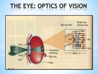
9. OPTICS OF THE EYE THE EYE AS A CAMERA The eye is optically equivalent to the usual photographic camera. It has a lens system, a variable aperture system (pupil), and a retina that corresponds to the film. LENS SYSTEM: Composed of four refractive interfaces: The interface between air and the anterior surface of the cornea The interface between the posterior surface of the cornea and aqueous humor
10. 3. The interface between the aqueous humor and the anterior surface of the lens 4. The interface between the posterior surface of the lens and the vitreous humor The internal index of air is 1; the cornea, 1.38; the aqueuoshumor, 1.33; the crystalline lens, 1.40 and the vitreous humor, 1.34. REDUCED EYE If all the refractive surfaces of the eye are algebraically added together and then considered to be one single lens, the optics of the normal eye may be simplfied and represented schematically as a “reduced eye”. This is useful in simple calculatons.
11. FORMATION OF AN IMAGE ON THE RETINA In the same manner that a glass lens can focus an image on a sheet of paper, the lens system of the eye can focus an image on the retina. The image is inverted and reversed with respect to the object. However, the mind perceives objects in the upright position despite the upside-down orientation on the retina because the brain is trained to consider an inverted image as normal.
12.
13. When the lens is in relaxed state with no tension on its capsule, it assumes an almost spherical shape, owing mainly to the elastic retraction of the lens capsule.
14. Located at the attachments of the lens ligaments to the eyeball is the ciliary muscle, which itself has two separate sets of smooth muscle fibers, meridonalfibers and circular fibers.
15.
16.
17.
18.
19.
20.
21.
22.
23.
24.
25. VISUAL ACUITY The normal visual acuity of the human eye for discriminating between point sources of the light is about 25 seconds of arc. NORMAL VISION- 20/20 DETERMINATION OF DISTANCE OF AN OBJECT FROM THE EYE – DEPTH PERCEPTION Determination of Distance by Sizes of Retinal Images of Known Objects Determination of Distance by Moving Parallax Determination of Distance by Stereopsis- Binocular Vision
26. OPHTHALMOSCOPE The ophthalmoscope is an instrument through which an observer can look into another person’s eye and see the retina with clarity.
27.
28.
29. INTRAOCULAR PRESSURE AVERAGE NORMAL IOP – 15mm Hg with a range from 12 to 20 TONOMETRY: The measurement of tension or pressure inside the eye (IOP)
30. GLAUCOMA- A PRINCIPAL CAUSE OF BLINDNESS GLAUCOMA – is a disease of the eye in which the IOP becomes pathologically high , sometimes rising acutely to 60 to 70 mm Hg. Pressures rising above 20 to 30 mm Hg can cause loss of vision when maintained for long periods. In most cases of glaucoma, the abnormally high pressures results from increased resistance to fluid outflow through the trabecular spaces into the canal of Schlemm at the iridocorneal junction. Treatment: Drops that reduce secretion or increases absorption of aqueous humor Surgery

![THE EYE: OPTICS OF VISION REFRACTION OF LIGHT ,[object Object],LIGHT RAYS TRAVEL: 300,000 km/sec – air 200,000km/sec – particular type of glass REFRACTIVE INDEX OF AIR – 1.00 ,[object Object],“THE DIRECTION IN WHICH LIGHT TRAVELS IS ALWAYS PERPENDICULAR TO THE PLANE OF THE WAVE FRONT , THE DIRECTION OF TRAVEL OF LIGHT BEAM BENDS DOWNWARD” REFRACTON- the bending of light rays at an angulated interface](data:image/gif;base64,R0lGODlhAQABAIAAAAAAAP///yH5BAEAAAAALAAAAAABAAEAAAIBRAA7)