WNT4 Role in Bone Formation and PTH Signaling

Wnt proteins are a family of conserved signaling proteins that regulate various processes including cell proliferation, fate determination, and embryonic development. They act through both canonical and non-canonical pathways. Wnt4 specifically promotes female development and suppresses male development. It also contributes to kidney and adrenal cortex development. Studies show Wnt4 enhances stem cell osteogenic differentiation by activating non-canonical pathways like P38MAPK. The NASA-Orthofix project examines the effects of pulsed electromagnetic fields on osteoblast proliferation, differentiation and mineralization using rat primary osteoblasts. Genes analyzed include osteocalcin, BAP, collagen I and GAPDH. Results show PEMF stimulation increases cell count, differentiation markers and mineralization
![WNT PROTEIN FAMILY: ,[object Object]](data:image/gif;base64,R0lGODlhAQABAIAAAAAAAP///yH5BAEAAAAALAAAAAABAAEAAAIBRAA7)
Recommended
More Related Content
What's hot
What's hot (20)
Viewers also liked
Viewers also liked (10)
Similar to WNT4 Role in Bone Formation and PTH Signaling
Similar to WNT4 Role in Bone Formation and PTH Signaling (20)
WNT4 Role in Bone Formation and PTH Signaling
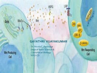
- 1. GAYATHRI VIJAYAKUMAR Dr. Nicola C. Partridge Dept of Basic Science & Craniofacial Biology NYU-CD
- 3. They do this by regulating and stimulating proliferation along the Notch-Wnt signaling pathway.
- 4. Wntprotiens grouped into canonical and non-canonical.
- 5. Wnt molecules can bind to the receptors LRP5, Frizzleds, Ror2 and RYK at cell surfaces to activate intracellular signaling pathways.TS19 mouse embryo with areas of Wnt signaling visible in red Targeted knockout of the Wnt4 genes in a female mouse shows that the kidney fails to develop. Wnt signals induce cancerous changes.
- 10. Cancer
- 12. Ca 2+ PCP B-catenin
- 14. As a mature molecule, human Wnt-4 is a 46 kDa, 329 aa glycoprotein that contains 24 cysteine residues and two possible N-linked glycosylation sites.
- 15. Wnt-4 expression in mesonephric and gonadalmesenchyme promotes the development of the Müllerianduct (a structure present in the embryo that develops into the uterus, fallopian tubes, cervix, and the upper part of the vagina) and blocks the development of Leydig cells.
- 16. In the same region, Wnt-4 expressing mesenchyme induces a mesenchyme-to-epithelium transition that promotes nephrondevelopment.
- 17. In the embryonic adrenal cortex mesoderm, Wnt-4 expression contributes to the proper formation of the zonaglomerulosa, a region that secretes aldosterone.
- 18. Wnt-4 is also found in virgin mammary epithelium. Progesterone increases Wnt-4 expression, leading to an enhanced side-branching (but not elongation) of mammary ducts.
- 19. Wnt-4 is detected in fibroblasts in areas of wound healing where fibrin degradation products are abundant.The WNT4 gene is located on the short (p) arm of chromosome 1 between positions 36.23 and 35.1.
- 20. WNT4 – A PTH ANABOLIC MEDIATOR PTH induces WNT4 mRNA expression in osteoblastic cells 8 hours after PTH injection and maximum expression is observed in the mineralization phase. Serum Ionized Ca2+ level decreases PTH Release Parathyroid Gland Enhance Osteoblast Differentiation Through Non-canonical Wnt/Ca2+ and Wnt/PCP pathway PKC Cell Proliferation & CFU-F numbers Regulates WNT4 PKA PTH binds to PTHR-1 in Bone, Kidney and other target tissues Expressed in chondrocytes & mature osteobalsts By inhibiting BMSSCs apoptosis & stimulating their rate of growth
- 21. WNT4-AN ENHANCER OF STEM CELL OSTEOGENIC DIFFERENTIATION WNT4 , a noncanonical member, was found to potently enhance osteogenic differentiation of MSCs isolated from craniofacial tissues in vitro and bone formation in vivo. Interestingly, Wnt4 did not increase the cytosolic level of B-catenin. But activated a novel noncanonical signaling pathway, P38MAPK, which is known to positively regulate osteogenic differentiation induced by BMPs and other growth factors Wnt 4 Stimulates osteocalcin mRNA Primary osteoblasts PCP & Cgmp/Ca2+ Pathway Some control in osteoblast differentiation Proliferating mBMSSCs B-catenin Pathway Wnt 4 Mouse BMSSCs Affects proliferation & differentiation Differentiating mBMSSCs Ca2+ Pathway
- 23. In committed osteoblasts, it stimulates non-canonical pathways responsible for its effect on differentiation.
- 24. Duality of Wnt4’s action depends on Development stage of the cell Active proliferatio of the cell + WNT4 mBMSSCs Both Osteogenic & non-osteogenicconditions Osteocalcin Bone Marker gene expression Adipocyte Marker Genes
- 25. Specific Aims Aim 1 : Assess the Wnt-4 effect on proliferation of human bone marrow stromal stem cells. Aim 2 : Assess the Wnt-4 effect on differentiation of human bone marrow stromal stem cells. Experimental Design Control and WNT-4 conditioned media collection: Nih3t3 Control Cells grown in DMEM supplemented with 10%FBS & 1%p/s Medium collected @ 80% confluency After syringe filtration Nih3t3 Wnt4 Producing Cells grown in DMEM supplemented with 10%FBS & 1%p/s Medium collected @ 80% confluency After syringe filtration Stored @-80C
- 26. Culturing Human BMSSCs with Recombinant WNT4 Medium: 10%FBS Alpha-MEM+ 1% p/s+1%Glu+Dex Isolate hBMSSCs 24ng/ml Wnt4 added to the wnt4 wells Human Cancellous Bone Specimen Plate cells at a conc of 1 million to 3 million cells/well in a 12 well plate Left untouched for 5 days for the cells to adhere Day 12: Viable Cell Count & Picture Taken Day 5: Medium changed to contain 1 μg/ml Ascorbic Acid Medium Changed every 2-3 days
- 27. Control hBMSSCS hBMSSCs treated with Wnt4
- 29. Sirtuin 1 is a member of the sirtuin family of proteins, homologsof the Sir2 gene in S. cerevisiae.
- 30. Sirt 1 is highly expressed in the foetal and adult brain .
- 31. Sirt1 not only deacetylateshistones H1, H3 and H4, but also deacetylates many non-histone proteins including p53, FOXO, Ku70, p300, Rb, E2F1, NF-kB, p73 and PGC-1α .
- 32. In virtue of these important targets, SIRT1 is linked to regulatory control of diverse normal and abnormal cellular processes ranging from stress responses, aging, and metabolism to cancer.
- 33. It is down-regulated in human senescent cells, suggesting that SIRT1 may be required to extend replicative life span
- 35. HDACs play a role in deacetylation of lysine residues in the tails of core histones.
- 36. They are classified into 4 groups.
- 38. It is activated through double phosphorylation by the JNK pathway but has also a phosphorylation-independent function.
- 40. HDAC-4 Under Basal Condition Runx2 RD AP-1 MMP-13 Promoter PTH Effect HDAC-4 Runx2 PKA dependent phosphorylation PTH RD AP-1 MMP-13 Promoter Cytoplasm Runx2 JUN & FOS pcaf p300 HAT pcaf p300 RD AP-1 HAT Nucleus
- 41. Sirt-1 HDAC-4 pcaf p300 JUN & FOS HAT Runx2 RD AP-1 MMP-13 Promoter
- 43. Whether there is association of Sirt-1 with Fos or Jun in Osteoblastic cells.
- 44. Whether there is an interaction between HDAC4 and Sirt1 as well as between HDAC4 , Fos and Jun.EXPERIMENT: Preparation of cell lysate PTH treatment for 4 and 8 hours UMR cells cultured till 80-90% confluent Immunopercipitated for Sirt1, Fos, Jun or HDAC4 Immunoblot for Sirt-1, HDAC4, Fosor Jun
- 45. Immunoblot Sirt-1 IgG jun Fos IP C4 P4 C8 P8 C4 P4 C8 P8 C4 P4 C8 P8 Immunoblot Sirt-1 IgG Sirt-1 HDAC4 IP C4 P4 C8 P8 C4 P4C8 P8 C4 P4 C8 P8
- 46. Immunoblot HDAC4 IgG jun Fos IP C4 P4 C8 P8 C4 P4 C8 P8 C4 P4 C8 P8 Immunoblot HDAC4 IgG Sirt-1 HDAC4 IP C4 P4 C8 P8 C4 P4 C8 P8 C4 P4 C8 P8
- 48. A decade later, FDA allowed the use of pulsed radiofrequency electromagnetic field (PRF) for treatment of pain and edema in superficial soft tissues.Three biological windows have been identified by analyzing the response of cells to a range of amplitudes and frequencies. These are: PEMF 50–100 lT (5–10 Gauss) 15–20 mT (150–200 Gauss) and 45–50 mT (450–500 Gauss) (Markov 2005).
- 49. NASA Signal Driver (Hydra) Project Protocol Rat Primary osteoblasts isolated from postnatal day 1 rat calvariae Plated with MEM+10% FBS. Treated with the PEMF signal 4h a day. Control not treated. Day 6 (proliferation stage) : Cell number counted Day 8: Induction of differentiation RT-PCR Day 12 & Day 18: Total RNA isolated from cells & von Kossa Staining Medium switch with BGJ + 10%FBS + Ascorbic Acid + ß-glycerophosphate
- 50. Genes Analyzed in the NASA-ORTHOFIX Project 1.Osteocalcin also known as bone gamma-carboxyglutamic acid-containing protein (BGLAP), is a noncollagenous protein secreted solely by osteoblasts.It is used as a biomarker for bone formation 2.Bone Alkaline Phosphatase (BAP) It is secreted by osteoblasts. Alkaline Phosphatase (ALP) is a ubiquitous enzyme associated with cell membranes.. It is produced by osteoblasts to provide a high PO4 concentration at the osteoblast cell surface during bone mineralization and is a marker of bone formation. 3.Type I Collagen formed by osteoblasts; reflects rate of collagen and bone formation; most sensitive marker of bone formation and particularly useful for monitoring bone formation therapies and antiresorptive therapies. 4.Glyceraldehyde 3-phosphate dehydrogenase (GAPDH ) is an enzyme that catalyzes the sixth step of glycolysis besides transcription activation, initiation of apoptosis, and ER to Golgi vesicle shuttling. Because the GAPDH gene is often stably and constitutively expressed at high levels in most tissues and cells, it is considered a housekeeping gene.
- 51. Osteoblast Mineralization – von Kossa Staining Cells fixed with 95% Ethanol (15 min at 37°C) Rinsed with 80% Ethanol Rinsed with 50% Ethanol Day 12 &18 Incubated with 5% silver nitrate solution (1 hr at 37°C) Rinsed with 20% Ethanol Washed with water Washed with water Measured by Image Quant UV Light (10 mins) Dried and Photographed
- 52. Overall Fold Stimulation in Cell Count during Proliferation Stage
- 53. Overall Fold Stimulation for Differentiation Stage
- 54. Overall Fold Stimulation for Mineralization Stage
- 55. Von Kossa Staining – Differentiation Stage Control PEMF Treated
- 56. Image Quant Data for von Kossa Staining- Differentiation Stage
- 57. Von Kossa Staining – Mineralization Stage Control PEMF Treated
- 58. Image Quant Data for von Kossa Staining- Mineralization Stage