Cerebellum by dr.gourav thakre 20 03-2012
•
75 likes•12,350 views

detailed anatomy of cerebellum

Recommended
More Related Content
What's hot
What's hot (20)
Similar to Cerebellum by dr.gourav thakre 20 03-2012
Similar to Cerebellum by dr.gourav thakre 20 03-2012 (20)
More from Gourav Thakre
More from Gourav Thakre (8)
Recently uploaded
Recently uploaded (20)
Cerebellum by dr.gourav thakre 20 03-2012
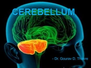
- 1. - Dr. Gourav D. Thakre
- 2. Introduction External Features Subdivisions Of Cerebellum Cytoarchitecture / Histology Connections Of Cerebellum Intrinsic Cerebellar Circuitry Cerebellar Peduncles Development Of Cerebellum Blood Supply Functions Applied Anatomy
- 3. Introduction Cerebellum (Latin) - little brain Weight- 150 gm Location- post. Cranial fossa Connected with brainstem by 3 peduncles Imp function of maintenance of posture, tone , coordination of voluntary motor activity.
- 4. External Features Parts – hemispheres vermis- superior inferior Surfaces Notches Fissures- horizontal Posterolateral Fissura prima
- 5. Subdivisions Of Cerebellum A. Anatomical subdivisions B. Morphological subdivisions C. Functional subdivisions
- 6. Anatomical Subdivisions Primary fissure Anterior lobe corpus of cerebellum Posterior lobe Posterolateral fissure Flocculonodular lobe
- 8. Some neuroscientist map out cerebellum by Ten Roman numericals
- 9. Morphological Subdivisions Archicerebellum / vestibular cerebellum Paleocerebellum / spinal cerebellum Neocerebellum / cerebro-pontine cerebellum
- 10. Archicerebellum / vestibular cerebellum First to appear Aquatic vertebrates Flocculonodular lobe& lingula Maintenance of equilibrium, tone & posture of Trunk muscles Paleocerebellum / spinal cerebellum Next to Archicerebellum in evolution Terrestrial vertebrates Anterior lobe except lingula, pyramid & uvula Muscle tone and posture &crude movements of limbs
- 11. Neocerebellum / cerebro-pontine cerebellum Developmentally last Max. development in mammals Rest of the cerebellum Smooth performance of the skilled acts by coordination of movements
- 12. Functional Subdivisions Corpus cerebelli (cerebellum except flocculo-nodular lobe) Vermal / median Nucleus fastigii Movement of the trunk & extensor muscle tone Pair of Para-vermal / intermediate Nucleus interpositus ( globosus , emboliformis) Flexor muscle tone Pair of hemisphere/ lateral zones Nucleus dentatus Distal limb muscles for skillful prehensile act.
- 13. Spinocerebellum: Vermis Intermediate hem Cerebrocerebellum: Lateral hem. Spinocerebellum: IVth vent Vermis Intermediate hem Cerebrocerebellum: Lateral hem. Floculo-nodular lobe
- 14. Cytoarchitecture / Histology Outer gray matter / cerebellar cortex Inner white matter Intracerebellar nuclei Fissures Folia Arbor vitae cerebelli
- 15. Cerebellar Cortex Uniform thought/ homotypical 3 layers a) Outer molecular layer b) intermediate Purkinje cell layer c) Inner granular layer
- 16. The molecular layer 300-400 μm thick sparse population of neurones, dendritic arborizations, non-myelinated axons & radial fibres of neuroglial cells stellate neurones (superficially) short processes , axons arborise with dendritic spines of purkinje cells basket cells (deeper) small in size, little cytoplasm & extensive processes axon runs transversely parallel to cortex & rt. Angle to longitudinal axis of folia synapse with >500 Purkinje cell
- 17. Purkinje cell dendritic trees Climbing fibres Radiating branches from large epithelial (Bergmann) glial cells give off processes -surround all neuronal elements, except at the synapses. At the surface of the cerebellum their conical expansions join to form an external limiting membrane.
- 18. Purkinje cell layer large, pear-shaped somata smaller somata of epithelial (Bergmann) glial cells. Clumps of granule cells and occasional Golgi cells penetrate between the Purkinje cell somata.
- 19. Dendrites synapse with Collaterals from basket cells Axons of granule cells Climbing fibers Axon pass through granular layer into white matter i.e to intracerebellar nuclei Sole output from cerebellar cortex Inhibitory to the intracerebellar nuclei Individual Purkinje cells are separated by 50 μm transversely & 50-100 μm longitudinally. Their somata measure 50-70 μm vertically & 30-35 μm transversely total number of dendritic spines per Purkinje neurone 180,000.
- 20. Granular Layer 100 μm thick in the fissures and 400-500 μm on foliar summits 2.7 million granular neurones per cubic millimetre 3000 granule cells for each Purkinje cell. granule cell - 5-8 μm in diameter 3 to 5 short dendrites- end in claw-like terminals within the synaptic glomeruli Axon bifurcates at a “T” junction , branches runs parallel to long axis of folium (Parallel fibers) Also contains few larger Golgi cells , dendrites ramify in molecular layer
- 21. Intrinsic neurons of cortex 5 types- Purkinje cells, granule cells , basket and stellate cells, Golgi cells All are inhibitory except granule cells.
- 23. A rosette of mossy fiber in centre ( pre-synaptic element) Claw like dendritic expansions of granule cells envelops rosette ( post synaptic element) Axon terminals of golgi cells come in contact with granule cell dendrites as pre-synaptic elements
- 24. Cerebellar Nuclei On each side- 4 nuclei Lat.- med. A. Dentate nucleus B. Emboliform nucleus C. Globose nucleus D. Fastigial nucleus Fastigial nucleus Cerebellar cortex Globose nucleus Emboliform nucleus Dentate nucleus medullary center
- 25. Dentate nucleus Most prominent Largest in primates Nucleus of neocerebellum Crumpled bag shape, hilum facing anteromedially Interior filled with white matter Fibers mainly forms superior cerebellar peduncle Dentorubral(red nucleus- spinal cord), dentothalamic fibers( ventral lateral nucleus of thalamus- cerebral cortex)
- 26. Emboliform nucleus Oval, medial to dentate nucleus Nucleus of paleocerebellum Red nucleus through sup. Cerebellar peduncle Red nucleus spinal cord (rubrospinal tract) Flexor muscle tone Globose nucleus Rounded Between emboliform & fastigial nucleus Similar connection as emboliform nucleus Globose & Emboliform nucleus collectively called nucleus interpositus
- 27. Fastigial nucleus Near midline in vermis, close to roof of 4th ventricle Nucleus of archicerebellum Afferents from flocculonodular lobe Efferents to vestibular & reticular nuclei Extensor muscle tone
- 28. Connections Of Cerebellum Afferent fibers- cerebral cortex, spinal cord, vestibular apparatus Also from red nucleus, tectum of midbrain Cortex- cortico-ponto-cerebellar cerebro-olivo-cerebellar cerebro-reticulo-cerebellar pathways Spinal cord- post. Spinocerebellar ant. Spinocerebellar cuneocerebellar tracts Afferent mainly enters through inferior & middle peduncles
- 29. Two types of fibers Climbing fibers Originate in inf. Olivary nucleus Collateral to intracerebellar nucleus Monosynaptic contact with purkinje cell. Mossy fibers Ends by forming 30-40 rosette Synapse with dendrites of granule cells & axons of golgi cells Forms cerebellar glomerulus. Both fibers are excitatory to the purkinje cells All afferent fibers are mossy except olivocerebellar & parolivocerebellar
- 30. Efferent fibers- to the red nucleus, thalamus, vestibular complex & reticular formation Entirely from axons of purkinje cells . Fibers from dentate, emboliform & globose nuclei- sup. Cerebellar peduncle Fastigial nucleus- inf. Cerebellar peduncle
- 31. Intrinsic Cerebellar Circuitry Feed forward inhibition Neural sharpening- row of excited purkinje cells is surrounded by zone of inhibition at periphery.
- 33. Inferior cerebellar peduncle Afferent fibers Posterior spinocerebellar fibers- ipsilateral thorasic nucleus (Clarke’s column) Olivocerebellar – opposite inf. Olivary nucleus Parolivocerebellar fibers- opposite medial & dorsal accessory olivary nucleus Cuneocerebellar- ipsilateral cuneate nucleus Anterior external arcuate fibers- arcuate nuclei of both sides Vestibulocerebellar- primarily from vestibular nerve. Secondary from medial & inf. Vestibular nuclei Reticulocerebellar- lateral & paramedian reticular nuclei of medulla oblongeta
- 34. Efferent fibers Cerebellovestibular fibers- ipsilateral flocculonodular lobe & fastigial nuclei of both sides Cerebelloreticular fibers- fastigial nuclei of both sides to pontine & medullary reticular formation Cerebello-olivary fibers- to inf. Olivary nucleus
- 35. Middle cerebellar peduncle Afferents Pontocerebellar fibers- forms the bulk of peduncle, from pontine nuclei to opposite neocerebellar cortex. (cortico-ponto-cerebellar pathway) Reticulocerebellar fibers- reticular formation to ipsilateral vermal region of cerebellum Seratogenic fibers- from raphe nuclei of pons. No efferent fibers
- 36. Superior cerebellar peduncle Afferent fibers Anterior spinocerebellar tract Tectocerebellar fibers- tectum of midbrain (sup. & inf. Colliculi of both sides) Trigeminocerebellar fibers- sup. Sensory & spinal nuclei of trigeminal nerve Ceruleocerebellar fibers- noradrenergic fibers from locus ceruleus Hypothalamocerebellar fibers- cholinergic fibers from hypothalamus
- 37. Efferent fibers Cerebellorubral- globose & emboliform nuclei to contralateral red nucleus Dentatorubral & dentatothalamic fibers- dentate nucleus to opposite red nucleus & thalamus Cerebello-olivary fibers- dentate nucleus to opposite inf.olivary nucleus Cerebelloreticular fibers-nucleus fastigius to reticular nucleus.
- 38. Development Of Cerebellum The cerebellum develops from the rhombic lip, the dorsal part of the alar plate of the metencephalon Two rounded swellings develop which at first project partly into the ventricle forming the rudimentary cerebellar hemispheres
- 39. With growth, a number of transverse grooves appear on the dorsal aspects of the cerebellar rudiment(precursors of the numerous fissures )
- 40. early in cerebellar development a layer of cells derived exclusively from the metencephalic rhombic lip cerebellum has an intraventricular portion (cells proliferating from the ventricular zone) and an extraventricular portion (cells proliferating from the external germinative layer) during development Before the end of the third month the main mass of the cerebellum is extraventricular.
- 41. Four stages in the histogenesis of the cerebellar cortex and the cerebellar nuclei Purkinje cells and cells of the cerebellar nuclei are produced by the ventricular epithelium and are in the process of migration to their future positions. The cells of the superficial matrix (the external granular layer) take their origin from the ventricular epithelium at the caudal pole of the cerebellar anlage and migrate rostrally over its surface.
- 42. After migration the Purkinje cells constitute a multicellular layer beneath the external granular layer. Cell production in the ventricular epithelium has stopped. The remaining cells transform into ependymal cells.
- 43. Granule cells are produced by the external granular layer and migrate inwards through the Purkinje cell layer to their position in the granular layer. Purkinje cells spread into a monolayer.
- 44. Adult position of cortical and nuclear neurones.
- 45. Blood Supply
- 46. Cerebellar veins arranged in 2 groups- superior & inferior Sup. Cerebellar vein partly in straight sinus & great cerebral vein, partly in transverse & superior petrosal sinus Inf. Cerebellar vein sigmoid, inferior petrosal, occipital sinus
- 47. Functions ANTICIPATORY FUNCTION The vermis of the cerebellum is involved in taking anticipatory action POSTURAL FIXATION The posterolateral region of the cerebellum is required to prevent oscillation of distal limb parts caused by the viscoelastic properties of the muscles in response to sudden movements MOTOR LEARNING when a novel motor skill is being learned, the olivocerebellar climbing fibre system becomes active when errors are made. The inferior olivary complex appears to be involved in correction HIGHER FUNCTIONS
- 48. Applied Anatomy Are usually vascular, may be traumatic or tumour. Manifestations of unilateral cerebellar lesions : 1-ipsilateral incoordination of (U.L) arm = intention tremors : it is a terminal tremors at the end of movement as in touching nose or button the shirt. 2-Or ipsilateral cerebellar ataxia affects (L.L.) leg, causing wide-based unsteady gait. Manifestations of bilateral cerebellar lesions (caused by alcoholic intoxication, hypothyrodism, cerebellar degeneration & multiple sclerosis) : 1-dysarthria : slowness & slurring of speech. 2-Incoordination of both arms.= intention tremors.
- 49. 3-Cerebellar ataxia : intermittent jerky movements or staggering , wide-based, unsteady gait. 4-Nystagmus : is a very common feature of multiple sclerosis. It is due to impairment coordination of eye movements /so, incoordination of eye movements occurs and eyes exhibit a to-and-fro motion. Combination of nystagmus+ dysarthria + intension tremors constitutes Chacot’triad, which is highly diagnostic of the disease.
- 50. For purpose of localization, cerebellum can be viewed as a saggitally-oriented structure containing 3 zones on each side: Midline Intermediate Lateral
- 51. Midline zone Consists of the anterior and posterior parts of the vermis, fastigial nucleus and associated input and output projections concerned with posture, locomotion, position of head relative to trunk, control of EOM’s Cerebellar signs resulting from midline cerebellar disease disorders of stance/gait, truncal postural disturbances, rotated postures of the head, disturbances of eye movements Rhomberg Test Tests of gait- tandem, toe + heel walking, walking backward Hop on each foot
- 52. Intermediate zone Consists of paravermal region of cerebellum and interposed nuclei (emboliform, globose) concerned with control of velocity, force and pattern of muscle activity Clinical disorders related to disease of this zone not clearly delineated
- 53. Lateral zone cerebellar hemisphere and dentate nucleus on each side concerned with the planning of movement in connection with neurons in the Rolandic region of the cerebral cortex (fine, skilled) Lesions result in abnormalities of skilled voluntary movements: hypotonia, dysarthria, dysmetria, dysdiadochokinesia, excessive rebound, impaired check, kinetic and static tremors, past-pointing Finger-to-nose test Rapidly alternating movements Heel-to-shin test
- 54. Hypotonia Ataxia Dysarthria Tremor Ocular Motor Dysfunction Depending on extent, an individual may have one symptom or a combination In all cases, symptoms from unilateral damage appear on the side ipsilateral to the injury Ascending spinocerebellar pathways are uncrossed and descending corticoopontocerebellar fibers are crossed; thus motor deficits from cerebellar damage are ipsilateral to the lesion whereas motor deficits from damage to motor areas of the cerebral cortex are contralateral to the lesion
- 55. postural instability delayed initiation and termination of motor actions inability to perform continuous, repetitive movements errors in smoothness and direction of a movement lack of coordingation or synergy of movement, especially complex movements lack of motor plasticity or learning
- 57. Cerebellar Medulloblastoma Cerebellar tumors on vermis - Truncal Ataxia - Frequent Falling The child in this picture: - would not try to stand unsupported - would not let go of the bed rail if she was stood on the floor.