Haliunaa subvesical bile duct
•Download as PPTX, PDF•
5 likes•2,737 views

ppt for surgeon

Recommended
Recommended
More Related Content
What's hot
What's hot (20)
Similar to Haliunaa subvesical bile duct
Similar to Haliunaa subvesical bile duct (20)
More from Haliunaa Battulga
Recently uploaded
Recently uploaded (20)
Haliunaa subvesical bile duct
- 1. Accessory cystic duct identification in laparoscopic cholecystectomy Duct of Luschka U. Parampalli, S. Helme, G. Asal and P. Sinha Department of General Surgery, Princess Royal University Hospital, Orpington, Kent, BR6 8ND, UK Corresponding address: Mr Prakash Sinha, Consultant, Department of General Surgery Princess Royal University Hospital, Orpington, Kent, BR6 8ND, UK. E- mail: p.sinha@btinternet.com Date accepted for publication 8 September 2008
- 2. Abstract We report the case of a 53-year-old lady who underwent laparoscopic cholecystectomy and was found to have an accessory cystic duct close to the fundus. Careful dissection of the liver bed was done and the duct clipped preventing a bile leak. The presence of such ducts though rare should be identified during surgery to prevent potential complications.
- 3. Case report A 53-year-old lady presented with symptoms suggestive of acute pancreatitis. Investigations confirmed the same and it was found to be gall stone related. She was managed conservatively until her symptoms subsided and biochemical parameters returned to normal. An abdominal ultrasound scan revealed multiple stones within the gallbladder and common bile duct (CBD) measuring 8 mm in diameter. Magnetic resonance cholangiopancreatography (MRCP) showed multiple stones in the gallbladder, common bile duct measuring 7.3 mm with the possibility of tiny non-obstructing calculi at the distal end. Radiological investigations did not reveal any anatomical abnormalities.
- 4. Case report Following this she was discharged. She returned for an interval laparoscopic cholecystectomy. At surgery normal anatomy was identified in the Calot’s triangle. The cystic duct and artery were isolated, double clipped and divided. On careful dissection of the gallbladder from the liver bed, a tubular structure was found running from the liver bed to the body of the gallbladder close to the fundus. The tubular structure was clipped close to the liver bed and the gall bladder end was divided. Bile was seen leaking from the gall bladder end confirming this was an accessory cystic duct (Fig. 1). The rest of the dissection was uneventful and the gallbladder was removed safely. She made a good recovery and was later discharged.
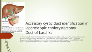
- 5. Discussion Ten percent of biliary collecting systems have variations in anatomy as a result of their complex embryological development[1]. Ninety percent of bile duct anomalies are found close to Calot’s triangle. Failure to identify these may result in bile leak. This causes significant morbidity and has been reported to occur in 0.2–2% of cases after laparoscopic cholecystectomy[2]. Accessory bile ducts in the gallbladder fossa have been classified into two types: the bile duct of Luschka and the cystohepatic duct. The bile duct of Luschka (subvesical duct) is a slender duct 1–2 mm in diameter which runs in the submucosa of the posterior gall bladder wall. It drains subsegmental areas of the liver into the right hepatic duct (Fig. 2). Its prevalence has been estimated at one-third of the population[3].
- 6. The cystohepatic duct drains the subsegmental parenchymal distribution of the right lobe of the liver to the right hepatic duct or the cystic duct (Fig. 2). Its prevalence has been estimated at 1–2% of surgical cases. Double cystic ducts are also classified into, ‘Y’ type where both ducts meet and form a single duct, ‘H’ type where an accessory duct joins the right, left or common hepatic duct, and the trabecular type in which the accessory duct directly enters the liver substance[4]. Fig. 2. (a) Duct of Luschka; (b) Cystohepatic duct.
- 7. Types of subvesical bile ducts
- 8. Preoperative identification of such anomalies can be difficult but helps in planning surgery and avoiding duct damage or bile leaks. Studies report that magnetic resonance cholangiography has 66% sensitivity to show accessory bile ducts, whereas helical computed tomography (CT) cholangiography has up to 100% sensitivity[5]. Routine use of intra-operative cholangiography is emphasized but not practiced widely. In the above case, the accessory duct was identified close to the fundus only after meticulous dissection of the liver bed.
- 9. Management of bile leaks Seventy-five percent of bile leaks are classified as major injuries occurring from common bile ducts, biliary confluence and hepatic ducts. Primary end to end repair over T-tube, direct closure and Roux-en-Y hepaticojejunostomies are used for the treatment of major injuries[6]. Accessory cystic duct identification in laparoscopic cholecystectomy 41 leaks occur from cystic ducts and ducts of Luschka. Sclerotherapy and embolization have been tried but surgery is the treatment of choice.