
Recommended
More Related Content
What's hot
What's hot (20)
Similar to Pelvic organ prolapse
Similar to Pelvic organ prolapse (20)
More from hemnathsubedii
More from hemnathsubedii (13)
Recently uploaded
Recently uploaded (20)
Pelvic organ prolapse
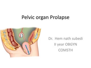
- 1. Pelvic organ Prolapse Dr. Hem nath subedi II year OBGYN COMSTH
- 2. DEFINITION • The entity includes descent of the vaginal wall and/or the uterus. • It is infact a form of hernia. • The uterus is normally placed in anteverted and anteflexed position.
- 3. SUPPORTS OF THE UTERUS • The external os lies at the level of ischial spines. • Apart from the normal position- anteverted anteflexed there is a three tier system • consisting of the following: – Upper – Middle (strongest and important) – Lowers
- 4. • Upper Tier – Weak – Mostly by maintaining the uterus in anteverted position – Endopelvic fascia – Round ligaments – Broad ligaments with intervening pelvic cellular tissues • Middle Tier – Strongest support of uterus – Cervico vaginal junction – Pelvic cellular tissue • Inferior Tier – Indirect support to the uterus – Principally given by the musculofascial tone of the hollow vagina which is amply supported by the fascial condensation at the vault and by the pelvic floor at the lower end.
- 10. • Nine specific sites are considered. Hymen is taken as the fixed point. The plane of hymen is defined as the zero level. Leading point of prolapse may be above (proximal) or below (distal) to the plane of hymen. Prolapse measurements (cm) are recorded as negative numbers when above and positive numbers when lies below the plane of hymen. Organ prolapse is measured with a wooden PAP spatula with markings. The woman may be examined in lithotomy or standing position (or even under anesthesia). She may be asked to do some maneuvers (valsalva) to demonstrate the prolapse maximally. Total vaginal length (TVL) is measured after reducing the prolapse while rest of the measurements are done when the prolapse is seen maximally.
- 12. Symptoms following symptoms are usually associated: (a) Feeling of something coming down per vaginum, especially while she is moving about. There may (b) Backache or dragging pain in the pelvis. (c) Dyspareunia. (e) Bowel symptom (in presence of rectocele). – Difficulty in passing stool. The patient has to push back the posterior vaginal wall in position to complete the evacuation of feces. Fecal incontinence may be associated.
- 13. (d) Urinary symptoms (in presence of cystocele). – Difficulty in passing urine. – Incomplete evacuation. – Urgency and frequency of micturition. – Painful micturition is due to infection. – Stress incontinence – Retention of urine may rarely occur. (f) Excessive white or blood-stained discharge per vaginum is due to associated vaginitis or decubitus ulcer.
- 14. Complications • Keratinization of the vagina • Decubital ulceration • Elongation of supravaginal cervix • Congestion and edema of cervix • Glandular hypertrophy • Ureteric obstruction • Renal failure • Incarcerationof the prolapse • Carcinoma of the cervix or vagina
- 15. Differential diagnosis • Cystocele – Gartner’s cyst • Uterine Prolapse – Congenital elongation of the cervix. – Chronic inversion – Fibroid polyp
- 16. MANAG EMENT OF PROLA PSE – Preventive – Conservative – Surgery
- 17. PREVENTIVE The following guidelines may be prescribed to prevent or minimize genital prolapse. Adequate antenatal and intranatal care • To avoid injury to the supporting structures during the time of vaginal delivery either spontaneous or instrumental. Adequate postnatal care • To encourage early ambulance. • To encourage pelvic floor exercises by squeezing the pelvic floor muscles in the puerperium. General measures • To avoid strenuous activities, chronic cough, constipation and heavy weight lifting. • To avoid future pregnancy too soon and too many by contraceptive practice.
- 18. CONSERVATIVE • Indications of conservative management are: – Asymptomatic women. – Mild degree prolapse. – POP in early pregnancy. • Meanwhile, following measures may be taken : Improvement of general measures (see above). Estrogen replacement therapy may improve minordegree prolapse in postmenopausal women. Pelvic floor exercises in an attempt to strengthen the muscles (Kegel exercises). Pessary treatment.
- 20. Anterior colporrhaphy • Preliminaries • The operation is done under general or epidural anesthesia. • The patient is placed in lithotomy position. • Vulva and vagina are to be swabbed with antiseptic solution. • The perineum is to be draped with sterile towel and legs with leggings. • Bladder is to be emptied by metal catheter. • Vaginal examination is done to assess the type anddegree of prolapse.
- 21. Steps of anterior colporrhaphy
- 22. Periniorrhaphy
- 24. Composite steps of forthergill’s operation Preliminary D and C Amputation of cervix Plication of Mackenrodt’s ligaments in front of cervix Anterior colporrhaphy Colpoperineorrhaphy
- 25. Vaginal hysterectomy Principles of the operation in prolapse • Removal of the uterus through vaginal route. • Correction of enterocele, if any. • Approximation of the pedicles in the midline to have a good buttress. • Fixation of the uterosacral ligaments to the vault to prevent vault prolapse. • Bladder support is reconstituted utilizing the broad ligaments and round ligaments as buttress. • Repair of cystocele. • Reconstruction of the perineum.
- 31. Thank you Take home message