SlideShare a Scribd company logo
1 of 132
Evaluation of the Relationship between Rest Position and
Habitual Occlusion in the Transverse Plane:
 
The position of the midline of the mandible is observed while the jaw is moved 
from the postural rest to habitual occlusion. This analysis is particularly relevant 
for the differential diagnosis of cases with unilateral cross­ bite. Depending on 
the  functional  analysis  two  types  of  skeletal  mandibular  deviation  can  be 
differentiated: laterognathy and lateroclusion.
 
Laterognathy: The center of the mandible is not aligned with the facial midline in 
rest  and  in  occlusion.  These  dysplasias  constitute  true  neuromuscular  or 
anatomical asymmetry. A lateral cross­bite with laterognathy is termed true cross­
bite. The prognosis is unfavorable for causal therapy.
 
Lateroclusion: The skeletal midline shift of the mandible can be observed only in 
occlusal position; in postural rest both midlines are well aligned. The deviation is 
due to tooth guidance (functional non­true malocclusion).
www.indiandentalacademy.com
Laterognathy:
Right: The center of the mandible is displaced in habitual occlusion. The skeletal 
midline of the lower jaw (mental spine) is shifted laterally in relation to the facial 
midsagittal plane (vertical line of reference).
 
Left: The  lateral  deviation  of  the  mandibular  midline  in  relation  to  the  facial 
midline persists in the postural rest position.www.indiandentalacademy.com
Laterocclusion:
Right: When  in  occlusal  position,  the  midline  of  the  mandible  is  displaced 
laterally from the facial mid­sagittal plane (vertical reference lines).
 
Left: In postural rest both midlines are coincident and well centered 
www.indiandentalacademy.com
Relationship of postural rest to occlusal position:
Postural resting position 
In the rest position the center of the mandible is aligned with the upper midline. (The 
mesial contact point of the lower central incisors coincide with the skeletal midline of 
the mandible).
Initial tooth contact position: 
The mandible is in the initial tooth contact position, thus terminating the first phase of 
the closing movement, which started from the rest position. The patient's right lateral 
incisors contact prematurely.
 Habitual occlusion: 
In the final phase of the closing action, after initial tooth contact, the mandible slides to 
the left.
Mandibular  deviation  in  habitual  occlusion  is  caused  by  tooth  interferences,  i.e.  the 
skeletal midline shift of the mandible which is only present in maximum intercuspation 
signifies a functional malocclusion (Lateroclusion )
www.indiandentalacademy.com
Examination of the Temporomandibular Joint:
The main objective of the clinical examination is to assess the severity of the clicking, pain, 
and dysfunction, which are characteristic of pathologic TMJ symptoms.
 
When auscultation is carried out with a, stethoscope, clicking and crepitus in the joint may 
be diagnosed during anteroposterior and eccentric movements of the mandible 
www.indiandentalacademy.com
Joint clicking is differentiated as follows: 
 
Initial clicking is a sign of retruded condyle in relation to the disc.
Intermediate clicking is a sign of unevenness of the condylar surfaces and of the 
articular disc, which slide over one another during the movements.
Terminal clicking occurs  most  commonly  and  is  an  effect  of  the  condyle  being 
moved too far anteriorly, in relation to the disc, on maximum jaw opening.
Reciprocal clicking occurs  during  opening  and  closing,  and  expresses  an 
incoordination between displacement of the condyle and disc. Clicking of the joint 
is rare in children.
www.indiandentalacademy.com
Palpation of the temporomandibular joint during  opening  maneuvers 
will reveal possible pain on pressure of the condylar areas. Besides the right and 
left condyles can thus be checked for synchrony of action. 
Lateral palpation of the temporomandibular joints:
Exert slight pressure on the condyloid process with the index fingers. Palpate both sides 
simultaneously.
Register  any  tenderness  to  palpation  of  the  joints  and  any  irregularities  in  condylar 
movement during opening and closing maneuvers. The coordination of action between 
the left and right condylar heads should be assessed at the same time.
www.indiandentalacademy.com
Posterior palpation of the temporomandibular joints: 
Position the little fingers in the external auditory meatus and palpate the posterior 
surface of the condyle during opening and closing movements of the mandible.
Palpation should be carried out in such a way that the condyle displaces the little 
finger when closing in full occlusion.www.indiandentalacademy.com
Palpation of the musculature involved in mandibular movements is a 
considerable part of the examination.
 
Palpation of the lateral pterygoid muscle:
The  pain  projection  area  of  the  lateral  pterygoid  muscle  is  palpated  in  close 
proximity to the neck of the condyle and the joint capsule, cranially behind the 
maxillary tuberosity. The examination is carried out with the mouth open and the 
mandible displaced laterally.
In the initial stages of TMJ dysfunction, the muscle often hurts upon palpation 
on one side only. In the advanced stage the pain is usually bilateral.
www.indiandentalacademy.com
Palpation of the temporalis muscle: 
The temporalis muscle is palpated bilaterally and extraorally. The anterior, medial, 
and posterior portions of the muscle are examined separately.
The palpation is carried out while the muscle is contracted isometrically
 
Left: The  temporal  tendinous  attachment  on  the  coronoid  process,  in  the 
posterolateral  region  of  the  upper  vestibule,  is  palpated.  The  patient's  mouth 
should be half open for the examination.www.indiandentalacademy.com
Palpation of the masseter muscle: 
The superficial masseter muscle is palpated beneath the eye, inferior to the zygomatic arch.
The deep portion is palpated on the same level, approximately 2 finger widths in front of tragus.
Left: During maximum isometric muscle­contractions the width of the superficial masseter and 
its direction of pull can be registered around the gonial angle. This muscle attachment should be 
examined for pain on to pressure. Occasional trigger spots may occur which can be quite painful.
www.indiandentalacademy.com
Recording the maximum interincisal distance:
On maximum jaw opening, the distance between the incisal edges of the upper 
and lower central incisors is measured with a Boley gauge.
In overbite cases this amount is added to the obtained value whereas in open 
bite it is subtract­ed. The extent of maximum jaw opening between the incisal 
edges  is  usually  40­45  mm.  ln  cases  with  TMJ  dysfunction,  hypermobility  is 
often registered in the initial stages and limitation in the later stages.
www.indiandentalacademy.com
Opening and Closing Movements of the Mandible:
The  opening  and  closing  movements  of  the  mandible  as  well  as  its  protrusive, 
retrusive and lateral excursions are examined as part of the functional analysis. 
The size and  direction of  these  actions  are  recorded  during  the  clinical 
examination. Deviations in speed can only be registered with electronic devices 
(e.g. kinesiograph).
 
The first signs of initial temporomandibular joint problems include deviations of 
the mandibular opening and closing paths in the sagittal and frontal planes. The 
characteristic  movement  deviations  include  incongruency  of  the  opening  and 
closing curves and uncoordinated zigzag movements. The "C" and "S" types of 
deviation are typical signs of functional disturbances. 
 
Occlusal  analysis  on  an  articulator  is  mostly  not  necessary  in  adolescents.  It  is 
only  indicated  in­patient  with  manifest  symptoms  of  temporomandibular  joint 
disease.
www.indiandentalacademy.com
Interference­free registration of mandibular opening and closing 
movements:
Head frame of the electronic recording unit
 
Frontal and lateral view of the head frame in position. A permanent magnet is 
secured  intraorally  on  the  mandible  to  produce  a  three­  dimensional  magnetic 
field. The head frame consists of a system of antennas, which record the changes 
in position of the magnetic field during movements of the mandible.www.indiandentalacademy.com
Pattern of mandibular movements during opening and closing
maneuvers:
Left: Opening and closing paths in the sagittal plane (XZ plane).
The opening and closing arcs crossover inconsistently.
The opening movements show greater deviations.
The closure pattern is straighter and more constant
 
Center: Opening and closing arcs in the horizontal plane (XY plane).
The opening path is pathologically C­shaped.
           At the end of the closing movement, the mandible shifts slightly toward the left.
 
Right: Opening and closing paths in the frontal plane (YZ plane).
The extent of maximum jaw opening is normal. During the final stages of closing, the mandible slides to 
the left due to occlusal interferences (1 millimeter = 0.5 mm movement).
www.indiandentalacademy.com
Temporomandibular Joint ­ Radiographic Examination:
Several radiographic techniques, which are taken in habitual occlusion and/or in 
open­mouth  position,  are  suitable  for  examination  of  the  temporomandibular 
joints.
 
 When analyzing the radiographs, the following findings are registered: 
 
Position of the condyle in relation to the fossa, width of the joint space, 
changes  in  shape  and  structure  of  the  condylar  head  and/or  the  mandibular 
fossa.
 
Adolescents with Class II, Division 1 malocclusions and lip dysfunction (lip­
biting  or  sucking)  are  most  frequently  affected  by  TMJ  disorders.  For  this 
reason, orofacial dysfunctions must also be assessed as a part of the functional 
analysis as they may lead to unbalanced loading of the joints and thus trigger off 
temporomandibular joint disturbances in adolescents.
www.indiandentalacademy.com
Tomograms of the temporomandibular joint in habitual occlusion: 
Tomographic film of the right and left temporomandibular joints in 
full intercuspation (projected along the sagittal plane). 
There are noticeable differences between the left and right sides regarding the 
shape of the condyles, the roof of the fossa, and the width of the joint space.
Right: The condyle and the roof of the fossa are flattened severely; the condyle 
is dislocated anteriorly in the glenoid cavity.www.indiandentalacademy.com
Tomograms  of  the  temporomandibular  joint  in  maximum  open­
mouth position:
Left: The right condyle is subluxated when the jaw is maximally opened.
 
Right: The vertex of the left condyle is positioned beneath the articular tubercle 
www.indiandentalacademy.com
     Examination of Orofacial Dysfunctions: 
•Swallowing 
•Tongue
•Speech
•Lips
Respiration 
Swallowing:
 
Normal mature swallowing takes place without contracting the muscles of facial 
expression. The teeth are momentarily in contact and the tongue remains inside the 
mouth. 
www.indiandentalacademy.com
1    Abnormal swallowing is caused by tongue-thrust, either as a simple thrusting 
action or as  "tongue-thrust syndrome". The following symptoms distinguish 
this syndrome: 
     Protrusion of the tip of the tongue,
2      No tooth contact of the molars, 
    Contraction of the perioral muscles during the deglutitional cycle.
During  their  first  few  years,  infants  swallow  viscerally, i.e.  with  the  tongue 
between  the  teeth.  As  the  deciduous  dentition  is  completed,  the  visceral 
swallowing is gradually replaced by somatic swallowing.
Should  visceral  swallowing  persist  after  the  fourth  year  of  age,  it  is  then 
considered an orofacial dysfunction.
 Infantile swallowing is seldom found in older children and, even if it occurs, then 
only as a mixed type of visceral/somatic swallowing.
 
www.indiandentalacademy.com
Tongue peristalsis during somatic swallowing - Collecting stage: 
During the first stage of swallowing, the food is collected in the fore­most part of 
the  mouth,  in  front  of  the  retracted  tongue.  The  posterior  arched  part  of  the 
dorsum is in con­tact with the soft palate.
The lips are not in contact and the teeth are not occluding. www.indiandentalacademy.com
Transporting stage -1st part of movement: 
During the second phase of swallowing, i. e. the transporting stage, the tip of the 
tongue first moves up­ward and the anterior section of the dorsum is depressed 
(according to Graber, 1972). 
www.indiandentalacademy.com
Transporting stage -2nd part of movement: 
The  entire  anterior  section  of  the  tongue  then  moves  upward  and  the  central 
section of the dorsum is depressed.
This peristalsis transports the bolus rearward www.indiandentalacademy.com
Transporting stage -3rd part of movement: 
At the end of the transporting stage, the soft palate is displaced upward and 
rearward.
The lip musculature contracts simultaneously, the lips are together, the mandible 
is raised and the teeth come into contact.www.indiandentalacademy.com
Third swallowing stage: 
The dorsum of the tongue is depressed even further during the third stage so that 
the  bolus  can  pass  through  the  oropharyngeal  isthmus;  simultaneously  the 
anterior part of the tongue is pressed against the hard palate, thus forcing more 
food rearward.
Passavant's  pad  and  soft  palate  form  the  palatopharyngeal  seal  and  close  the 
nasopharynx. The teeth are in full occlusion and the lips in contactwww.indiandentalacademy.com
Fourth swallowing stage: 
During the fourth stage of the swallowing act, the dorsum of the tongue is 
moved further upward and rearward against the soft pal­ate and squeezes the 
remaining food bolus out of the oropharyngeal area www.indiandentalacademy.com
Final stage of swallowing cycle:
Once the swallowing act has been completed, the mandible 
returns to its rest position. 
www.indiandentalacademy.com
Visceral (infantile) swallow in the neonate: 
The jaws are apart during swallowing. The tongue is pushed for­ward and placed 
between the gum pads. The tip of the tongue protrudes.
The  mandible  is  stabilized  by  the  contraction  of  the  tongue  and  the  orofacial 
musculature as well as by the tongue contact with the lips.
Swallowing  is  triggered  off  and,  to  a  large  extent,  carried  out  by  sensory 
interchange between the lips and the tongue. Peristalsis already commences in 
the vestibule.
Right: The  transverse  section  shows  that  the  tongue  is  positioned  low  in  the 
mouth and that the central furrow is depressed (according to Graber, 1972).
www.indiandentalacademy.com
Somatic swallow:
As swallowing is triggered off by contraction of the mandibular elevators (masseter 
muscle), the teeth occlude momentarily during the swallowing act and the tip of the 
tongue is enclosed in the oral cavity.
Right: The transverse section shows that the dorsum of the tongue is less concave 
and approaches the palate during swallowing (according to Graber, 1972).
www.indiandentalacademy.com
Tongue-Thrust:
Tongue­thrust has an important effect on the etiopathogenesis of 
malocclusions.
 
      Primary or secondary.
      Anterior, lateral or complex
      Endogenous, habitual or adaptive.
 
 
Cases with an anterior open bite during childhood  are often self­compensating. 
Complex or skeletal open bites do not regulate themselves spontaneously, but 
rather persist.
In the first case, the dysfunction is significant during the development of an 
anterior open bite. In the second case during the development of a lateral open 
bite  or a deep overbite. In case of a complex tongue­thrust, the occlusion is 
supported only in the molar region.
www.indiandentalacademy.com
Anterior open bite: 
Occlusion: Open bite in a deciduous dentition, caused by a tongue dysfunction 
as a residuum of a sucking habit.
 
Habitual position: The tongue is positioned forward during functioning, thus 
impeding the vertical development of the dentoalveolar structures around the 
upper and lower anterior teeth [Same patient].www.indiandentalacademy.com
Lateral open bite: 
Occlusion:  In this type of open bite the occlusion on both sides is    
                   Supported only anteriorly and by the first permanent molars.
 
Habitual position: The tongue thrusts between the teeth laterally.
The tongue dysfunction occurs in conjunction with a disturbance in the 
physiologic growth processes around the first and second deciduous molars.
www.indiandentalacademy.com
Complex open bite: 
Severe vertical malocclusion. The teeth occlude only on the second 
molars. Tongue thrusting occurs during function. 
www.indiandentalacademy.com
Tongue dysfunction and malocclusion:
In mandibular prognathism, the downward forward displacement of
the tongue often causes an anterior tongue-thrust habit.
www.indiandentalacademy.com
Primary - Secondary Dysfunctions:
From the etiologic point of view, tongue-thrust may be considered
primary or secondary. Principally speaking, all dysfunctions can be
divided into
 Primary, [i.e. causal]
 Secondary, [i.e. adaptive malfunctions].
The primary dysfunctions cause malocclusions and the treatment
must concentrate on eliminating the orofacial dysfunction.
Secondary dysfunctions can be considered an adaptive
phenomenon to an existing skeletal or dentoalveolar deviation in the
vertical development. These secondary abnormalities usually
correct spontaneously while the morphological discrepancies are
being treated (homeostasis).
www.indiandentalacademy.com
Primary tongue dysfunction in conjunction with hyperplastic tonsils
A retracted tongue would touch infected, swollen tonsils if these were to protrude
far out of the surrounding structures. In order to avoid painful sensations and to
keep the oral airway open the mandible is dropped and the tongue postured forward
(according to Moyers).
www.indiandentalacademy.com
Adaptive tongue dysfunction with tooth mal positions
After loss of teeth, the tongue is used to fill the gaps, thus sealing the oral cavity,
i. e. compensatory dysfunction.
In cases with premature extraction of deciduous teeth, this primarily physiologic
displacement of the tongue may persist as a functional abnormality even after
the permanent teeth have eruptedwww.indiandentalacademy.com
Configuration of the Craniofacial Skeleton and Dysfunctions:
The morphology of the facial skeleton and the effects of tongue-
thrusting are correlated to a certain degree.
Whereas a horizontal growth pattern in conjunction with tongue-
thrust usually results in a bimaxillary dental protrusion. In a vertical
growth pattern with tongue-thrust the lower incisors are often in
lingual inclination.
From the differential diagnostic point of view, it is important to
clarify both the skeletal relationships and the tongue dysfunction in
order to localize the results of the abnormal tongue functioning.
www.indiandentalacademy.com
Horizontal growth pattern associated with anterior tongue
dysfunction
In most cases with this type of growth pattern, tongue-thrust causes bimaxillary
dental protrusion, i.e. labial tipping of upper and lower anterior teeth.
Schematic illustration of the incisor relationships in a case with an anterior open
bite, tongue-thrust, and horizontal growth pattern.
www.indiandentalacademy.com
Vertical growth pattern associated with anterior tongue
dysfunction
In cases with this type of growth pattern, tongue-thrust tends to tip the upper
incisors to the labial and the lower incisors to the lingual.
Schematic illustration of the incisor relationships in a case with an anterior open
bite, tongue-thrust, and vertical growth pattern (over eruption of posterior teeth
and steeper than normal mandibular plane).www.indiandentalacademy.com
Methods of Examining tongue dysfunctions:
The different types of clinical examination are:
 Electronic recordings,
 Electromyographic examination,
 Roentgenocephalometric analysis,
 Cine-radiographic,
 Palatographic,
 Neurophysiologic examinations.
www.indiandentalacademy.com
Roentgenocephalometric analysis:
[Assessment of tongue position on the lateral cephalogram]
Is T = Incisal edge of the lower central incisor.
Mc
= Cervical distal third of the last erupted molar.
V = The most inferior point of the uvula, respectively its projection on the reference line
(Connecting line between Is T and Mc
).
0 = midpoint on the reference line between Is T and V.
A line is drawn through 0, perpendicular to the horizontal base-line, and extended to the
palate. A further four lines are drawn, at 30° to each other, resulting in a total of seven lines.
www.indiandentalacademy.com
Tracing of the analysis on the lateral cephalogram:
Marking of the contours of the bony palate and dorsum of the tongue. Horizontal
and vertical reference lines for metric evaluation are illustrated
Left: The morphologic relation-ships in case of a retracted, elevated tongue.
Right: Relationships in case of a downward forward tongue-posture.www.indiandentalacademy.com
Template for metric analysis of tongue position:
Transparent plastic template with an inscribed millimeter scale for
analyzing the position of the tongue on the lateral cephalogram.
The template is oriented on the point 0 shownwww.indiandentalacademy.com
Palatography:
Palatography involves recording the contact surfaces of the tongue with the
palate and teeth while the patient produces speech sounds or performs certain
tongue functions
A palatogram is an illustration of these contact areas.
Palatographic examination
A thin, uniform layer of contrasting, precise impression material is applied to the patient's
tongue with a spatula.
Once the consonant has been pronounced or the tongue movement carried out (e.g.
swallowing), the palatogram can be documented photographically using a surface mirror.
www.indiandentalacademy.com
Palatogram during accurate pronunciation of the "s"
During articulation, the mandible is lowered slightly and pushed forwards. The tongue
rests on the teeth and the alveolar processes, and a groove is formed in the center through
which the air stream is directed onto the central incisors.
Interdental sigmatism (lisping):
During this defective pronunciation of the "S" sound, the tongue is usually protruded and
clearly visible between the anterior teeth.
www.indiandentalacademy.com
Palatal sigmatism:
This abnormal pronunciation is caused by an unphysiologic friction noise between
tongue and hard palate.
Lateral sigmatism on the left side:
The tongue rests on the anterior teeth. The column of air escapes on the left side.
www.indiandentalacademy.com
Bilateral sigmatism:
Palatogram of this type of defective articulation in a patient with micro-glossia.
Sigmatism due to lateroflexion to the left side:
www.indiandentalacademy.com
Lip Habits:
The various habits of the lips can be divided into
Lip-sucking
Lip-thrust
Lip insufficiency
Lip dysfunctions can be observed while the patient is speaking and swallowing.
The lower lip often shows variations of dysfunction with regard to the tip of the
tongue. The lower lip and the tip of the tongue are often in contact. In such cases,
the lower lip is sucked in and pressed against the tip of the tongue. Any lip activity
during swallowing - apart from closing the lips - is unphysiologic and a symptom
of an orofacial dysfunction. Visual evidence of mentalis muscle activity is also
abnormal.
www.indiandentalacademy.com
Lip-sucking:
Extra oral findings. The lower lip is positioned behind the upper incisors.ln many
patients, malpositioning of the lips occurs in conjunction with hyperactivity of the
mentalis muscle.
Right: The lateral cephalogram indicates that the dysfunction of the lower lip
causes further protrusion of the upper incisors and impedes the forward
development of the lower anterior alveolar process.
www.indiandentalacademy.com
Lip-thrust:
Characteristic profile of the lower third of the face in a case with hyperactivity
of the mentalis muscle.
Right: In many patients, this type of lip habit is combined with lingual
inclination of the Incisors
www.indiandentalacademy.com
Cheek Dysfunctions:
In case of cheek sucking or cheek-biting the soft tissues are interposed between
the occlusal surfaces of the teeth, which promotes the formation of a lateral open
bite or a deep overbite.
Increased lateral pressure by the cheek musculature on, for example, the
mandible impedes the transverse development of the jaw. This type of cheek
dysfunction is common in cases with buccal non-occlusion.
Cheek dysfunction
Extra oral findings in a case with hyper function of buccinator muscle and cheek-
sucking
www.indiandentalacademy.com
Cheek-biting
This female patient shows a weal like horizontal swelling of the buccal mucosa
caused by the dysfunction.
Cheek dysfunction and malocclusion
Buccal nonocclusion in the deciduous dentition combined with a cheek dysfunction.
www.indiandentalacademy.com
Hyperactivity of Mentalis Muscle
The deep mentolabial sulcus is characteristic of a hyperactive mentalis muscle.
This habitual pattern of muscle behavior impedes the forward development of the
anterior alveolar process in the mandible.
The abnormal mentalis function often occurs together with lip sucking or lip-
thrust. Cases of hyperactivity of the mentalis muscle, which occur in the same
family, are usually hereditary.
Deep mentolabial sulci and hyperactivity of mentalis muscle:
Profile view with the clinical appearance of the abnormal muscle function.
Right: The same dysfunction is diagnosed in the sister, who is 2 years older
www.indiandentalacademy.com
Cephalometric findings in case of hyperfunction of the mentalis
muscle and the lower lip
Right: The hyperactive mentalis muscle pulls the lower lip upward and rearward
and presses it against the lingual surfaces of the upper incisors. The upper lip
remains relatively motionless. The normal lip seal is disturbed and the tongue
displaced downward.
This type of soft-tissue morphology aggravates the dentoalveolar malocclusion.
www.indiandentalacademy.com
Mouth-Breathing:
The mode of respiration is examined to establish whether the nasal breathing is
impeded or not. Chronically disturbed nasal respiration represents a dysfunction
of the orofacial musculature; it can restrict development of the dentition and
hinders the orthodontic treatment. The following are the clinical findings
Adenoid facies:
6-year-old female patient with chronically restricted nasal respiratory function.www.indiandentalacademy.com
Occlusal and dental findings in case of oronasal respiration:
The upper jaw is markedly constricted, the "tooth germ position" of the upper
incisors has persisted, and the mandibular arch is well formed. Due to the
incongruence in arch width a bilateral cross-bite exists
Configuration of the maxilla in oronasal respiration:
The high palate and narrow upper arch are characteristic featureswww.indiandentalacademy.com
Examination of Breathing Mode:
When interpreting the findings during clinical examination it must be taken into
account that the respiratory mode is controlled by the nasal cycle, which changes
approximately every 6 hours. This is a physiologic protective mechanism that
prevents the nasal membranes from drying out (Eccles, 1978; Masing and Wolf
1969). Due to the nasal cycle, one nasal airway is always more constricted than the
other, i.e. an apparent unilaterally obstructed nasal passage during the crude clinical
examination is not necessarily a pathologic finding.
Mirror test
The mirrors are held in front of both nostrils. In nasal-breathers the mirror will cloud with
condensed moisture during expiration as shown on the right.www.indiandentalacademy.com
Examination of alar musculature: [Nasal respiration]
The size and shape of the external nares of a patient with nasal respiration during inspiration
(left) and expiration (right).
The very noticeable changes in the cross-section of the nasal orifices are typical for nasal-
breathers.
Oronasal respiration
The cross-section of the external nares of a patient with prevailing oral respiration during
inhaling (left) and exhaling (right).
The alar muscles are inactive -nares do not change their size -, which is a clinical feature of
in-creased oral respiration.
www.indiandentalacademy.com
Differential Diagnosis:
Differential diagnosis must be used to determine whether the problems in nasal
respiration are due to an obstruction of the upper nasal passages or to habitual oral
respiration. In the first case, an operation by an ENT specialist is indicated; Should
the nose not be obstructed, pre-orthodontic therapy should be carried out to treat the
restricted nasal breathing. This may include breathing exercises or incorporation of
a perforated oral screen
Myofunctional exercises for patients with habitual respiration. {The cardboard should be
held loosely in a horizontal position with the lips to improve the lip seal}.
Changing habitual oral respiration with the help of custom made, perforated oral screenwww.indiandentalacademy.com
Photographic Analysis:
The clinical value of the photographic picture is that it is more realistic
and gives a better record of any changes in the soft-tissue profile
during the course of treatment, which is of great advantage.
This is done with the patient sitting upright in habitual occlusion and
with relaxed lips and mentalis muscles.
A precondition for obtaining comparable photographs, which can be
evaluated by measurement, is a reproducable position of the patient.
Such profile and frontal photographic views can be achieved in
various ways:
(1)
www.indiandentalacademy.com
Extra oral photographs:
In orthodontics, lateral (left) and frontal views (center) are taken as a rule.
An oblique facial view to assess the smile line can be taken in addition (right).
www.indiandentalacademy.com
Profile View:
For the profile exposure the camera is placed parallel to the facial
mid sagittal plane. The patient's head is oriented in accordance
with the Frankfurt horizontal plane. The patient's eyes should be
looking straight ahead, unstrained, and the ears should be
uncovered.
A. M. Schwarz (1958) compiled a detailed classification of the
variations of the facial profile.
The evaluation is based upon the construction of three reference
planes:
1. Eye-ear plane (Frankfurt horizontal plane);
2. Skin nasion perpendicular, according to Dreyfuss
3. Orbital perpendicular, according to Simon.
The perpendiculars delimit the "jaw-profile field" (JPF). In children this is 13-
14 mm wide, in adults 15-17 mm.
www.indiandentalacademy.com
Photographic analysis according to A. M. Schwarz:
N = Skin nasion
Sn = Subnasale
Gn = Skin gnathion
Pog = Skin pogonion
P = Porion (uppermost point of tragus)
Or = Orbitale (a point, located below the pupil, at a distance equivalent to the gap between the
eyelids, with the eyes relaxed and looking straight ahead)
H = Frankfurt horizontal plane
Po = Orbital perpendicular
Pn = Skin nasion perpendicular
JPF = Jaw profile field
www.indiandentalacademy.com
Slanting profile:
In a slanting profile there is a discrepancy between the subnasal point
and the soft-tissue pogonion in relation to the anteroposterior
position. This disturbs the harmonious appearance of the facial
profile.
www.indiandentalacademy.com
Depending on the location of the subnasal point relative to the skin nasion
perpendicular, there are typical profile variations:
 Average face= Subnasale lying on the skin nasion perpendicular;
 Anteface = Subnasale lying in front of the skin nasion perpendicular;
 Retroface= Subnasale lying behind the skin nasion perpendicular.
In straight-jawed, ante and retrofaces the chin is displaced to the same extent as
the subnasal point. For each of the above profiles two further facial types can
be differentiated, depending on the changed location of the "soft-tissue pogonion"
relative to the Subnasale. There are
 Forward-slanting
 Backward-slanting faces
That means nine different types of profile in all.
www.indiandentalacademy.com
The nine possible profile variants according to the classification
by A.M. Schwarz:
 
 
Straight-jawed profile
 
A  straight-jawed  profile  -  whether  an  average  face,  an  anteface  or  a  retroface 
always  looks  harmonious.  The  straight  average  face  (or  biometric  face)  is 
considered ideal.
www.indiandentalacademy.com
Backward-slanting profile 
The soft-tissue pogonion is displaced too far posteriorly relative to the subnasal point.
Left: Backward-slanting average face.
Center: Backward-slanting ante-face.
Right: Backward-slanting retro-face 
www.indiandentalacademy.com
Forward-slanting profile
The soft tissue of the chin is too far anterior in relation to the sub-
nasal point.


Left: Forward-slanting average face.
Center: Forward-slanting anteface
Right: Forward-slanting retroface     
www.indiandentalacademy.com
Facial Divergence:
 
Another analysis of the lateral photograph is based upon evaluation of the divergence of
the face. The inclination between the' following two reference lines is here analyzed:
 
 (1) The line joining the forehead and the border of the upper lip; 
 (2) The line joining the border of the upper lip and the soft-tissue pogonion.
The following three profile types are differentiated according to the relationship between 
these two lines: 
www.indiandentalacademy.com
Frontal View:
An  analysis  of  the  frontal  picture  is  important  in  assessing  major 
disproportions  and  asymmetries of  the  face  in  the  transverse  and 
vertical planes. Even a slight rotation of the head from the plane of 
the  film  can  result  in  major  discrepancies  between  the  relative 
patterns  of  the  right  and  left  facial  contours.  It  is,  therefore, 
absolutely essential for the camera to be placed perpendicular to the 
facial midline during the exposure.
For clinical analysis it has proven practical to mark the two orbital 
points and to construct the skin nasion perpendicular. During the 
evaluation of the measurements the diagnostician should bear in 
mind that a mild degree of physiologic asymmetry between the two 
sides of the face exists in nearly all normal individuals. 
www.indiandentalacademy.com
Facial symmetry:
Vertical reference plane = Facial midsagittal plane (joins the skin nasion point   
                
                                           to the subnasal point);
 Upper horizontal plane = Bipupillary plane;
Lower horizontal plane = Parallel to the Bipupillary plane through the stomion.
www.indiandentalacademy.com
The smile arc
. The ideal smile arc has the curvature of the maxillary incisal edges parallel to the 
curvature of the lower lip upon smile, and the term consonant is used to describe 
this parallel relationship. 
Nonconsonant, or flat, smile arc is characterized by the maxillary incisal curvature 
being flatter than the curvature of the lower lip on smile.  
 
It is quite possible that in the realm of dentofacial esthetics, orthodontists recently 
have concentrated so intently on not creating "flat faces" that the esthetic importance 
of smile arcs has been overlooked. 
www.indiandentalacademy.com
Bracket placement based on tooth measurements
Placing  brackets  solely  based  on  tooth  measurements,  as  traditionally  has  been 
taught, often is not appropriate for maxi-mum esthetics. It is important to assess 
and visualize the incisor-smile arc relationships and place brackets so as to extrude 
the  maxillary  incisors  in  flat  smiles  and  maintain  the  smile  arc  where  it  is 
appropriate. 
Placing  the  lower  incisor  brackets  close  to  the  gingival  margins  in  an  effort  to 
avoid occlusal interferences that might cause loss of brackets results in extrusion 
of the lower incisors . If this requires vertical compensation of the upper incisors 
to open the bite, flattening of the smile arc is likely. 
www.indiandentalacademy.com
Attrition 
Wear of the maxillary incisors, with more shortening of the central 
than the lateral incisors, would flatten the smile arc 
A, Flattening of the smile arc also can occur from attrition of the incisors, as shown by this 
individual with severe wear of these teeth. 
B, Restoration of the missing tooth structure (in this case, with laminates on the facial surface 
of the incisors) can restore the smile arc.
www.indiandentalacademy.com
The transverse dimension of the smile
This  characteristic  is  referred  to  in  terms  of  "broadness  to  the  smile"  and  the 
presence  and  amount  of  "buccal  corridors."  Recently,  excessively  wide  buccal 
corridors  have  been  referred  to  by  some  orthodontists  as  "negative  space;'  to  be 
eliminated  by  transverse  expansion  of  the  maxilla.  It  is  well  documented  in  the 
prosthodontic  literature  that  one  of  the  characteristics  of  an  unrealistic  "denture 
smile" is a lack of buccal corridors. 
Although  this  smile  feature  has  been  thought  of  primarily  in  terms  of  maxillary 
width, there is evidence that the buccal corridors are also heavily influenced by the 
anteroposterior  position  of  the  maxilla  relative  to  the  lip  drape.  This  means,  that 
moving  the  maxilla  forward  also  reduces  the  size  of  the  buccal  corridors  and 
decreases negative space . 
www.indiandentalacademy.com
Radiologic Examination:
Radiologic Examination is absolutely essential in orthodontic diagnosis. The type 
and number of radiographs should, therefore, subject the patient to as little 
radiation exposure as possible while providing maximal information at the same 
time. In some cases, depending on the nature of the problems, a routine 
examination may necessitate supplemental radiographs.
Broadly there are 2 kinds of photographs required for an orthodontic diagnosis.
1. Those taken to provide information regarding the condition of the teeth, the   
    Periodontium, and the bony structures.
2. The objective of the others is an assessment of the malocclusion in relation to 
the facial skeletal structure. 
Radiographs of the hands and TMJ are not among the routine requirements of 
orthodontic diagnosis. It is only necessary to take radiographs of the TMJ in those 
cases where TMJ symptoms of dysfunction are either present or where changes 
are reasonably suspected, that are not clearly seen on the panoramic view.
www.indiandentalacademy.com
Panoramic view: 
For orthodontic diagnosis it provides in one single film a total survey of the dental 
status and adjacent bony structures of both jaws, but at the same time it also includes 
the TMJ. It involves the least exposure to radiation. A disadvantage of this rotatory 
laminographic technique is a possible distortion in the anterior region. some cases  
will, therefore, require supplementary radiographs, such as an enlarged panoramic 
view or periapical view.   www.indiandentalacademy.com
Enlarged panoramic view:
Its advantage is the accurate imaging of the region of the anterior teeth; its 
disadvantage is distortion in the posterior region. 
www.indiandentalacademy.com
Periapical view (small intraoral film):
A full series of intraoral radiographs [10-16 films] is required for assessment of the 
periodontal state in adults. Otherwise periapical films are only indicated where the 
panoramic view suggests possible pathologic conditions [e.g. congenitally missing 
teeth or malposed tooth germs]. www.indiandentalacademy.com
Occlusal view: 
This radiograph is indicated as a supplementary projection to determine the 3-D 
location of malposed teeth. The oblique occlusal film is an alternative view to 
determine the width of as yet unerupted permanent teeth in the mixed dentition. 
www.indiandentalacademy.com
Study Cast Analysis:
Study cast analysis is a three-dimensional assessment of the maxillary 
and mandibular dental arches and the occlusal relationships.
 A certain correlation between arch length, width, and mesiodistal 
tooth material does exist, however. These relationships are defined as 
indices by various authors.
In these cast analyses, the actual value of individual case is compared 
with the standard values of the "normal arch". With present-day 
knowledge, this method is often considered to have minimal 
diagnostic value. Nevertheless, this procedure is still widely used in 
orthodontic practice.
www.indiandentalacademy.com
Orientation of study cast models 
Midpalatal raphe plane = mid-sagittal plane, which is defined by anatomical points on the 
palatine raphe. It is the reference plane for assessment of transverse discrepancies.
 
Tuberosity plane = Para frontal plane which runs through the maxillary tuberosities 
respectively through the distal-most tuberosity. It is the reference plane for analysis of 
anteroposterior dental malpositions.
 
Occlusal plane = horizontal plane through the tips of the buccal cusps of the premolars or the 
tips of the mesiobuccal cusps of the first molars and first premolars. This plane allows 
vertical malpositions to be assessed. 
www.indiandentalacademy.com
Measuring the overjet:
 
 Determination of the overjet with a graduated ruler.
 
The  overjet  is  defined  as  the  distance  between  the  labial  surface  of  the  lower 
central incisor and the upper incisal edge. The measurement is performed parallel 
to the occlusal plane
www.indiandentalacademy.com
Determination of overbite :
 
 The upper incisal edge is projected with a pencil mark on the
 labial surface of the lower central incisor parallel to the occlusal plane. 
www.indiandentalacademy.com
Measurement of the curve of Spee:
The depth of the curve of Spee is defined as the distance from the vertex of the 
curvature  to  the  side  of  a  plastic  template  placed  over  the  lower  arch.  The 
template  touches  anteriorly  the  incisal  edges  and  posteriorly  the  distal-most 
molar cusps. The measurement is carried out separately on both the left and right 
sides of the dental arch.
www.indiandentalacademy.com
Dental midline shift:
Dental midline shifts are the result of tooth migration. (according to 
Reichenbach and Bruckel,1967).  
www.indiandentalacademy.com
Differentiation between dental and skeletal midline shift in the
mandible:
Left: Mandibular arches with dental midline deviation in opposite direction in 
conjunction with tooth mal positioning in the respective anterior region.
 
Right: Skeletal mandibular midline shift, as a result of displacement of the whole 
mandible to the left.  www.indiandentalacademy.com
Model Analysis in the Permanent Dentition:
For  patients  with  malalignment  of  teeth  resulting  from  lack  of  space,  it  is 
important  to  determine  from  the  study  casts  the  amount  of  crowding  in  the 
maxillary  and  mandibular  arches.  The  purpose  is  to  determine  the  difference 
between space available and space required for tooth alignment. This means that 
two measurements are required in each arch for intramaxillary analysis of space 
requirement:
1) Calculation of space required and
2) Calculation of space available.
 
The analysis can be carried out by two methods:
 
www.indiandentalacademy.com
The Nance Analysis
The steps are as follows:
 
1 Recording the mesiodistal width of each tooth mesial to the first permanent 
molar. The sum total of the width corresponds to the necessary space required 
(ideal dental arch length).
The maximal mesiodistal width of each tooth is measured and the single values 
are added together 
www.indiandentalacademy.com
     Recording the actual arch length using a soft wire.
  This  is  contoured  to  the  individual  arch  shape  and  placed  on  the  occlusal 
surfaces over the contact points of the posterior teeth and the incisal edges of 
the  anteriors.  The  distance  between  the  mesial  contact  points  of  the  first 
permanent  molars  -  recorded  from  the  straightened  wire  -  is  the  amount  of 
space available in the dental arch (actual arch length).
3) The assessment of space relationship is the result of the difference between the 
ideal and actual arch length (negative value = space deficiency, positive value = 
space excess)  www.indiandentalacademy.com
The Lundstrom Segmental Analysis:
 
The segmental analysis involves an indirect assessment of the dental arch 
perimeter, which can be carried out in the following way
1.      Division of the dental arch into six straight-line segments of two   
         teeth per segment, including the first permanent molars.
1.      Recording the mesiodistal width of the twelve teeth.
2.      Summing the individual tooth width of each segment.
3.      Recording the available mesiodistal space on the study cast     
         separately for each segment.
4.      The sum of the difference between ideal and actual length of    
          each segment expresses the space relationship.
www.indiandentalacademy.com
Bolton Analysis:
 
The  Bolton  analysis  (Bolton,  1958)  determines  the  ratio  of  the 
mesiodistal  widths  of  the  maxillary  versus  the  mandibular  teeth 
(i.e., tooth size discrepancy). 
In  the  analysis  of  the  overall  ratio the  relationship  of  the  12 
mandibular teeth to the 12 maxillary teeth is assessed (second and 
third molars are excluded).
 On account of the importance for the canine relations as well as for 
overbite and overjet relationships, a further analysis is performed to 
evaluate  the  ratio  between  the  six  upper  and  lower  anterior  teeth 
(anterior ratio).
www.indiandentalacademy.com
Index of overall ratio
Formula to determine the intermaxillary mesiodistal congruence of overall tooth 
widths, including the first permanent molars. If the calculated ratio is greater than 
91.3 %, the mandibular teeth are too wide compared to the maxillary teeth. If the 
ratio is reduced, the maxillary teeth are relatively too large. 
Sum mand12 (m-d) X 100 = 91.3%
Sum max12 (m-d)www.indiandentalacademy.com
Index of anterior ratio
 
Formula to determine the intermaxillary tooth width congruence in the anterior 
region. If the ratio is greater than 77.2% the total width of the lower six anterior 
teeth is relatively too large. If the index value is reduced, the discrepancy is due 
to an excess in maxillary tooth material. 
www.indiandentalacademy.com
Excessive mesiodistal tooth material
 
In the maxillary arch
 
1. Increased overbite 
2. Increased overjet
3. Crowding in the maxillary arch 
4. Spacing in the mandibular arch
5. Linguoversion of upper incisors
6. Labioversion of lower incisors
 
In the mandibular arch
 
1. Reduced overbite
2. Reduced overjet
3. Crowding in the mandibular arch 
4. Spacing in the maxillary arch
5. Labioversion of upper incisors
6. Linguoversion of lower incisors
www.indiandentalacademy.com
Ideal relationship of maxillary and mandibular tooth widths
according to Bolton: 
After calculation of the Bolton ratio, the arch with the relatively smaller tooth material is 
determined and the actual figure corresponding to the arch tooth size located in the table. The 
ideal value for the size of the opposing teeth is read off from the accompanying column. 
The difference between the actual value and the ideal value (according to the table) for the 
relatively enlarged tooth material represents in mm the amount of excess tooth size in this 
arch.
www.indiandentalacademy.com
Ashley Howe’s analysis:
Ashley Howe’s devised a formula for determining the whether the apical bases 
could accommodate the patient’s teeth. The procedure is as follows.
 
Determination of total tooth material:
The mesiodistal width of all the teeth mesial to the second permanent molars is 
measured with the help of divider and the values are summed up.
www.indiandentalacademy.com
Determination of Premolar diameter (PMD):
The premolar diameter refers to the arch width from the tip of the
buccal cusps of one first premolar to the tip of the buccal cusp of the
opposite first premolar.
Premolar diameter to tooth material ratio is obtained by dividing the
premolar diameter by the sum of widths of 12 teeth.
www.indiandentalacademy.com
Determination of Premolar basal arch width (PMBAW):
This is also called as canine fossa width. The measurement of the width from the
canine fossa (Distal to the canine eminence on the casts at the apices of the first
premolars) of one side to the other gives the width of the dental arch at the apical
base .
Premolar basal arch width to tooth material ratio is obtained by dividing the
premolar basal arch width by the sum of widths of 12 teeth.
www.indiandentalacademy.com
Determination of Basal Arch Length
It is measured at the midline from the estimated anterior limits of the apical base
to a perpendicular that is tangent to the distal surfaces of the two first molars.
Basal arch length to tooth material ratio is obtained by dividing the basal arch
length by the sum of widths of 12 teeth.
www.indiandentalacademy.com
INFERENCE:
Premolar basal arch width (PMBAW) should equal approximately
44% of the MD width of 12 teeth in the maxilla if it is to be
sufficiently large to accommodate all the teeth. If it is less than
37% it is considered to be basal arch deficiency-necessitating
extraction of premolars. If it is more than 44% expansion of the
premolars can be undertaken safely.
Since this method was introduced, rapid palatal expansion has
come into more common use.
www.indiandentalacademy.com
Carey’s / Arch perimeter Analysis
Many malocclusions occur as a result of discrepancy between the arch length
and tooth material. Carey’s analysis helps in determining the extent of
discrepancy on the lower cast and the same analysis on the upper cast is called
arch perimeter analysis.
Determination of arch length
www.indiandentalacademy.com
The arch length anterior to the first perm molar is measured using a soft brass wire. The wire
is placed contacting the mesial surface of the first perm molar of one side and is passed over
the buccal cusps of the premolars and along the incisal edges of the anteriors and is continued
on the opposite side in the same way upto the mesial surface of the opposite first perm molar.
In case of proclined anteriors, the wire is passed along the cingulum of anterior teeth. If the
anterior teeth are retroclined, the wire passes labial to the teeth.
Determination of tooth material
The MD width of the teeth anterior to the first molars is measured and summed up.
Determination of discrepancy
The discrepancy refers to the difference between the arch length and tooth material.
Discrepancy Inference
0-2.5 mm Proximal stripping
2.5-5 mm Extraction of second premolars
> 5 Extraction of first premolarswww.indiandentalacademy.com
SPACE ANALYSIS
The objective of space analysis is to quantify the space required within each dental
arch for the correction of a malocclusion to an aligned Class I occlusion with
normal axial inclination of the teeth.
The valuable information can be gained to help judge the need for extraction,
choice of extraction, and to help plan anchorage and mechanics.
The process of space analysis is carried out in three stages. The first is an
assessment of space requirement, the second is an assessment of any additional
space to be created or utilized during treatment, and the third is a prediction of
anteroposterior molar movements required for occlusal correction.
www.indiandentalacademy.com
The dental analysis presented here “A Dental Visualized Treatment
Objective”—is designed to provide organized and simplified information to
help in diagnosis, treatment planning, and the extraction/non extraction
decision. It should be used as an adjunct to, but not a substitute for,
conventional cephalometric analyses. Progress can be checked by referring to
the dental VTO at the patient’s regular adjustment appointments.
Method
The dental VTO consists of three charts:
Chart 1
Records the initial midline and first molar positions with the mandible in
centric relation.
www.indiandentalacademy.com
Chart 2
Measures the lower arch discrepancy, similarly to the Steiner analysis. The four
primary factors in each case are:
1. Space required for relief of crowding, measured from canine to midline and from
first molar to midline on each side.
2. Space required for the desired correction of protrusion or retrusion of the
mandibular incisors.
3. Space required for leveling the curve of Spee.
4. Space required for midline correction
www.indiandentalacademy.com
Four secondary factors that can sometimes provide additional space are
listed, if applicable, below the primary chart:
1. Additional space from interproximal enamel reduction.
2. Additional space from uprighting or distal movement of mandibular first molars.
3. Additional space from buccal uprighting of mandibular canines and posterior teeth.
4. Additional leeway or “E” space.
The primary and secondary factors are added together at the bottom of the chart to
determine the total lower arch discrepancy from canine to midline and from first
molar to midline on each side.
Chart 3 records the anticipated treatment change in terms of dental
movements of the first molars, canines, and midline.
www.indiandentalacademy.com
Discrepancy calculation:
Limiting the assessment of space relationships to the analysis of
study casts is insufficient in itself. The difference between space
required and the amount of space available for alignment of the teeth
is determined by two different parameters:
1) Amount of dental crowding
2) Anteroposterior position of the incisors in relation to the facial
skeleton.
Comprehensive space analysis must therefore consist of a
combined analysis including measurements from the cephalogram
and study casts. The steps in this overall discrepancy calculation in
upper and lower arches are:
www.indiandentalacademy.com
1. Determination of dental discrepancy (calculated on study cast)
a) the difference between the actual and ideal dental arch length
b) the amount of curve of Spee separately on the left and right side (To level the
curve of Spee by 1 mm requires 1 mm of arch length).
The sum of the measurements of a) and b) is known as the dental discrepancy (DD).
2. Determination of sagittal discrepancy (calculated on cephalogram)
The distance of the incisal edge of the central incisors to N-Pog-line is measured
on the lateral cephalogram. The degree to which incisor position varies from the
standard value represents the sagittal discrepancy (SD).
A forward position of the incisors signifies a need of dental arch length,
retroposition signifies an increase in dental arch length (1 mm change of incisor
position in the lateral cephalogram = 1 mm arch length).
www.indiandentalacademy.com
3. Determination of total discrepancy
Dental discrepancy (TD) is the sum of the dental and 19ittal
discrepancy and - since the measurement which is for both sides of
the dental arch on the study cast but only on one side on the
radiograph - is calculated as follows:
TD per arch side = SD + 1/2 DD
www.indiandentalacademy.com
Cephalometric Analysis:
In 1895, Roentgen discovered X-rays.
In 1931, Broadbent in US and Hofrath in Germany simultaneously published methods
to obtain standardized head radiography.
Cephalometric analyses of skeletal, dental and soft tissues are merely aids in
determining diagnosis.
For accurate information, the various readings must not be assessed independently. To
interpret the data, all readings must be correlated with other clinical and diagnostic
criteria before arriving at the diagnosis and treatment planning.
Cephalomety must not be regarded as number game in which the measured
parameters of the tracing must appropriate those of normal occlusions or the
dentofacial skeletal pattern that will be regarded as being imbalanced.
www.indiandentalacademy.com
Variation in biology is a rule rather than exception. Normal is never a point it is a
range. Because of this clinicians developed a set of figures as mean.
Compared to dental growth pattern orthodontists have little control over skeletal
pattern, because during growth there are varying degrees of downward and
forward growth of the face relative to the cranial base. It is better to recognize
skeletal disharmony by means of ceph before treatment and alert the patient than
to be embarrassed by the discovery of difficulties in the later part of the
treatment.
www.indiandentalacademy.com
CEPHLOMETRIC ANALYSIS – Explanation
1. Go – Gn: SN ratio: Normal is 1, that is cranial base is same length as
mandible. In pre-pubertal period cranial base may be more by 0-5 mm and
post-pubertal mandible may be greater by 0-5mm.
2. Max. To Mand. ANS-PNS is about half of mandible Ar-Go. This
measurement with the previous one will help to determine whether Mandible
is Short/Normal/Long.
3. Wits: If wits is 0-1, it is normal, -ve in Cl-III higher +ve value – more Cl. II.
4. If width of symphysis is less – clockwise rotation – vert. growth. More –
Anti-clockwise rotation – horizontal Growth.
5. Saddle, Gonial, Articulare angle – If sum is less then 396 – Horizontal, if it is
more vertical. Saddle & Ar angle increase one degree each year from 12 – 20
yrs. During the same period, gonial angle decrease by 2 degree. Hence the
total is maintained.
www.indiandentalacademy.com
6. In Gonial angle if upper angle is more then 75% of lower, it
indicates horizontal growth. If ratio is lower vertical growth.
7. Ramus to post. Cranial base: PCB is 75% of Ramus height, if
the ratio is higher it means the Ramus is shorter indicating a
more clock-wise rotation.
8. Post – Ant face height- Post is 65%: If it is higher – horizontal
growth p. & vise-versa.
9. Lower face ht to total face ht (lower face height is 60% of total
face ht). If lower face ht. is more – vertical growth pattern &
vise versa.
10. Basal angle: will be less in deep bite & high in open bite, lower
basal angle is high- indicates easy bite opening.
www.indiandentalacademy.com
Soft Tissue Cephalometric Analysis:
This analysis is an attempt to express quantatively those soft tissue relationships
which are pleasing and harmonious as well as those which are not, to
differentiate one from the other and to explain how this information is used in
ortho treatment planning.
METHODS:
The eleven measurements used in the analysis are;
 Soft-tissue facial angle
 Nose prominence
 Superior sulcus depth
 Soft-tissue subnasale to H line.
 Skeletal profile convexity.
 Basic upper lip thickness
 Upper lip strain measurement.
 H angle
 Lower lip to H line
 Inferior sulcus to the H line.
 Soft-tissue chin thicknesswww.indiandentalacademy.com
Soft-tissue facial angle:
Angular measurement of a line drawn from soft-tissue nasion where the sella-
nasion line crosses the soft-tissue profile, to the soft-tissue chin at a point
overlying the hard-tissue suprapogonion of Ricketts measured to the Frankfort
horizontal plane.
. A measurement of 91 degrees is ideal, with an acceptable range of ±7 degrees.
High angle- prognathic chin
Low angle - retrognathic chin
www.indiandentalacademy.com
Nose prominence:
 Nose prominence can be measured by means of a line perpendicular to
Frankfort horizontal and running tangent to the vermilion border of the upper lip.
This measures the nose from its tip in front of the line and the depth of the
incurvation of the upper lip to the line
Balanced face has a nose prominence measurement of 16 mm.
Arbitrarily, those noses under 14 mm are considered small, while those above
24 mm. are in the large or prominent range.www.indiandentalacademy.com
Superior sulcus depth:
 Superior sulcus depth measured from inward curvature of upper lip to a
perpendicular from FH and tangent to the vermilion border to the upper lip.
A range of 1 to 4 mm. is acceptable in certain types of faces, with 3 mm being
ideal.
During orthodontic treatment or surgical orthodontic procedures, we should
strive never to allow this measurement to become less than 1.5 mm
Long faces: thin upper lip: 1 mm
Short faces: thick upper lip: 4 mmwww.indiandentalacademy.com
soft-tissue subnasale to H line
. Here the ideal is 5 mm., with a range of 3 to 7 mm.
. With short and/or thin lips, 3 mm. will be adequate .
 In longer and/or thicker lips, 7 mm. may be in excellent balance
The upper lip form is considered to be of such importance in the study of facial
lines that its perspective in relation to both lines (the line perpendicular to
Frankfort and the H line) is needed for the decision as to where the denture should
be oriented to provide the best possible lip supportwww.indiandentalacademy.com
Skeletal profile convexity:
Measurement from point A to the hard-tissue line Na-Pog or facial plane.
This is not really a soft-tissue measurement, but convexity is directly interrelated to
harmonious lip positions and, therefore, has a bearing on the dental relationships
needed to produce harmony of the features of the human face.www.indiandentalacademy.com
Basic upper lip thickness:
This is near the base of the alveolar process, measured about 3 mm below point A. It is at a
level just below where the nasal structures influence the drape of the upper lip. This
measurement is useful, when compared to the lip thickness overlying the incisor crowns at
the level of the vermilion border, in determining the amount of lip strain or incompetency
present as the patient closes his or her lips over protrusive teeth.
Upper lip strain measurement: (see above Fig)
The usual thickness at the vermilion border level is 13 to 14 mm.
Excessive taper is indicative of the thinning of the upper lip as it is stretched over
protrusive teeth;
Excessive vertical height may produce more than 1 mm. of taper due to lip stretching.
When the lip thickness at the vermilion border is larger than the basic thickness
measurement; this identifies a lack of vertical growth of the lower face with a deep overbite
and resulting lip redundancy.
www.indiandentalacademy.com
H angle:
Angular measurement of the H line to the soft-tissue Na-Po line.
This angle measures the prominence of the upper lip in relation to the over-all soft-tissue
profile.
 Ten degrees is ideal when the convexity measurement is 0 mm. However,
measurements of 7 to 15 degrees are all in the best range as dictated by the convexity
Ideally, as the skeletal convexity increases, the H angle must also increase if a
harmonious drape of soft tissues is to be realized in varying degrees of profile convexity.
H angle considered along with the basic skeletal convexity and sulcus depth
measurements can be used in planning where the denture should be oriented to provide the
best possible lip support.
www.indiandentalacademy.com
Lower lip to H line :
 The ideal position of the lower lip to the H line is 0 to 0.5 mm. anterior,
but individual variations from 1 mm. behind to 2 mm. in front of the H
line are considered to be in a good range..
A lower lip measurement of much more than – 1 mm. when other profile
measurements are only reasonably good is indicative of lower incisors that are
positioned too far lingually. www.indiandentalacademy.com
Inferior sulcus to the H line:
This is measured at the point of greatest incurvation between the vermilion
border of the lower lip and the soft-tissue chin and is measured to the H line.
The contour in the inferior sulcus area should fall into harmonious lines with
the superior sulcus form.
It is an indicator of how well we manage axial inclinations of the lower
anterior teeth. Leveling procedures on round arch wires may cause a lingual
tipping of the lower incisor roots with point B following and thus exaggerate an
already excessive labiomental furrow and a prominent chin.
www.indiandentalacademy.com
Soft-tissue chin thickness (10 to 12 mm. average):
The distance between the two vertical lines representing the hard-tissue
and soft-tissue facial planes at the level of Ricketts' suprapogonion.
 Large variations, such as 19 mm. of thickness need to be recognized,
and in such cases it is essential to leave the lower incisorswww.indiandentalacademy.com
Variations in response
Responses vary with type of lip structure, patient's age and sex.
If lip strain is present in the malocclusion, this must be taken into consideration in
treatment planning with the VTO.
The upper lip will follow the tooth movement with two exceptions.
The first exception is found in those patients who have or who are developing
very thick lips. Upper lip thickness measuring at the vermilion border exceeds 18
mm., the upper lip usually changes very little if at all when the upper incisors are
retracted.
Upper lip thickness measuring at the vermilion border is 16 or 17mm, will be very
slow in acquiring its final adaptation to or drape over the teeth.
Upper lip thickness measuring at the vermilion border is 13 to 15 mm, it usually
follows the tooth movement quite well, but some of this group still shows a thicker
lip measurement at the vermilion border at retention than at the beginning of
treatment.
www.indiandentalacademy.com
Upper lip thickness measuring at the vermilion border is 12mm or below, and it
is not due to stretching of upper lip over protrusive teeth (lip-strain factor) the lip
usually follows the tooth movement faster
One need be concerned only about those in the thick-measurement group and
older patients with excessive taper of the upper lip.
. In the others the tooth movement is planned for the final lip position visualized
as being the most desirable for that patient. Patients will not all be at that point in
the adaptation process at the time of retention, but they will get there.
 This is much better than overtreating the dentition and showing a balanced lip
position at retention and then watching it deteriorate after treatment as the lips,
especially the upper lip, finally catch up.
www.indiandentalacademy.com
Cephalometrics for orthognathic surgery(COGS):
The first step in the diagnosis of the orthognathic surgical patient is to determine the
nature of the dental and skeletal defects.
Patients who require orthognathic surgery usually have facial bones as well as tooth
positions that must be modified by a combined orthodontic and surgical treatment.
For this reason, a specialized cephalometric appraisal system, called Cephalometrics
for Orthognathic Surgery (COGS), was developed at the University of Connecticut.
The COGS system describes the horizontal and vertical position of facial bones by
use of a constant coordinate system; the sizes of bones are represented by linear
dimensions and their shapes, by angular measurements.
The baseline for comparison of most of the data in this analysis is a constructed plane
called the horizontal plane (HP), which is a surrogate Frankfort plane, constructed by
drawing a line 7° from the line S to N. Most measurements will be made from
projections either parallel to HP (11 HP) or perpendicular to HP ( 1 HP).
www.indiandentalacademy.com
CRANIAL BASE
First, it is necessary to establish the length of the cranial base, which is a
measurement parallel to HP from Ar to N. This measurement should not be
considered an absolute value but a skeletal baseline to be correlated to other
measurements, such as maxillary and mandibular length, to obtain a diagnosis of
proportional dysplasia.
Ar-pterygomaxill1 fissure (Ar-PTM) is measured parallel to HP to
determine the horizontal distance between the posterior aspects of the mandible and
maxilla. The greater the distance between Ar-PTM, the more the mandible will lie
posterior to the maxilla, assuming that all other facial dimensions are normal.
Therefore, one causal factor for prognathism or retrognathism can be evaluated by
this measurement of the cranial base.
www.indiandentalacademy.com

More Related Content

What's hot

Chin cup for treatment of growing class III patient
Chin cup for treatment of growing class III patientChin cup for treatment of growing class III patient
Chin cup for treatment of growing class III patientbilal falahi
 
Splints in orthodontics /certified fixed orthodontic courses by Indian denta...
Splints in orthodontics  /certified fixed orthodontic courses by Indian denta...Splints in orthodontics  /certified fixed orthodontic courses by Indian denta...
Splints in orthodontics /certified fixed orthodontic courses by Indian denta...Indian dental academy
 
Biomechanics of Headgears
Biomechanics of HeadgearsBiomechanics of Headgears
Biomechanics of HeadgearsKunaal Agrawal
 
Pre & post surgical orthodontics /certified fixed orthodontic courses by Indi...
Pre & post surgical orthodontics /certified fixed orthodontic courses by Indi...Pre & post surgical orthodontics /certified fixed orthodontic courses by Indi...
Pre & post surgical orthodontics /certified fixed orthodontic courses by Indi...Indian dental academy
 
Basic concepts of functional appliances ashok
Basic concepts of functional appliances ashokBasic concepts of functional appliances ashok
Basic concepts of functional appliances ashokAshok Kumar
 
hygenic rapid maxillary expansion in orthodontics
hygenic rapid maxillary expansion in orthodonticshygenic rapid maxillary expansion in orthodontics
hygenic rapid maxillary expansion in orthodonticsDhanyabhiram Chowdary
 
Tongue /certified fixed orthodontic courses by Indian dental academy
Tongue  /certified fixed orthodontic courses by Indian   dental academy Tongue  /certified fixed orthodontic courses by Indian   dental academy
Tongue /certified fixed orthodontic courses by Indian dental academy Indian dental academy
 
Interproximal Enamel Reduction (stripping)
Interproximal Enamel Reduction (stripping)Interproximal Enamel Reduction (stripping)
Interproximal Enamel Reduction (stripping)Maher Fouda
 
orthodontic bracket prescription 1
orthodontic bracket prescription 1 orthodontic bracket prescription 1
orthodontic bracket prescription 1 Maher Fouda
 
Extrusion arches of Nanda by Dr Maher Fouda
Extrusion arches of Nanda by Dr Maher FoudaExtrusion arches of Nanda by Dr Maher Fouda
Extrusion arches of Nanda by Dr Maher FoudaMaher Fouda
 
Molar distalisation
Molar distalisationMolar distalisation
Molar distalisationTony Pious
 
Edgewise appliance
Edgewise applianceEdgewise appliance
Edgewise applianceAstha Patel
 

What's hot (20)

Chin cup for treatment of growing class III patient
Chin cup for treatment of growing class III patientChin cup for treatment of growing class III patient
Chin cup for treatment of growing class III patient
 
Utility arch
Utility archUtility arch
Utility arch
 
Splints in orthodontics /certified fixed orthodontic courses by Indian denta...
Splints in orthodontics  /certified fixed orthodontic courses by Indian denta...Splints in orthodontics  /certified fixed orthodontic courses by Indian denta...
Splints in orthodontics /certified fixed orthodontic courses by Indian denta...
 
Burstone’s T Loop
Burstone’s T LoopBurstone’s T Loop
Burstone’s T Loop
 
Alexander discipline
Alexander disciplineAlexander discipline
Alexander discipline
 
Biomechanics of Headgears
Biomechanics of HeadgearsBiomechanics of Headgears
Biomechanics of Headgears
 
Vertical maxillary excess
Vertical maxillary excessVertical maxillary excess
Vertical maxillary excess
 
Pre & post surgical orthodontics /certified fixed orthodontic courses by Indi...
Pre & post surgical orthodontics /certified fixed orthodontic courses by Indi...Pre & post surgical orthodontics /certified fixed orthodontic courses by Indi...
Pre & post surgical orthodontics /certified fixed orthodontic courses by Indi...
 
Basic concepts of functional appliances ashok
Basic concepts of functional appliances ashokBasic concepts of functional appliances ashok
Basic concepts of functional appliances ashok
 
hygenic rapid maxillary expansion in orthodontics
hygenic rapid maxillary expansion in orthodonticshygenic rapid maxillary expansion in orthodontics
hygenic rapid maxillary expansion in orthodontics
 
Tongue /certified fixed orthodontic courses by Indian dental academy
Tongue  /certified fixed orthodontic courses by Indian   dental academy Tongue  /certified fixed orthodontic courses by Indian   dental academy
Tongue /certified fixed orthodontic courses by Indian dental academy
 
Arnetts analysis
Arnetts analysisArnetts analysis
Arnetts analysis
 
Interproximal Enamel Reduction (stripping)
Interproximal Enamel Reduction (stripping)Interproximal Enamel Reduction (stripping)
Interproximal Enamel Reduction (stripping)
 
High angle -low angle cases
High angle -low angle casesHigh angle -low angle cases
High angle -low angle cases
 
orthodontic bracket prescription 1
orthodontic bracket prescription 1 orthodontic bracket prescription 1
orthodontic bracket prescription 1
 
Extrusion arches of Nanda by Dr Maher Fouda
Extrusion arches of Nanda by Dr Maher FoudaExtrusion arches of Nanda by Dr Maher Fouda
Extrusion arches of Nanda by Dr Maher Fouda
 
Molar distalisation
Molar distalisationMolar distalisation
Molar distalisation
 
Bonding in orthodontics
Bonding in orthodonticsBonding in orthodontics
Bonding in orthodontics
 
Orthodontic triage
Orthodontic triageOrthodontic triage
Orthodontic triage
 
Edgewise appliance
Edgewise applianceEdgewise appliance
Edgewise appliance
 

Viewers also liked

Functional craniofatial orthodontic analysis
Functional craniofatial orthodontic analysisFunctional craniofatial orthodontic analysis
Functional craniofatial orthodontic analysisAuday Altaai
 
Development of maxilla and mandible/prosthodontic courses
Development of maxilla and mandible/prosthodontic coursesDevelopment of maxilla and mandible/prosthodontic courses
Development of maxilla and mandible/prosthodontic coursesIndian dental academy
 
Section 2 Senses And Perception
Section 2 Senses And PerceptionSection 2 Senses And Perception
Section 2 Senses And Perceptiondgifford
 
Surgical analysis in orthodontics /certified fixed orthodontic courses by Ind...
Surgical analysis in orthodontics /certified fixed orthodontic courses by Ind...Surgical analysis in orthodontics /certified fixed orthodontic courses by Ind...
Surgical analysis in orthodontics /certified fixed orthodontic courses by Ind...Indian dental academy
 
Tongue seminar presentation (2) /certified fixed orthodontic courses by India...
Tongue seminar presentation (2) /certified fixed orthodontic courses by India...Tongue seminar presentation (2) /certified fixed orthodontic courses by India...
Tongue seminar presentation (2) /certified fixed orthodontic courses by India...Indian dental academy
 
Facial keys to orthodontic diagnosis and treatment planning
Facial keys to orthodontic diagnosis and treatment planningFacial keys to orthodontic diagnosis and treatment planning
Facial keys to orthodontic diagnosis and treatment planningIndian dental academy
 
Cross bite /certified fixed orthodontic courses by Indian dental academy
Cross bite /certified fixed orthodontic courses by Indian dental academy Cross bite /certified fixed orthodontic courses by Indian dental academy
Cross bite /certified fixed orthodontic courses by Indian dental academy Indian dental academy
 
Diagnosis define the problem /certified fixed orthodontic courses by Indian d...
Diagnosis define the problem /certified fixed orthodontic courses by Indian d...Diagnosis define the problem /certified fixed orthodontic courses by Indian d...
Diagnosis define the problem /certified fixed orthodontic courses by Indian d...Indian dental academy
 
Management of cross bite /certified fixed orthodontic courses by Indian dent...
Management of cross bite  /certified fixed orthodontic courses by Indian dent...Management of cross bite  /certified fixed orthodontic courses by Indian dent...
Management of cross bite /certified fixed orthodontic courses by Indian dent...Indian dental academy
 
Development of occlusion_kritika
Development of occlusion_kritikaDevelopment of occlusion_kritika
Development of occlusion_kritikaKritika Sarkar
 
Tongue and its importance in orthodontic treatment /certified fixed orthodont...
Tongue and its importance in orthodontic treatment /certified fixed orthodont...Tongue and its importance in orthodontic treatment /certified fixed orthodont...
Tongue and its importance in orthodontic treatment /certified fixed orthodont...Indian dental academy
 
Modelanalysis /certified fixed orthodontic courses by Indian dental academy
Modelanalysis /certified fixed orthodontic courses by Indian dental academy Modelanalysis /certified fixed orthodontic courses by Indian dental academy
Modelanalysis /certified fixed orthodontic courses by Indian dental academy Indian dental academy
 
D&g of orthognathic surgery
D&g of orthognathic surgeryD&g of orthognathic surgery
D&g of orthognathic surgeryMohammad Akheel
 
Growth assessment in orthodontics using radiograph by dr aghimien
Growth assessment in orthodontics using radiograph by dr aghimienGrowth assessment in orthodontics using radiograph by dr aghimien
Growth assessment in orthodontics using radiograph by dr aghimienAghimien Osaronse
 
Cross bite ppt
Cross bite pptCross bite ppt
Cross bite pptshaista173
 
Extra oral examination /certified fixed orthodontic courses by Indian dental ...
Extra oral examination /certified fixed orthodontic courses by Indian dental ...Extra oral examination /certified fixed orthodontic courses by Indian dental ...
Extra oral examination /certified fixed orthodontic courses by Indian dental ...Indian dental academy
 

Viewers also liked (20)

Functional craniofatial orthodontic analysis
Functional craniofatial orthodontic analysisFunctional craniofatial orthodontic analysis
Functional craniofatial orthodontic analysis
 
Development of maxilla and mandible/prosthodontic courses
Development of maxilla and mandible/prosthodontic coursesDevelopment of maxilla and mandible/prosthodontic courses
Development of maxilla and mandible/prosthodontic courses
 
Soft tissue
Soft tissueSoft tissue
Soft tissue
 
Dynamic evaluation of soft tissues
Dynamic evaluation of soft tissuesDynamic evaluation of soft tissues
Dynamic evaluation of soft tissues
 
Section 2 Senses And Perception
Section 2 Senses And PerceptionSection 2 Senses And Perception
Section 2 Senses And Perception
 
Cross bite correction
Cross bite correctionCross bite correction
Cross bite correction
 
Surgical analysis in orthodontics /certified fixed orthodontic courses by Ind...
Surgical analysis in orthodontics /certified fixed orthodontic courses by Ind...Surgical analysis in orthodontics /certified fixed orthodontic courses by Ind...
Surgical analysis in orthodontics /certified fixed orthodontic courses by Ind...
 
Tongue seminar presentation (2) /certified fixed orthodontic courses by India...
Tongue seminar presentation (2) /certified fixed orthodontic courses by India...Tongue seminar presentation (2) /certified fixed orthodontic courses by India...
Tongue seminar presentation (2) /certified fixed orthodontic courses by India...
 
Facial keys to orthodontic diagnosis and treatment planning
Facial keys to orthodontic diagnosis and treatment planningFacial keys to orthodontic diagnosis and treatment planning
Facial keys to orthodontic diagnosis and treatment planning
 
Cross bite /certified fixed orthodontic courses by Indian dental academy
Cross bite /certified fixed orthodontic courses by Indian dental academy Cross bite /certified fixed orthodontic courses by Indian dental academy
Cross bite /certified fixed orthodontic courses by Indian dental academy
 
Diagnosis define the problem /certified fixed orthodontic courses by Indian d...
Diagnosis define the problem /certified fixed orthodontic courses by Indian d...Diagnosis define the problem /certified fixed orthodontic courses by Indian d...
Diagnosis define the problem /certified fixed orthodontic courses by Indian d...
 
Malocclusion-Cross bite
Malocclusion-Cross biteMalocclusion-Cross bite
Malocclusion-Cross bite
 
Management of cross bite /certified fixed orthodontic courses by Indian dent...
Management of cross bite  /certified fixed orthodontic courses by Indian dent...Management of cross bite  /certified fixed orthodontic courses by Indian dent...
Management of cross bite /certified fixed orthodontic courses by Indian dent...
 
Development of occlusion_kritika
Development of occlusion_kritikaDevelopment of occlusion_kritika
Development of occlusion_kritika
 
Tongue and its importance in orthodontic treatment /certified fixed orthodont...
Tongue and its importance in orthodontic treatment /certified fixed orthodont...Tongue and its importance in orthodontic treatment /certified fixed orthodont...
Tongue and its importance in orthodontic treatment /certified fixed orthodont...
 
Modelanalysis /certified fixed orthodontic courses by Indian dental academy
Modelanalysis /certified fixed orthodontic courses by Indian dental academy Modelanalysis /certified fixed orthodontic courses by Indian dental academy
Modelanalysis /certified fixed orthodontic courses by Indian dental academy
 
D&g of orthognathic surgery
D&g of orthognathic surgeryD&g of orthognathic surgery
D&g of orthognathic surgery
 
Growth assessment in orthodontics using radiograph by dr aghimien
Growth assessment in orthodontics using radiograph by dr aghimienGrowth assessment in orthodontics using radiograph by dr aghimien
Growth assessment in orthodontics using radiograph by dr aghimien
 
Cross bite ppt
Cross bite pptCross bite ppt
Cross bite ppt
 
Extra oral examination /certified fixed orthodontic courses by Indian dental ...
Extra oral examination /certified fixed orthodontic courses by Indian dental ...Extra oral examination /certified fixed orthodontic courses by Indian dental ...
Extra oral examination /certified fixed orthodontic courses by Indian dental ...
 

Similar to Case history 2

Temporomandibular joint disorders I
Temporomandibular joint disorders ITemporomandibular joint disorders I
Temporomandibular joint disorders IIAU Dent
 
1 anatomy & physiology of tmj
1 anatomy & physiology of tmj1 anatomy & physiology of tmj
1 anatomy & physiology of tmjDrKamini Dadsena
 
Temporomandibular joints presntation by dr.ushma saini
Temporomandibular joints presntation by dr.ushma sainiTemporomandibular joints presntation by dr.ushma saini
Temporomandibular joints presntation by dr.ushma sainiushma Saini
 
functional examination
functional examinationfunctional examination
functional examinationKumar Adarsh
 
Jaw relationship records for orthognathic surgery
Jaw relationship records for orthognathic surgeryJaw relationship records for orthognathic surgery
Jaw relationship records for orthognathic surgeryIndian dental academy
 
Functional analysis /certified fixed orthodontic courses by Indian dental a...
Functional analysis   /certified fixed orthodontic courses by Indian dental a...Functional analysis   /certified fixed orthodontic courses by Indian dental a...
Functional analysis /certified fixed orthodontic courses by Indian dental a...Indian dental academy
 
01 occlusion in prosthodontics introduction - stomatognathic system- definiti...
01 occlusion in prosthodontics introduction - stomatognathic system- definiti...01 occlusion in prosthodontics introduction - stomatognathic system- definiti...
01 occlusion in prosthodontics introduction - stomatognathic system- definiti...Amal Kaddah
 
New Microsoft PowerPoint Presentation.pptx
New Microsoft PowerPoint Presentation.pptxNew Microsoft PowerPoint Presentation.pptx
New Microsoft PowerPoint Presentation.pptxmalti19
 
Tmj examination & imaging
Tmj examination & imagingTmj examination & imaging
Tmj examination & imagingChetan Basnet
 
Different mandibular movements /certified fixed orthodontic courses by Indian...
Different mandibular movements /certified fixed orthodontic courses by Indian...Different mandibular movements /certified fixed orthodontic courses by Indian...
Different mandibular movements /certified fixed orthodontic courses by Indian...Indian dental academy
 
Joints upper limb 2nd lecture10122010
Joints upper limb 2nd lecture10122010Joints upper limb 2nd lecture10122010
Joints upper limb 2nd lecture10122010Lawrence James
 
Joints upper limb 2nd lecture10122010
Joints upper limb 2nd lecture10122010Joints upper limb 2nd lecture10122010
Joints upper limb 2nd lecture10122010Lawrence James
 

Similar to Case history 2 (20)

Diseases of temporomandibular joint.ppt
Diseases of temporomandibular joint.pptDiseases of temporomandibular joint.ppt
Diseases of temporomandibular joint.ppt
 
Temporomandibular joint disorders I
Temporomandibular joint disorders ITemporomandibular joint disorders I
Temporomandibular joint disorders I
 
1 anatomy & physiology of tmj
1 anatomy & physiology of tmj1 anatomy & physiology of tmj
1 anatomy & physiology of tmj
 
Temporomandibular joints presntation by dr.ushma saini
Temporomandibular joints presntation by dr.ushma sainiTemporomandibular joints presntation by dr.ushma saini
Temporomandibular joints presntation by dr.ushma saini
 
Hypermobility of TMJ
Hypermobility of TMJHypermobility of TMJ
Hypermobility of TMJ
 
Occlusion ppt
Occlusion pptOcclusion ppt
Occlusion ppt
 
functional examination
functional examinationfunctional examination
functional examination
 
Jaw relationship records for orthognathic surgery
Jaw relationship records for orthognathic surgeryJaw relationship records for orthognathic surgery
Jaw relationship records for orthognathic surgery
 
Jaw relationship records
Jaw relationship recordsJaw relationship records
Jaw relationship records
 
Tmj prostho
Tmj prosthoTmj prostho
Tmj prostho
 
Functional analysis /certified fixed orthodontic courses by Indian dental a...
Functional analysis   /certified fixed orthodontic courses by Indian dental a...Functional analysis   /certified fixed orthodontic courses by Indian dental a...
Functional analysis /certified fixed orthodontic courses by Indian dental a...
 
01 occlusion in prosthodontics introduction - stomatognathic system- definiti...
01 occlusion in prosthodontics introduction - stomatognathic system- definiti...01 occlusion in prosthodontics introduction - stomatognathic system- definiti...
01 occlusion in prosthodontics introduction - stomatognathic system- definiti...
 
Tmj dislocation
Tmj dislocationTmj dislocation
Tmj dislocation
 
New Microsoft PowerPoint Presentation.pptx
New Microsoft PowerPoint Presentation.pptxNew Microsoft PowerPoint Presentation.pptx
New Microsoft PowerPoint Presentation.pptx
 
Imaging of TMJ
Imaging of  TMJImaging of  TMJ
Imaging of TMJ
 
Functional analysis
Functional analysisFunctional analysis
Functional analysis
 
Tmj examination & imaging
Tmj examination & imagingTmj examination & imaging
Tmj examination & imaging
 
Different mandibular movements /certified fixed orthodontic courses by Indian...
Different mandibular movements /certified fixed orthodontic courses by Indian...Different mandibular movements /certified fixed orthodontic courses by Indian...
Different mandibular movements /certified fixed orthodontic courses by Indian...
 
Joints upper limb 2nd lecture10122010
Joints upper limb 2nd lecture10122010Joints upper limb 2nd lecture10122010
Joints upper limb 2nd lecture10122010
 
Joints upper limb 2nd lecture10122010
Joints upper limb 2nd lecture10122010Joints upper limb 2nd lecture10122010
Joints upper limb 2nd lecture10122010
 

More from Indian dental academy

Indian Dentist - relocate to united kingdom
Indian Dentist - relocate to united kingdomIndian Dentist - relocate to united kingdom
Indian Dentist - relocate to united kingdomIndian dental academy
 
1ST, 2ND AND 3RD ORDER BENDS IN STANDARD EDGEWISE APPLIANCE SYSTEM /Fixed ort...
1ST, 2ND AND 3RD ORDER BENDS IN STANDARD EDGEWISE APPLIANCE SYSTEM /Fixed ort...1ST, 2ND AND 3RD ORDER BENDS IN STANDARD EDGEWISE APPLIANCE SYSTEM /Fixed ort...
1ST, 2ND AND 3RD ORDER BENDS IN STANDARD EDGEWISE APPLIANCE SYSTEM /Fixed ort...Indian dental academy
 
Invisalign -invisible aligners course in india
Invisalign -invisible aligners course in india Invisalign -invisible aligners course in india
Invisalign -invisible aligners course in india Indian dental academy
 
Invisible aligners for your orthodontics pratice
Invisible aligners for your orthodontics praticeInvisible aligners for your orthodontics pratice
Invisible aligners for your orthodontics praticeIndian dental academy
 
Development of muscles of mastication / dental implant courses
Development of muscles of mastication / dental implant coursesDevelopment of muscles of mastication / dental implant courses
Development of muscles of mastication / dental implant coursesIndian dental academy
 
Corticosteriods uses in dentistry/ oral surgery courses  
Corticosteriods uses in dentistry/ oral surgery courses  Corticosteriods uses in dentistry/ oral surgery courses  
Corticosteriods uses in dentistry/ oral surgery courses  Indian dental academy
 
Cytotoxicity of silicone materials used in maxillofacial prosthesis / dental ...
Cytotoxicity of silicone materials used in maxillofacial prosthesis / dental ...Cytotoxicity of silicone materials used in maxillofacial prosthesis / dental ...
Cytotoxicity of silicone materials used in maxillofacial prosthesis / dental ...Indian dental academy
 
Diagnosis and treatment planning in completely endntulous arches/dental courses
Diagnosis and treatment planning in completely endntulous arches/dental coursesDiagnosis and treatment planning in completely endntulous arches/dental courses
Diagnosis and treatment planning in completely endntulous arches/dental coursesIndian dental academy
 
Properties of Denture base materials /rotary endodontic courses
Properties of Denture base materials /rotary endodontic coursesProperties of Denture base materials /rotary endodontic courses
Properties of Denture base materials /rotary endodontic coursesIndian dental academy
 
Use of modified tooth forms in complete denture occlusion / dental implant...
Use of modified  tooth forms  in  complete denture occlusion / dental implant...Use of modified  tooth forms  in  complete denture occlusion / dental implant...
Use of modified tooth forms in complete denture occlusion / dental implant...Indian dental academy
 
Dental luting cements / oral surgery courses  
Dental   luting cements / oral surgery courses  Dental   luting cements / oral surgery courses  
Dental luting cements / oral surgery courses  Indian dental academy
 
Dental casting alloys/ oral surgery courses  
Dental casting alloys/ oral surgery courses  Dental casting alloys/ oral surgery courses  
Dental casting alloys/ oral surgery courses  Indian dental academy
 
Dental casting investment materials/endodontic courses
Dental casting investment materials/endodontic coursesDental casting investment materials/endodontic courses
Dental casting investment materials/endodontic coursesIndian dental academy
 
Dental casting waxes/ oral surgery courses  
Dental casting waxes/ oral surgery courses  Dental casting waxes/ oral surgery courses  
Dental casting waxes/ oral surgery courses  Indian dental academy
 
Dental ceramics/prosthodontic courses
Dental ceramics/prosthodontic coursesDental ceramics/prosthodontic courses
Dental ceramics/prosthodontic coursesIndian dental academy
 
Dental implant/ oral surgery courses  
Dental implant/ oral surgery courses  Dental implant/ oral surgery courses  
Dental implant/ oral surgery courses  Indian dental academy
 
Dental perspective/cosmetic dentistry courses
Dental perspective/cosmetic dentistry coursesDental perspective/cosmetic dentistry courses
Dental perspective/cosmetic dentistry coursesIndian dental academy
 
Dental tissues and their replacements/ oral surgery courses  
Dental tissues and their replacements/ oral surgery courses  Dental tissues and their replacements/ oral surgery courses  
Dental tissues and their replacements/ oral surgery courses  Indian dental academy
 

More from Indian dental academy (20)

Indian Dentist - relocate to united kingdom
Indian Dentist - relocate to united kingdomIndian Dentist - relocate to united kingdom
Indian Dentist - relocate to united kingdom
 
1ST, 2ND AND 3RD ORDER BENDS IN STANDARD EDGEWISE APPLIANCE SYSTEM /Fixed ort...
1ST, 2ND AND 3RD ORDER BENDS IN STANDARD EDGEWISE APPLIANCE SYSTEM /Fixed ort...1ST, 2ND AND 3RD ORDER BENDS IN STANDARD EDGEWISE APPLIANCE SYSTEM /Fixed ort...
1ST, 2ND AND 3RD ORDER BENDS IN STANDARD EDGEWISE APPLIANCE SYSTEM /Fixed ort...
 
Invisalign -invisible aligners course in india
Invisalign -invisible aligners course in india Invisalign -invisible aligners course in india
Invisalign -invisible aligners course in india
 
Invisible aligners for your orthodontics pratice
Invisible aligners for your orthodontics praticeInvisible aligners for your orthodontics pratice
Invisible aligners for your orthodontics pratice
 
online fixed orthodontics course
online fixed orthodontics courseonline fixed orthodontics course
online fixed orthodontics course
 
online orthodontics course
online orthodontics courseonline orthodontics course
online orthodontics course
 
Development of muscles of mastication / dental implant courses
Development of muscles of mastication / dental implant coursesDevelopment of muscles of mastication / dental implant courses
Development of muscles of mastication / dental implant courses
 
Corticosteriods uses in dentistry/ oral surgery courses  
Corticosteriods uses in dentistry/ oral surgery courses  Corticosteriods uses in dentistry/ oral surgery courses  
Corticosteriods uses in dentistry/ oral surgery courses  
 
Cytotoxicity of silicone materials used in maxillofacial prosthesis / dental ...
Cytotoxicity of silicone materials used in maxillofacial prosthesis / dental ...Cytotoxicity of silicone materials used in maxillofacial prosthesis / dental ...
Cytotoxicity of silicone materials used in maxillofacial prosthesis / dental ...
 
Diagnosis and treatment planning in completely endntulous arches/dental courses
Diagnosis and treatment planning in completely endntulous arches/dental coursesDiagnosis and treatment planning in completely endntulous arches/dental courses
Diagnosis and treatment planning in completely endntulous arches/dental courses
 
Properties of Denture base materials /rotary endodontic courses
Properties of Denture base materials /rotary endodontic coursesProperties of Denture base materials /rotary endodontic courses
Properties of Denture base materials /rotary endodontic courses
 
Use of modified tooth forms in complete denture occlusion / dental implant...
Use of modified  tooth forms  in  complete denture occlusion / dental implant...Use of modified  tooth forms  in  complete denture occlusion / dental implant...
Use of modified tooth forms in complete denture occlusion / dental implant...
 
Dental luting cements / oral surgery courses  
Dental   luting cements / oral surgery courses  Dental   luting cements / oral surgery courses  
Dental luting cements / oral surgery courses  
 
Dental casting alloys/ oral surgery courses  
Dental casting alloys/ oral surgery courses  Dental casting alloys/ oral surgery courses  
Dental casting alloys/ oral surgery courses  
 
Dental casting investment materials/endodontic courses
Dental casting investment materials/endodontic coursesDental casting investment materials/endodontic courses
Dental casting investment materials/endodontic courses
 
Dental casting waxes/ oral surgery courses  
Dental casting waxes/ oral surgery courses  Dental casting waxes/ oral surgery courses  
Dental casting waxes/ oral surgery courses  
 
Dental ceramics/prosthodontic courses
Dental ceramics/prosthodontic coursesDental ceramics/prosthodontic courses
Dental ceramics/prosthodontic courses
 
Dental implant/ oral surgery courses  
Dental implant/ oral surgery courses  Dental implant/ oral surgery courses  
Dental implant/ oral surgery courses  
 
Dental perspective/cosmetic dentistry courses
Dental perspective/cosmetic dentistry coursesDental perspective/cosmetic dentistry courses
Dental perspective/cosmetic dentistry courses
 
Dental tissues and their replacements/ oral surgery courses  
Dental tissues and their replacements/ oral surgery courses  Dental tissues and their replacements/ oral surgery courses  
Dental tissues and their replacements/ oral surgery courses  
 

Recently uploaded

Full Stack Web Development Course for Beginners
Full Stack Web Development Course  for BeginnersFull Stack Web Development Course  for Beginners
Full Stack Web Development Course for BeginnersSabitha Banu
 
Integumentary System SMP B. Pharm Sem I.ppt
Integumentary System SMP B. Pharm Sem I.pptIntegumentary System SMP B. Pharm Sem I.ppt
Integumentary System SMP B. Pharm Sem I.pptshraddhaparab530
 
ENG 5 Q4 WEEk 1 DAY 1 Restate sentences heard in one’s own words. Use appropr...
ENG 5 Q4 WEEk 1 DAY 1 Restate sentences heard in one’s own words. Use appropr...ENG 5 Q4 WEEk 1 DAY 1 Restate sentences heard in one’s own words. Use appropr...
ENG 5 Q4 WEEk 1 DAY 1 Restate sentences heard in one’s own words. Use appropr...JojoEDelaCruz
 
ISYU TUNGKOL SA SEKSWLADIDA (ISSUE ABOUT SEXUALITY
ISYU TUNGKOL SA SEKSWLADIDA (ISSUE ABOUT SEXUALITYISYU TUNGKOL SA SEKSWLADIDA (ISSUE ABOUT SEXUALITY
ISYU TUNGKOL SA SEKSWLADIDA (ISSUE ABOUT SEXUALITYKayeClaireEstoconing
 
HỌC TỐT TIẾNG ANH 11 THEO CHƯƠNG TRÌNH GLOBAL SUCCESS ĐÁP ÁN CHI TIẾT - CẢ NĂ...
HỌC TỐT TIẾNG ANH 11 THEO CHƯƠNG TRÌNH GLOBAL SUCCESS ĐÁP ÁN CHI TIẾT - CẢ NĂ...HỌC TỐT TIẾNG ANH 11 THEO CHƯƠNG TRÌNH GLOBAL SUCCESS ĐÁP ÁN CHI TIẾT - CẢ NĂ...
HỌC TỐT TIẾNG ANH 11 THEO CHƯƠNG TRÌNH GLOBAL SUCCESS ĐÁP ÁN CHI TIẾT - CẢ NĂ...Nguyen Thanh Tu Collection
 
What is Model Inheritance in Odoo 17 ERP
What is Model Inheritance in Odoo 17 ERPWhat is Model Inheritance in Odoo 17 ERP
What is Model Inheritance in Odoo 17 ERPCeline George
 
Transaction Management in Database Management System
Transaction Management in Database Management SystemTransaction Management in Database Management System
Transaction Management in Database Management SystemChristalin Nelson
 
Student Profile Sample - We help schools to connect the data they have, with ...
Student Profile Sample - We help schools to connect the data they have, with ...Student Profile Sample - We help schools to connect the data they have, with ...
Student Profile Sample - We help schools to connect the data they have, with ...Seán Kennedy
 
Virtual-Orientation-on-the-Administration-of-NATG12-NATG6-and-ELLNA.pdf
Virtual-Orientation-on-the-Administration-of-NATG12-NATG6-and-ELLNA.pdfVirtual-Orientation-on-the-Administration-of-NATG12-NATG6-and-ELLNA.pdf
Virtual-Orientation-on-the-Administration-of-NATG12-NATG6-and-ELLNA.pdfErwinPantujan2
 
Grade 9 Quarter 4 Dll Grade 9 Quarter 4 DLL.pdf
Grade 9 Quarter 4 Dll Grade 9 Quarter 4 DLL.pdfGrade 9 Quarter 4 Dll Grade 9 Quarter 4 DLL.pdf
Grade 9 Quarter 4 Dll Grade 9 Quarter 4 DLL.pdfJemuel Francisco
 
4.16.24 Poverty and Precarity--Desmond.pptx
4.16.24 Poverty and Precarity--Desmond.pptx4.16.24 Poverty and Precarity--Desmond.pptx
4.16.24 Poverty and Precarity--Desmond.pptxmary850239
 
Barangay Council for the Protection of Children (BCPC) Orientation.pptx
Barangay Council for the Protection of Children (BCPC) Orientation.pptxBarangay Council for the Protection of Children (BCPC) Orientation.pptx
Barangay Council for the Protection of Children (BCPC) Orientation.pptxCarlos105
 
Visit to a blind student's school🧑‍🦯🧑‍🦯(community medicine)
Visit to a blind student's school🧑‍🦯🧑‍🦯(community medicine)Visit to a blind student's school🧑‍🦯🧑‍🦯(community medicine)
Visit to a blind student's school🧑‍🦯🧑‍🦯(community medicine)lakshayb543
 
Activity 2-unit 2-update 2024. English translation
Activity 2-unit 2-update 2024. English translationActivity 2-unit 2-update 2024. English translation
Activity 2-unit 2-update 2024. English translationRosabel UA
 
THEORIES OF ORGANIZATION-PUBLIC ADMINISTRATION
THEORIES OF ORGANIZATION-PUBLIC ADMINISTRATIONTHEORIES OF ORGANIZATION-PUBLIC ADMINISTRATION
THEORIES OF ORGANIZATION-PUBLIC ADMINISTRATIONHumphrey A Beña
 
4.16.24 21st Century Movements for Black Lives.pptx
4.16.24 21st Century Movements for Black Lives.pptx4.16.24 21st Century Movements for Black Lives.pptx
4.16.24 21st Century Movements for Black Lives.pptxmary850239
 
Earth Day Presentation wow hello nice great
Earth Day Presentation wow hello nice greatEarth Day Presentation wow hello nice great
Earth Day Presentation wow hello nice greatYousafMalik24
 
AUDIENCE THEORY -CULTIVATION THEORY - GERBNER.pptx
AUDIENCE THEORY -CULTIVATION THEORY -  GERBNER.pptxAUDIENCE THEORY -CULTIVATION THEORY -  GERBNER.pptx
AUDIENCE THEORY -CULTIVATION THEORY - GERBNER.pptxiammrhaywood
 
ENGLISH 7_Q4_LESSON 2_ Employing a Variety of Strategies for Effective Interp...
ENGLISH 7_Q4_LESSON 2_ Employing a Variety of Strategies for Effective Interp...ENGLISH 7_Q4_LESSON 2_ Employing a Variety of Strategies for Effective Interp...
ENGLISH 7_Q4_LESSON 2_ Employing a Variety of Strategies for Effective Interp...JhezDiaz1
 

Recently uploaded (20)

Full Stack Web Development Course for Beginners
Full Stack Web Development Course  for BeginnersFull Stack Web Development Course  for Beginners
Full Stack Web Development Course for Beginners
 
Integumentary System SMP B. Pharm Sem I.ppt
Integumentary System SMP B. Pharm Sem I.pptIntegumentary System SMP B. Pharm Sem I.ppt
Integumentary System SMP B. Pharm Sem I.ppt
 
ENG 5 Q4 WEEk 1 DAY 1 Restate sentences heard in one’s own words. Use appropr...
ENG 5 Q4 WEEk 1 DAY 1 Restate sentences heard in one’s own words. Use appropr...ENG 5 Q4 WEEk 1 DAY 1 Restate sentences heard in one’s own words. Use appropr...
ENG 5 Q4 WEEk 1 DAY 1 Restate sentences heard in one’s own words. Use appropr...
 
ISYU TUNGKOL SA SEKSWLADIDA (ISSUE ABOUT SEXUALITY
ISYU TUNGKOL SA SEKSWLADIDA (ISSUE ABOUT SEXUALITYISYU TUNGKOL SA SEKSWLADIDA (ISSUE ABOUT SEXUALITY
ISYU TUNGKOL SA SEKSWLADIDA (ISSUE ABOUT SEXUALITY
 
HỌC TỐT TIẾNG ANH 11 THEO CHƯƠNG TRÌNH GLOBAL SUCCESS ĐÁP ÁN CHI TIẾT - CẢ NĂ...
HỌC TỐT TIẾNG ANH 11 THEO CHƯƠNG TRÌNH GLOBAL SUCCESS ĐÁP ÁN CHI TIẾT - CẢ NĂ...HỌC TỐT TIẾNG ANH 11 THEO CHƯƠNG TRÌNH GLOBAL SUCCESS ĐÁP ÁN CHI TIẾT - CẢ NĂ...
HỌC TỐT TIẾNG ANH 11 THEO CHƯƠNG TRÌNH GLOBAL SUCCESS ĐÁP ÁN CHI TIẾT - CẢ NĂ...
 
What is Model Inheritance in Odoo 17 ERP
What is Model Inheritance in Odoo 17 ERPWhat is Model Inheritance in Odoo 17 ERP
What is Model Inheritance in Odoo 17 ERP
 
Transaction Management in Database Management System
Transaction Management in Database Management SystemTransaction Management in Database Management System
Transaction Management in Database Management System
 
Student Profile Sample - We help schools to connect the data they have, with ...
Student Profile Sample - We help schools to connect the data they have, with ...Student Profile Sample - We help schools to connect the data they have, with ...
Student Profile Sample - We help schools to connect the data they have, with ...
 
Virtual-Orientation-on-the-Administration-of-NATG12-NATG6-and-ELLNA.pdf
Virtual-Orientation-on-the-Administration-of-NATG12-NATG6-and-ELLNA.pdfVirtual-Orientation-on-the-Administration-of-NATG12-NATG6-and-ELLNA.pdf
Virtual-Orientation-on-the-Administration-of-NATG12-NATG6-and-ELLNA.pdf
 
Grade 9 Quarter 4 Dll Grade 9 Quarter 4 DLL.pdf
Grade 9 Quarter 4 Dll Grade 9 Quarter 4 DLL.pdfGrade 9 Quarter 4 Dll Grade 9 Quarter 4 DLL.pdf
Grade 9 Quarter 4 Dll Grade 9 Quarter 4 DLL.pdf
 
YOUVE GOT EMAIL_FINALS_EL_DORADO_2024.pptx
YOUVE GOT EMAIL_FINALS_EL_DORADO_2024.pptxYOUVE GOT EMAIL_FINALS_EL_DORADO_2024.pptx
YOUVE GOT EMAIL_FINALS_EL_DORADO_2024.pptx
 
4.16.24 Poverty and Precarity--Desmond.pptx
4.16.24 Poverty and Precarity--Desmond.pptx4.16.24 Poverty and Precarity--Desmond.pptx
4.16.24 Poverty and Precarity--Desmond.pptx
 
Barangay Council for the Protection of Children (BCPC) Orientation.pptx
Barangay Council for the Protection of Children (BCPC) Orientation.pptxBarangay Council for the Protection of Children (BCPC) Orientation.pptx
Barangay Council for the Protection of Children (BCPC) Orientation.pptx
 
Visit to a blind student's school🧑‍🦯🧑‍🦯(community medicine)
Visit to a blind student's school🧑‍🦯🧑‍🦯(community medicine)Visit to a blind student's school🧑‍🦯🧑‍🦯(community medicine)
Visit to a blind student's school🧑‍🦯🧑‍🦯(community medicine)
 
Activity 2-unit 2-update 2024. English translation
Activity 2-unit 2-update 2024. English translationActivity 2-unit 2-update 2024. English translation
Activity 2-unit 2-update 2024. English translation
 
THEORIES OF ORGANIZATION-PUBLIC ADMINISTRATION
THEORIES OF ORGANIZATION-PUBLIC ADMINISTRATIONTHEORIES OF ORGANIZATION-PUBLIC ADMINISTRATION
THEORIES OF ORGANIZATION-PUBLIC ADMINISTRATION
 
4.16.24 21st Century Movements for Black Lives.pptx
4.16.24 21st Century Movements for Black Lives.pptx4.16.24 21st Century Movements for Black Lives.pptx
4.16.24 21st Century Movements for Black Lives.pptx
 
Earth Day Presentation wow hello nice great
Earth Day Presentation wow hello nice greatEarth Day Presentation wow hello nice great
Earth Day Presentation wow hello nice great
 
AUDIENCE THEORY -CULTIVATION THEORY - GERBNER.pptx
AUDIENCE THEORY -CULTIVATION THEORY -  GERBNER.pptxAUDIENCE THEORY -CULTIVATION THEORY -  GERBNER.pptx
AUDIENCE THEORY -CULTIVATION THEORY - GERBNER.pptx
 
ENGLISH 7_Q4_LESSON 2_ Employing a Variety of Strategies for Effective Interp...
ENGLISH 7_Q4_LESSON 2_ Employing a Variety of Strategies for Effective Interp...ENGLISH 7_Q4_LESSON 2_ Employing a Variety of Strategies for Effective Interp...
ENGLISH 7_Q4_LESSON 2_ Employing a Variety of Strategies for Effective Interp...
 

Case history 2

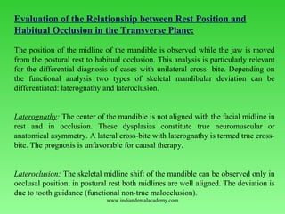
  • 1. Evaluation of the Relationship between Rest Position and Habitual Occlusion in the Transverse Plane:   The position of the midline of the mandible is observed while the jaw is moved  from the postural rest to habitual occlusion. This analysis is particularly relevant  for the differential diagnosis of cases with unilateral cross­ bite. Depending on  the  functional  analysis  two  types  of  skeletal  mandibular  deviation  can  be  differentiated: laterognathy and lateroclusion.   Laterognathy: The center of the mandible is not aligned with the facial midline in  rest  and  in  occlusion.  These  dysplasias  constitute  true  neuromuscular  or  anatomical asymmetry. A lateral cross­bite with laterognathy is termed true cross­ bite. The prognosis is unfavorable for causal therapy.   Lateroclusion: The skeletal midline shift of the mandible can be observed only in  occlusal position; in postural rest both midlines are well aligned. The deviation is  due to tooth guidance (functional non­true malocclusion). www.indiandentalacademy.com
  • 3. Laterocclusion: Right: When  in  occlusal  position,  the  midline  of  the  mandible  is  displaced  laterally from the facial mid­sagittal plane (vertical reference lines).   Left: In postural rest both midlines are coincident and well centered  www.indiandentalacademy.com
  • 4. Relationship of postural rest to occlusal position: Postural resting position  In the rest position the center of the mandible is aligned with the upper midline. (The  mesial contact point of the lower central incisors coincide with the skeletal midline of  the mandible). Initial tooth contact position:  The mandible is in the initial tooth contact position, thus terminating the first phase of  the closing movement, which started from the rest position. The patient's right lateral  incisors contact prematurely.  Habitual occlusion:  In the final phase of the closing action, after initial tooth contact, the mandible slides to  the left. Mandibular  deviation  in  habitual  occlusion  is  caused  by  tooth  interferences,  i.e.  the  skeletal midline shift of the mandible which is only present in maximum intercuspation  signifies a functional malocclusion (Lateroclusion ) www.indiandentalacademy.com
  • 5. Examination of the Temporomandibular Joint: The main objective of the clinical examination is to assess the severity of the clicking, pain,  and dysfunction, which are characteristic of pathologic TMJ symptoms.   When auscultation is carried out with a, stethoscope, clicking and crepitus in the joint may  be diagnosed during anteroposterior and eccentric movements of the mandible  www.indiandentalacademy.com
  • 6. Joint clicking is differentiated as follows:    Initial clicking is a sign of retruded condyle in relation to the disc. Intermediate clicking is a sign of unevenness of the condylar surfaces and of the  articular disc, which slide over one another during the movements. Terminal clicking occurs  most  commonly  and  is  an  effect  of  the  condyle  being  moved too far anteriorly, in relation to the disc, on maximum jaw opening. Reciprocal clicking occurs  during  opening  and  closing,  and  expresses  an  incoordination between displacement of the condyle and disc. Clicking of the joint  is rare in children. www.indiandentalacademy.com
  • 7. Palpation of the temporomandibular joint during  opening  maneuvers  will reveal possible pain on pressure of the condylar areas. Besides the right and  left condyles can thus be checked for synchrony of action.  Lateral palpation of the temporomandibular joints: Exert slight pressure on the condyloid process with the index fingers. Palpate both sides  simultaneously. Register  any  tenderness  to  palpation  of  the  joints  and  any  irregularities  in  condylar  movement during opening and closing maneuvers. The coordination of action between  the left and right condylar heads should be assessed at the same time. www.indiandentalacademy.com
  • 8. Posterior palpation of the temporomandibular joints:  Position the little fingers in the external auditory meatus and palpate the posterior  surface of the condyle during opening and closing movements of the mandible. Palpation should be carried out in such a way that the condyle displaces the little  finger when closing in full occlusion.www.indiandentalacademy.com
  • 9. Palpation of the musculature involved in mandibular movements is a  considerable part of the examination.   Palpation of the lateral pterygoid muscle: The  pain  projection  area  of  the  lateral  pterygoid  muscle  is  palpated  in  close  proximity to the neck of the condyle and the joint capsule, cranially behind the  maxillary tuberosity. The examination is carried out with the mouth open and the  mandible displaced laterally. In the initial stages of TMJ dysfunction, the muscle often hurts upon palpation  on one side only. In the advanced stage the pain is usually bilateral. www.indiandentalacademy.com
  • 10. Palpation of the temporalis muscle:  The temporalis muscle is palpated bilaterally and extraorally. The anterior, medial,  and posterior portions of the muscle are examined separately. The palpation is carried out while the muscle is contracted isometrically   Left: The  temporal  tendinous  attachment  on  the  coronoid  process,  in  the  posterolateral  region  of  the  upper  vestibule,  is  palpated.  The  patient's  mouth  should be half open for the examination.www.indiandentalacademy.com
  • 11. Palpation of the masseter muscle:  The superficial masseter muscle is palpated beneath the eye, inferior to the zygomatic arch. The deep portion is palpated on the same level, approximately 2 finger widths in front of tragus. Left: During maximum isometric muscle­contractions the width of the superficial masseter and  its direction of pull can be registered around the gonial angle. This muscle attachment should be  examined for pain on to pressure. Occasional trigger spots may occur which can be quite painful. www.indiandentalacademy.com
  • 12. Recording the maximum interincisal distance: On maximum jaw opening, the distance between the incisal edges of the upper  and lower central incisors is measured with a Boley gauge. In overbite cases this amount is added to the obtained value whereas in open  bite it is subtract­ed. The extent of maximum jaw opening between the incisal  edges  is  usually  40­45  mm.  ln  cases  with  TMJ  dysfunction,  hypermobility  is  often registered in the initial stages and limitation in the later stages. www.indiandentalacademy.com
  • 13. Opening and Closing Movements of the Mandible: The  opening  and  closing  movements  of  the  mandible  as  well  as  its  protrusive,  retrusive and lateral excursions are examined as part of the functional analysis.  The size and  direction of  these  actions  are  recorded  during  the  clinical  examination. Deviations in speed can only be registered with electronic devices  (e.g. kinesiograph).   The first signs of initial temporomandibular joint problems include deviations of  the mandibular opening and closing paths in the sagittal and frontal planes. The  characteristic  movement  deviations  include  incongruency  of  the  opening  and  closing curves and uncoordinated zigzag movements. The "C" and "S" types of  deviation are typical signs of functional disturbances.    Occlusal  analysis  on  an  articulator  is  mostly  not  necessary  in  adolescents.  It  is  only  indicated  in­patient  with  manifest  symptoms  of  temporomandibular  joint  disease. www.indiandentalacademy.com
  • 14. Interference­free registration of mandibular opening and closing  movements: Head frame of the electronic recording unit   Frontal and lateral view of the head frame in position. A permanent magnet is  secured  intraorally  on  the  mandible  to  produce  a  three­  dimensional  magnetic  field. The head frame consists of a system of antennas, which record the changes  in position of the magnetic field during movements of the mandible.www.indiandentalacademy.com
  • 15. Pattern of mandibular movements during opening and closing maneuvers: Left: Opening and closing paths in the sagittal plane (XZ plane). The opening and closing arcs crossover inconsistently. The opening movements show greater deviations. The closure pattern is straighter and more constant   Center: Opening and closing arcs in the horizontal plane (XY plane). The opening path is pathologically C­shaped.            At the end of the closing movement, the mandible shifts slightly toward the left.   Right: Opening and closing paths in the frontal plane (YZ plane). The extent of maximum jaw opening is normal. During the final stages of closing, the mandible slides to  the left due to occlusal interferences (1 millimeter = 0.5 mm movement). www.indiandentalacademy.com
  • 16. Temporomandibular Joint ­ Radiographic Examination: Several radiographic techniques, which are taken in habitual occlusion and/or in  open­mouth  position,  are  suitable  for  examination  of  the  temporomandibular  joints.    When analyzing the radiographs, the following findings are registered:    Position of the condyle in relation to the fossa, width of the joint space,  changes  in  shape  and  structure  of  the  condylar  head  and/or  the  mandibular  fossa.   Adolescents with Class II, Division 1 malocclusions and lip dysfunction (lip­ biting  or  sucking)  are  most  frequently  affected  by  TMJ  disorders.  For  this  reason, orofacial dysfunctions must also be assessed as a part of the functional  analysis as they may lead to unbalanced loading of the joints and thus trigger off  temporomandibular joint disturbances in adolescents. www.indiandentalacademy.com
  • 18. Tomograms  of  the  temporomandibular  joint  in  maximum  open­ mouth position: Left: The right condyle is subluxated when the jaw is maximally opened.   Right: The vertex of the left condyle is positioned beneath the articular tubercle  www.indiandentalacademy.com
  • 19.      Examination of Orofacial Dysfunctions:  •Swallowing  •Tongue •Speech •Lips Respiration  Swallowing:   Normal mature swallowing takes place without contracting the muscles of facial  expression. The teeth are momentarily in contact and the tongue remains inside the  mouth.  www.indiandentalacademy.com
  • 20. 1    Abnormal swallowing is caused by tongue-thrust, either as a simple thrusting  action or as  "tongue-thrust syndrome". The following symptoms distinguish  this syndrome:       Protrusion of the tip of the tongue, 2      No tooth contact of the molars,      Contraction of the perioral muscles during the deglutitional cycle. During  their  first  few  years,  infants  swallow  viscerally, i.e.  with  the  tongue  between  the  teeth.  As  the  deciduous  dentition  is  completed,  the  visceral  swallowing is gradually replaced by somatic swallowing. Should  visceral  swallowing  persist  after  the  fourth  year  of  age,  it  is  then  considered an orofacial dysfunction.  Infantile swallowing is seldom found in older children and, even if it occurs, then  only as a mixed type of visceral/somatic swallowing.   www.indiandentalacademy.com
  • 21. Tongue peristalsis during somatic swallowing - Collecting stage:  During the first stage of swallowing, the food is collected in the fore­most part of  the  mouth,  in  front  of  the  retracted  tongue.  The  posterior  arched  part  of  the  dorsum is in con­tact with the soft palate. The lips are not in contact and the teeth are not occluding. www.indiandentalacademy.com
  • 22. Transporting stage -1st part of movement:  During the second phase of swallowing, i. e. the transporting stage, the tip of the  tongue first moves up­ward and the anterior section of the dorsum is depressed  (according to Graber, 1972).  www.indiandentalacademy.com
  • 23. Transporting stage -2nd part of movement:  The  entire  anterior  section  of  the  tongue  then  moves  upward  and  the  central  section of the dorsum is depressed. This peristalsis transports the bolus rearward www.indiandentalacademy.com
  • 24. Transporting stage -3rd part of movement:  At the end of the transporting stage, the soft palate is displaced upward and  rearward. The lip musculature contracts simultaneously, the lips are together, the mandible  is raised and the teeth come into contact.www.indiandentalacademy.com
  • 25. Third swallowing stage:  The dorsum of the tongue is depressed even further during the third stage so that  the  bolus  can  pass  through  the  oropharyngeal  isthmus;  simultaneously  the  anterior part of the tongue is pressed against the hard palate, thus forcing more  food rearward. Passavant's  pad  and  soft  palate  form  the  palatopharyngeal  seal  and  close  the  nasopharynx. The teeth are in full occlusion and the lips in contactwww.indiandentalacademy.com
  • 27. Final stage of swallowing cycle: Once the swallowing act has been completed, the mandible  returns to its rest position.  www.indiandentalacademy.com
  • 28. Visceral (infantile) swallow in the neonate:  The jaws are apart during swallowing. The tongue is pushed for­ward and placed  between the gum pads. The tip of the tongue protrudes. The  mandible  is  stabilized  by  the  contraction  of  the  tongue  and  the  orofacial  musculature as well as by the tongue contact with the lips. Swallowing  is  triggered  off  and,  to  a  large  extent,  carried  out  by  sensory  interchange between the lips and the tongue. Peristalsis already commences in  the vestibule. Right: The  transverse  section  shows  that  the  tongue  is  positioned  low  in  the  mouth and that the central furrow is depressed (according to Graber, 1972). www.indiandentalacademy.com
  • 30. Tongue-Thrust: Tongue­thrust has an important effect on the etiopathogenesis of  malocclusions.         Primary or secondary.       Anterior, lateral or complex       Endogenous, habitual or adaptive.     Cases with an anterior open bite during childhood  are often self­compensating.  Complex or skeletal open bites do not regulate themselves spontaneously, but  rather persist. In the first case, the dysfunction is significant during the development of an  anterior open bite. In the second case during the development of a lateral open  bite  or a deep overbite. In case of a complex tongue­thrust, the occlusion is  supported only in the molar region. www.indiandentalacademy.com
  • 31. Anterior open bite:  Occlusion: Open bite in a deciduous dentition, caused by a tongue dysfunction  as a residuum of a sucking habit.   Habitual position: The tongue is positioned forward during functioning, thus  impeding the vertical development of the dentoalveolar structures around the  upper and lower anterior teeth [Same patient].www.indiandentalacademy.com
  • 32. Lateral open bite:  Occlusion:  In this type of open bite the occlusion on both sides is                        Supported only anteriorly and by the first permanent molars.   Habitual position: The tongue thrusts between the teeth laterally. The tongue dysfunction occurs in conjunction with a disturbance in the  physiologic growth processes around the first and second deciduous molars. www.indiandentalacademy.com
  • 34. Tongue dysfunction and malocclusion: In mandibular prognathism, the downward forward displacement of the tongue often causes an anterior tongue-thrust habit. www.indiandentalacademy.com
  • 35. Primary - Secondary Dysfunctions: From the etiologic point of view, tongue-thrust may be considered primary or secondary. Principally speaking, all dysfunctions can be divided into  Primary, [i.e. causal]  Secondary, [i.e. adaptive malfunctions]. The primary dysfunctions cause malocclusions and the treatment must concentrate on eliminating the orofacial dysfunction. Secondary dysfunctions can be considered an adaptive phenomenon to an existing skeletal or dentoalveolar deviation in the vertical development. These secondary abnormalities usually correct spontaneously while the morphological discrepancies are being treated (homeostasis). www.indiandentalacademy.com
  • 36. Primary tongue dysfunction in conjunction with hyperplastic tonsils A retracted tongue would touch infected, swollen tonsils if these were to protrude far out of the surrounding structures. In order to avoid painful sensations and to keep the oral airway open the mandible is dropped and the tongue postured forward (according to Moyers). www.indiandentalacademy.com
  • 37. Adaptive tongue dysfunction with tooth mal positions After loss of teeth, the tongue is used to fill the gaps, thus sealing the oral cavity, i. e. compensatory dysfunction. In cases with premature extraction of deciduous teeth, this primarily physiologic displacement of the tongue may persist as a functional abnormality even after the permanent teeth have eruptedwww.indiandentalacademy.com
  • 38. Configuration of the Craniofacial Skeleton and Dysfunctions: The morphology of the facial skeleton and the effects of tongue- thrusting are correlated to a certain degree. Whereas a horizontal growth pattern in conjunction with tongue- thrust usually results in a bimaxillary dental protrusion. In a vertical growth pattern with tongue-thrust the lower incisors are often in lingual inclination. From the differential diagnostic point of view, it is important to clarify both the skeletal relationships and the tongue dysfunction in order to localize the results of the abnormal tongue functioning. www.indiandentalacademy.com
  • 39. Horizontal growth pattern associated with anterior tongue dysfunction In most cases with this type of growth pattern, tongue-thrust causes bimaxillary dental protrusion, i.e. labial tipping of upper and lower anterior teeth. Schematic illustration of the incisor relationships in a case with an anterior open bite, tongue-thrust, and horizontal growth pattern. www.indiandentalacademy.com
  • 40. Vertical growth pattern associated with anterior tongue dysfunction In cases with this type of growth pattern, tongue-thrust tends to tip the upper incisors to the labial and the lower incisors to the lingual. Schematic illustration of the incisor relationships in a case with an anterior open bite, tongue-thrust, and vertical growth pattern (over eruption of posterior teeth and steeper than normal mandibular plane).www.indiandentalacademy.com
  • 41. Methods of Examining tongue dysfunctions: The different types of clinical examination are:  Electronic recordings,  Electromyographic examination,  Roentgenocephalometric analysis,  Cine-radiographic,  Palatographic,  Neurophysiologic examinations. www.indiandentalacademy.com
  • 42. Roentgenocephalometric analysis: [Assessment of tongue position on the lateral cephalogram] Is T = Incisal edge of the lower central incisor. Mc = Cervical distal third of the last erupted molar. V = The most inferior point of the uvula, respectively its projection on the reference line (Connecting line between Is T and Mc ). 0 = midpoint on the reference line between Is T and V. A line is drawn through 0, perpendicular to the horizontal base-line, and extended to the palate. A further four lines are drawn, at 30° to each other, resulting in a total of seven lines. www.indiandentalacademy.com
  • 43. Tracing of the analysis on the lateral cephalogram: Marking of the contours of the bony palate and dorsum of the tongue. Horizontal and vertical reference lines for metric evaluation are illustrated Left: The morphologic relation-ships in case of a retracted, elevated tongue. Right: Relationships in case of a downward forward tongue-posture.www.indiandentalacademy.com
  • 44. Template for metric analysis of tongue position: Transparent plastic template with an inscribed millimeter scale for analyzing the position of the tongue on the lateral cephalogram. The template is oriented on the point 0 shownwww.indiandentalacademy.com
  • 45. Palatography: Palatography involves recording the contact surfaces of the tongue with the palate and teeth while the patient produces speech sounds or performs certain tongue functions A palatogram is an illustration of these contact areas. Palatographic examination A thin, uniform layer of contrasting, precise impression material is applied to the patient's tongue with a spatula. Once the consonant has been pronounced or the tongue movement carried out (e.g. swallowing), the palatogram can be documented photographically using a surface mirror. www.indiandentalacademy.com
  • 46. Palatogram during accurate pronunciation of the "s" During articulation, the mandible is lowered slightly and pushed forwards. The tongue rests on the teeth and the alveolar processes, and a groove is formed in the center through which the air stream is directed onto the central incisors. Interdental sigmatism (lisping): During this defective pronunciation of the "S" sound, the tongue is usually protruded and clearly visible between the anterior teeth. www.indiandentalacademy.com
  • 47. Palatal sigmatism: This abnormal pronunciation is caused by an unphysiologic friction noise between tongue and hard palate. Lateral sigmatism on the left side: The tongue rests on the anterior teeth. The column of air escapes on the left side. www.indiandentalacademy.com
  • 48. Bilateral sigmatism: Palatogram of this type of defective articulation in a patient with micro-glossia. Sigmatism due to lateroflexion to the left side: www.indiandentalacademy.com
  • 49. Lip Habits: The various habits of the lips can be divided into Lip-sucking Lip-thrust Lip insufficiency Lip dysfunctions can be observed while the patient is speaking and swallowing. The lower lip often shows variations of dysfunction with regard to the tip of the tongue. The lower lip and the tip of the tongue are often in contact. In such cases, the lower lip is sucked in and pressed against the tip of the tongue. Any lip activity during swallowing - apart from closing the lips - is unphysiologic and a symptom of an orofacial dysfunction. Visual evidence of mentalis muscle activity is also abnormal. www.indiandentalacademy.com
  • 50. Lip-sucking: Extra oral findings. The lower lip is positioned behind the upper incisors.ln many patients, malpositioning of the lips occurs in conjunction with hyperactivity of the mentalis muscle. Right: The lateral cephalogram indicates that the dysfunction of the lower lip causes further protrusion of the upper incisors and impedes the forward development of the lower anterior alveolar process. www.indiandentalacademy.com
  • 51. Lip-thrust: Characteristic profile of the lower third of the face in a case with hyperactivity of the mentalis muscle. Right: In many patients, this type of lip habit is combined with lingual inclination of the Incisors www.indiandentalacademy.com
  • 52. Cheek Dysfunctions: In case of cheek sucking or cheek-biting the soft tissues are interposed between the occlusal surfaces of the teeth, which promotes the formation of a lateral open bite or a deep overbite. Increased lateral pressure by the cheek musculature on, for example, the mandible impedes the transverse development of the jaw. This type of cheek dysfunction is common in cases with buccal non-occlusion. Cheek dysfunction Extra oral findings in a case with hyper function of buccinator muscle and cheek- sucking www.indiandentalacademy.com
  • 53. Cheek-biting This female patient shows a weal like horizontal swelling of the buccal mucosa caused by the dysfunction. Cheek dysfunction and malocclusion Buccal nonocclusion in the deciduous dentition combined with a cheek dysfunction. www.indiandentalacademy.com
  • 54. Hyperactivity of Mentalis Muscle The deep mentolabial sulcus is characteristic of a hyperactive mentalis muscle. This habitual pattern of muscle behavior impedes the forward development of the anterior alveolar process in the mandible. The abnormal mentalis function often occurs together with lip sucking or lip- thrust. Cases of hyperactivity of the mentalis muscle, which occur in the same family, are usually hereditary. Deep mentolabial sulci and hyperactivity of mentalis muscle: Profile view with the clinical appearance of the abnormal muscle function. Right: The same dysfunction is diagnosed in the sister, who is 2 years older www.indiandentalacademy.com
  • 55. Cephalometric findings in case of hyperfunction of the mentalis muscle and the lower lip Right: The hyperactive mentalis muscle pulls the lower lip upward and rearward and presses it against the lingual surfaces of the upper incisors. The upper lip remains relatively motionless. The normal lip seal is disturbed and the tongue displaced downward. This type of soft-tissue morphology aggravates the dentoalveolar malocclusion. www.indiandentalacademy.com
  • 56. Mouth-Breathing: The mode of respiration is examined to establish whether the nasal breathing is impeded or not. Chronically disturbed nasal respiration represents a dysfunction of the orofacial musculature; it can restrict development of the dentition and hinders the orthodontic treatment. The following are the clinical findings Adenoid facies: 6-year-old female patient with chronically restricted nasal respiratory function.www.indiandentalacademy.com
  • 57. Occlusal and dental findings in case of oronasal respiration: The upper jaw is markedly constricted, the "tooth germ position" of the upper incisors has persisted, and the mandibular arch is well formed. Due to the incongruence in arch width a bilateral cross-bite exists Configuration of the maxilla in oronasal respiration: The high palate and narrow upper arch are characteristic featureswww.indiandentalacademy.com
  • 58. Examination of Breathing Mode: When interpreting the findings during clinical examination it must be taken into account that the respiratory mode is controlled by the nasal cycle, which changes approximately every 6 hours. This is a physiologic protective mechanism that prevents the nasal membranes from drying out (Eccles, 1978; Masing and Wolf 1969). Due to the nasal cycle, one nasal airway is always more constricted than the other, i.e. an apparent unilaterally obstructed nasal passage during the crude clinical examination is not necessarily a pathologic finding. Mirror test The mirrors are held in front of both nostrils. In nasal-breathers the mirror will cloud with condensed moisture during expiration as shown on the right.www.indiandentalacademy.com
  • 59. Examination of alar musculature: [Nasal respiration] The size and shape of the external nares of a patient with nasal respiration during inspiration (left) and expiration (right). The very noticeable changes in the cross-section of the nasal orifices are typical for nasal- breathers. Oronasal respiration The cross-section of the external nares of a patient with prevailing oral respiration during inhaling (left) and exhaling (right). The alar muscles are inactive -nares do not change their size -, which is a clinical feature of in-creased oral respiration. www.indiandentalacademy.com
  • 60. Differential Diagnosis: Differential diagnosis must be used to determine whether the problems in nasal respiration are due to an obstruction of the upper nasal passages or to habitual oral respiration. In the first case, an operation by an ENT specialist is indicated; Should the nose not be obstructed, pre-orthodontic therapy should be carried out to treat the restricted nasal breathing. This may include breathing exercises or incorporation of a perforated oral screen Myofunctional exercises for patients with habitual respiration. {The cardboard should be held loosely in a horizontal position with the lips to improve the lip seal}. Changing habitual oral respiration with the help of custom made, perforated oral screenwww.indiandentalacademy.com
  • 61. Photographic Analysis: The clinical value of the photographic picture is that it is more realistic and gives a better record of any changes in the soft-tissue profile during the course of treatment, which is of great advantage. This is done with the patient sitting upright in habitual occlusion and with relaxed lips and mentalis muscles. A precondition for obtaining comparable photographs, which can be evaluated by measurement, is a reproducable position of the patient. Such profile and frontal photographic views can be achieved in various ways: (1) www.indiandentalacademy.com
  • 62. Extra oral photographs: In orthodontics, lateral (left) and frontal views (center) are taken as a rule. An oblique facial view to assess the smile line can be taken in addition (right). www.indiandentalacademy.com
  • 63. Profile View: For the profile exposure the camera is placed parallel to the facial mid sagittal plane. The patient's head is oriented in accordance with the Frankfurt horizontal plane. The patient's eyes should be looking straight ahead, unstrained, and the ears should be uncovered. A. M. Schwarz (1958) compiled a detailed classification of the variations of the facial profile. The evaluation is based upon the construction of three reference planes: 1. Eye-ear plane (Frankfurt horizontal plane); 2. Skin nasion perpendicular, according to Dreyfuss 3. Orbital perpendicular, according to Simon. The perpendiculars delimit the "jaw-profile field" (JPF). In children this is 13- 14 mm wide, in adults 15-17 mm. www.indiandentalacademy.com
  • 64. Photographic analysis according to A. M. Schwarz: N = Skin nasion Sn = Subnasale Gn = Skin gnathion Pog = Skin pogonion P = Porion (uppermost point of tragus) Or = Orbitale (a point, located below the pupil, at a distance equivalent to the gap between the eyelids, with the eyes relaxed and looking straight ahead) H = Frankfurt horizontal plane Po = Orbital perpendicular Pn = Skin nasion perpendicular JPF = Jaw profile field www.indiandentalacademy.com
  • 65. Slanting profile: In a slanting profile there is a discrepancy between the subnasal point and the soft-tissue pogonion in relation to the anteroposterior position. This disturbs the harmonious appearance of the facial profile. www.indiandentalacademy.com
  • 66. Depending on the location of the subnasal point relative to the skin nasion perpendicular, there are typical profile variations:  Average face= Subnasale lying on the skin nasion perpendicular;  Anteface = Subnasale lying in front of the skin nasion perpendicular;  Retroface= Subnasale lying behind the skin nasion perpendicular. In straight-jawed, ante and retrofaces the chin is displaced to the same extent as the subnasal point. For each of the above profiles two further facial types can be differentiated, depending on the changed location of the "soft-tissue pogonion" relative to the Subnasale. There are  Forward-slanting  Backward-slanting faces That means nine different types of profile in all. www.indiandentalacademy.com
  • 67. The nine possible profile variants according to the classification by A.M. Schwarz:     Straight-jawed profile   A  straight-jawed  profile  -  whether  an  average  face,  an  anteface  or  a  retroface  always  looks  harmonious.  The  straight  average  face  (or  biometric  face)  is  considered ideal. www.indiandentalacademy.com
  • 70. Facial Divergence:   Another analysis of the lateral photograph is based upon evaluation of the divergence of the face. The inclination between the' following two reference lines is here analyzed:    (1) The line joining the forehead and the border of the upper lip;   (2) The line joining the border of the upper lip and the soft-tissue pogonion. The following three profile types are differentiated according to the relationship between  these two lines:  www.indiandentalacademy.com
  • 71. Frontal View: An  analysis  of  the  frontal  picture  is  important  in  assessing  major  disproportions  and  asymmetries of  the  face  in  the  transverse  and  vertical planes. Even a slight rotation of the head from the plane of  the  film  can  result  in  major  discrepancies  between  the  relative  patterns  of  the  right  and  left  facial  contours.  It  is,  therefore,  absolutely essential for the camera to be placed perpendicular to the  facial midline during the exposure. For clinical analysis it has proven practical to mark the two orbital  points and to construct the skin nasion perpendicular. During the  evaluation of the measurements the diagnostician should bear in  mind that a mild degree of physiologic asymmetry between the two  sides of the face exists in nearly all normal individuals.  www.indiandentalacademy.com
  • 72. Facial symmetry: Vertical reference plane = Facial midsagittal plane (joins the skin nasion point                                                                to the subnasal point);  Upper horizontal plane = Bipupillary plane; Lower horizontal plane = Parallel to the Bipupillary plane through the stomion. www.indiandentalacademy.com
  • 73. The smile arc . The ideal smile arc has the curvature of the maxillary incisal edges parallel to the  curvature of the lower lip upon smile, and the term consonant is used to describe  this parallel relationship.  Nonconsonant, or flat, smile arc is characterized by the maxillary incisal curvature  being flatter than the curvature of the lower lip on smile.     It is quite possible that in the realm of dentofacial esthetics, orthodontists recently  have concentrated so intently on not creating "flat faces" that the esthetic importance  of smile arcs has been overlooked.  www.indiandentalacademy.com
  • 74. Bracket placement based on tooth measurements Placing  brackets  solely  based  on  tooth  measurements,  as  traditionally  has  been  taught, often is not appropriate for maxi-mum esthetics. It is important to assess  and visualize the incisor-smile arc relationships and place brackets so as to extrude  the  maxillary  incisors  in  flat  smiles  and  maintain  the  smile  arc  where  it  is  appropriate.  Placing  the  lower  incisor  brackets  close  to  the  gingival  margins  in  an  effort  to  avoid occlusal interferences that might cause loss of brackets results in extrusion  of the lower incisors . If this requires vertical compensation of the upper incisors  to open the bite, flattening of the smile arc is likely.  www.indiandentalacademy.com
  • 76. The transverse dimension of the smile This  characteristic  is  referred  to  in  terms  of  "broadness  to  the  smile"  and  the  presence  and  amount  of  "buccal  corridors."  Recently,  excessively  wide  buccal  corridors  have  been  referred  to  by  some  orthodontists  as  "negative  space;'  to  be  eliminated  by  transverse  expansion  of  the  maxilla.  It  is  well  documented  in  the  prosthodontic  literature  that  one  of  the  characteristics  of  an  unrealistic  "denture  smile" is a lack of buccal corridors.  Although  this  smile  feature  has  been  thought  of  primarily  in  terms  of  maxillary  width, there is evidence that the buccal corridors are also heavily influenced by the  anteroposterior  position  of  the  maxilla  relative  to  the  lip  drape.  This  means,  that  moving  the  maxilla  forward  also  reduces  the  size  of  the  buccal  corridors  and  decreases negative space .  www.indiandentalacademy.com
  • 77. Radiologic Examination: Radiologic Examination is absolutely essential in orthodontic diagnosis. The type  and number of radiographs should, therefore, subject the patient to as little  radiation exposure as possible while providing maximal information at the same  time. In some cases, depending on the nature of the problems, a routine  examination may necessitate supplemental radiographs. Broadly there are 2 kinds of photographs required for an orthodontic diagnosis. 1. Those taken to provide information regarding the condition of the teeth, the        Periodontium, and the bony structures. 2. The objective of the others is an assessment of the malocclusion in relation to  the facial skeletal structure.  Radiographs of the hands and TMJ are not among the routine requirements of  orthodontic diagnosis. It is only necessary to take radiographs of the TMJ in those  cases where TMJ symptoms of dysfunction are either present or where changes  are reasonably suspected, that are not clearly seen on the panoramic view. www.indiandentalacademy.com
  • 80. Periapical view (small intraoral film): A full series of intraoral radiographs [10-16 films] is required for assessment of the  periodontal state in adults. Otherwise periapical films are only indicated where the  panoramic view suggests possible pathologic conditions [e.g. congenitally missing  teeth or malposed tooth germs]. www.indiandentalacademy.com
  • 83. Orientation of study cast models  Midpalatal raphe plane = mid-sagittal plane, which is defined by anatomical points on the  palatine raphe. It is the reference plane for assessment of transverse discrepancies.   Tuberosity plane = Para frontal plane which runs through the maxillary tuberosities  respectively through the distal-most tuberosity. It is the reference plane for analysis of  anteroposterior dental malpositions.   Occlusal plane = horizontal plane through the tips of the buccal cusps of the premolars or the  tips of the mesiobuccal cusps of the first molars and first premolars. This plane allows  vertical malpositions to be assessed.  www.indiandentalacademy.com
  • 84. Measuring the overjet:    Determination of the overjet with a graduated ruler.   The  overjet  is  defined  as  the  distance  between  the  labial  surface  of  the  lower  central incisor and the upper incisal edge. The measurement is performed parallel  to the occlusal plane www.indiandentalacademy.com
  • 85. Determination of overbite :    The upper incisal edge is projected with a pencil mark on the  labial surface of the lower central incisor parallel to the occlusal plane.  www.indiandentalacademy.com
  • 86. Measurement of the curve of Spee: The depth of the curve of Spee is defined as the distance from the vertex of the  curvature  to  the  side  of  a  plastic  template  placed  over  the  lower  arch.  The  template  touches  anteriorly  the  incisal  edges  and  posteriorly  the  distal-most  molar cusps. The measurement is carried out separately on both the left and right  sides of the dental arch. www.indiandentalacademy.com
  • 88. Differentiation between dental and skeletal midline shift in the mandible: Left: Mandibular arches with dental midline deviation in opposite direction in  conjunction with tooth mal positioning in the respective anterior region.   Right: Skeletal mandibular midline shift, as a result of displacement of the whole  mandible to the left.  www.indiandentalacademy.com
  • 89. Model Analysis in the Permanent Dentition: For  patients  with  malalignment  of  teeth  resulting  from  lack  of  space,  it  is  important  to  determine  from  the  study  casts  the  amount  of  crowding  in  the  maxillary  and  mandibular  arches.  The  purpose  is  to  determine  the  difference  between space available and space required for tooth alignment. This means that  two measurements are required in each arch for intramaxillary analysis of space  requirement: 1) Calculation of space required and 2) Calculation of space available.   The analysis can be carried out by two methods:   www.indiandentalacademy.com
  • 91.      Recording the actual arch length using a soft wire.   This  is  contoured  to  the  individual  arch  shape  and  placed  on  the  occlusal  surfaces over the contact points of the posterior teeth and the incisal edges of  the  anteriors.  The  distance  between  the  mesial  contact  points  of  the  first  permanent  molars  -  recorded  from  the  straightened  wire  -  is  the  amount  of  space available in the dental arch (actual arch length). 3) The assessment of space relationship is the result of the difference between the  ideal and actual arch length (negative value = space deficiency, positive value =  space excess)  www.indiandentalacademy.com
  • 92. The Lundstrom Segmental Analysis:   The segmental analysis involves an indirect assessment of the dental arch  perimeter, which can be carried out in the following way 1.      Division of the dental arch into six straight-line segments of two             teeth per segment, including the first permanent molars. 1.      Recording the mesiodistal width of the twelve teeth. 2.      Summing the individual tooth width of each segment. 3.      Recording the available mesiodistal space on the study cast               separately for each segment. 4.      The sum of the difference between ideal and actual length of               each segment expresses the space relationship. www.indiandentalacademy.com
  • 94. Bolton Analysis:   The  Bolton  analysis  (Bolton,  1958)  determines  the  ratio  of  the  mesiodistal  widths  of  the  maxillary  versus  the  mandibular  teeth  (i.e., tooth size discrepancy).  In  the  analysis  of  the  overall  ratio the  relationship  of  the  12  mandibular teeth to the 12 maxillary teeth is assessed (second and  third molars are excluded).  On account of the importance for the canine relations as well as for  overbite and overjet relationships, a further analysis is performed to  evaluate  the  ratio  between  the  six  upper  and  lower  anterior  teeth  (anterior ratio). www.indiandentalacademy.com
  • 95. Index of overall ratio Formula to determine the intermaxillary mesiodistal congruence of overall tooth  widths, including the first permanent molars. If the calculated ratio is greater than  91.3 %, the mandibular teeth are too wide compared to the maxillary teeth. If the  ratio is reduced, the maxillary teeth are relatively too large.  Sum mand12 (m-d) X 100 = 91.3% Sum max12 (m-d)www.indiandentalacademy.com
  • 96. Index of anterior ratio   Formula to determine the intermaxillary tooth width congruence in the anterior  region. If the ratio is greater than 77.2% the total width of the lower six anterior  teeth is relatively too large. If the index value is reduced, the discrepancy is due  to an excess in maxillary tooth material.  www.indiandentalacademy.com
  • 97. Excessive mesiodistal tooth material   In the maxillary arch   1. Increased overbite  2. Increased overjet 3. Crowding in the maxillary arch  4. Spacing in the mandibular arch 5. Linguoversion of upper incisors 6. Labioversion of lower incisors   In the mandibular arch   1. Reduced overbite 2. Reduced overjet 3. Crowding in the mandibular arch  4. Spacing in the maxillary arch 5. Labioversion of upper incisors 6. Linguoversion of lower incisors www.indiandentalacademy.com
  • 98. Ideal relationship of maxillary and mandibular tooth widths according to Bolton:  After calculation of the Bolton ratio, the arch with the relatively smaller tooth material is  determined and the actual figure corresponding to the arch tooth size located in the table. The  ideal value for the size of the opposing teeth is read off from the accompanying column.  The difference between the actual value and the ideal value (according to the table) for the  relatively enlarged tooth material represents in mm the amount of excess tooth size in this  arch. www.indiandentalacademy.com
  • 100. Determination of Premolar diameter (PMD): The premolar diameter refers to the arch width from the tip of the buccal cusps of one first premolar to the tip of the buccal cusp of the opposite first premolar. Premolar diameter to tooth material ratio is obtained by dividing the premolar diameter by the sum of widths of 12 teeth. www.indiandentalacademy.com
  • 101. Determination of Premolar basal arch width (PMBAW): This is also called as canine fossa width. The measurement of the width from the canine fossa (Distal to the canine eminence on the casts at the apices of the first premolars) of one side to the other gives the width of the dental arch at the apical base . Premolar basal arch width to tooth material ratio is obtained by dividing the premolar basal arch width by the sum of widths of 12 teeth. www.indiandentalacademy.com
  • 102. Determination of Basal Arch Length It is measured at the midline from the estimated anterior limits of the apical base to a perpendicular that is tangent to the distal surfaces of the two first molars. Basal arch length to tooth material ratio is obtained by dividing the basal arch length by the sum of widths of 12 teeth. www.indiandentalacademy.com
  • 103. INFERENCE: Premolar basal arch width (PMBAW) should equal approximately 44% of the MD width of 12 teeth in the maxilla if it is to be sufficiently large to accommodate all the teeth. If it is less than 37% it is considered to be basal arch deficiency-necessitating extraction of premolars. If it is more than 44% expansion of the premolars can be undertaken safely. Since this method was introduced, rapid palatal expansion has come into more common use. www.indiandentalacademy.com
  • 104. Carey’s / Arch perimeter Analysis Many malocclusions occur as a result of discrepancy between the arch length and tooth material. Carey’s analysis helps in determining the extent of discrepancy on the lower cast and the same analysis on the upper cast is called arch perimeter analysis. Determination of arch length www.indiandentalacademy.com
  • 105. The arch length anterior to the first perm molar is measured using a soft brass wire. The wire is placed contacting the mesial surface of the first perm molar of one side and is passed over the buccal cusps of the premolars and along the incisal edges of the anteriors and is continued on the opposite side in the same way upto the mesial surface of the opposite first perm molar. In case of proclined anteriors, the wire is passed along the cingulum of anterior teeth. If the anterior teeth are retroclined, the wire passes labial to the teeth. Determination of tooth material The MD width of the teeth anterior to the first molars is measured and summed up. Determination of discrepancy The discrepancy refers to the difference between the arch length and tooth material. Discrepancy Inference 0-2.5 mm Proximal stripping 2.5-5 mm Extraction of second premolars > 5 Extraction of first premolarswww.indiandentalacademy.com
  • 106. SPACE ANALYSIS The objective of space analysis is to quantify the space required within each dental arch for the correction of a malocclusion to an aligned Class I occlusion with normal axial inclination of the teeth. The valuable information can be gained to help judge the need for extraction, choice of extraction, and to help plan anchorage and mechanics. The process of space analysis is carried out in three stages. The first is an assessment of space requirement, the second is an assessment of any additional space to be created or utilized during treatment, and the third is a prediction of anteroposterior molar movements required for occlusal correction. www.indiandentalacademy.com
  • 107. The dental analysis presented here “A Dental Visualized Treatment Objective”—is designed to provide organized and simplified information to help in diagnosis, treatment planning, and the extraction/non extraction decision. It should be used as an adjunct to, but not a substitute for, conventional cephalometric analyses. Progress can be checked by referring to the dental VTO at the patient’s regular adjustment appointments. Method The dental VTO consists of three charts: Chart 1 Records the initial midline and first molar positions with the mandible in centric relation. www.indiandentalacademy.com
  • 108. Chart 2 Measures the lower arch discrepancy, similarly to the Steiner analysis. The four primary factors in each case are: 1. Space required for relief of crowding, measured from canine to midline and from first molar to midline on each side. 2. Space required for the desired correction of protrusion or retrusion of the mandibular incisors. 3. Space required for leveling the curve of Spee. 4. Space required for midline correction www.indiandentalacademy.com
  • 109. Four secondary factors that can sometimes provide additional space are listed, if applicable, below the primary chart: 1. Additional space from interproximal enamel reduction. 2. Additional space from uprighting or distal movement of mandibular first molars. 3. Additional space from buccal uprighting of mandibular canines and posterior teeth. 4. Additional leeway or “E” space. The primary and secondary factors are added together at the bottom of the chart to determine the total lower arch discrepancy from canine to midline and from first molar to midline on each side. Chart 3 records the anticipated treatment change in terms of dental movements of the first molars, canines, and midline. www.indiandentalacademy.com
  • 110. Discrepancy calculation: Limiting the assessment of space relationships to the analysis of study casts is insufficient in itself. The difference between space required and the amount of space available for alignment of the teeth is determined by two different parameters: 1) Amount of dental crowding 2) Anteroposterior position of the incisors in relation to the facial skeleton. Comprehensive space analysis must therefore consist of a combined analysis including measurements from the cephalogram and study casts. The steps in this overall discrepancy calculation in upper and lower arches are: www.indiandentalacademy.com
  • 111. 1. Determination of dental discrepancy (calculated on study cast) a) the difference between the actual and ideal dental arch length b) the amount of curve of Spee separately on the left and right side (To level the curve of Spee by 1 mm requires 1 mm of arch length). The sum of the measurements of a) and b) is known as the dental discrepancy (DD). 2. Determination of sagittal discrepancy (calculated on cephalogram) The distance of the incisal edge of the central incisors to N-Pog-line is measured on the lateral cephalogram. The degree to which incisor position varies from the standard value represents the sagittal discrepancy (SD). A forward position of the incisors signifies a need of dental arch length, retroposition signifies an increase in dental arch length (1 mm change of incisor position in the lateral cephalogram = 1 mm arch length). www.indiandentalacademy.com
  • 112. 3. Determination of total discrepancy Dental discrepancy (TD) is the sum of the dental and 19ittal discrepancy and - since the measurement which is for both sides of the dental arch on the study cast but only on one side on the radiograph - is calculated as follows: TD per arch side = SD + 1/2 DD www.indiandentalacademy.com
  • 113. Cephalometric Analysis: In 1895, Roentgen discovered X-rays. In 1931, Broadbent in US and Hofrath in Germany simultaneously published methods to obtain standardized head radiography. Cephalometric analyses of skeletal, dental and soft tissues are merely aids in determining diagnosis. For accurate information, the various readings must not be assessed independently. To interpret the data, all readings must be correlated with other clinical and diagnostic criteria before arriving at the diagnosis and treatment planning. Cephalomety must not be regarded as number game in which the measured parameters of the tracing must appropriate those of normal occlusions or the dentofacial skeletal pattern that will be regarded as being imbalanced. www.indiandentalacademy.com
  • 114. Variation in biology is a rule rather than exception. Normal is never a point it is a range. Because of this clinicians developed a set of figures as mean. Compared to dental growth pattern orthodontists have little control over skeletal pattern, because during growth there are varying degrees of downward and forward growth of the face relative to the cranial base. It is better to recognize skeletal disharmony by means of ceph before treatment and alert the patient than to be embarrassed by the discovery of difficulties in the later part of the treatment. www.indiandentalacademy.com
  • 115. CEPHLOMETRIC ANALYSIS – Explanation 1. Go – Gn: SN ratio: Normal is 1, that is cranial base is same length as mandible. In pre-pubertal period cranial base may be more by 0-5 mm and post-pubertal mandible may be greater by 0-5mm. 2. Max. To Mand. ANS-PNS is about half of mandible Ar-Go. This measurement with the previous one will help to determine whether Mandible is Short/Normal/Long. 3. Wits: If wits is 0-1, it is normal, -ve in Cl-III higher +ve value – more Cl. II. 4. If width of symphysis is less – clockwise rotation – vert. growth. More – Anti-clockwise rotation – horizontal Growth. 5. Saddle, Gonial, Articulare angle – If sum is less then 396 – Horizontal, if it is more vertical. Saddle & Ar angle increase one degree each year from 12 – 20 yrs. During the same period, gonial angle decrease by 2 degree. Hence the total is maintained. www.indiandentalacademy.com
  • 116. 6. In Gonial angle if upper angle is more then 75% of lower, it indicates horizontal growth. If ratio is lower vertical growth. 7. Ramus to post. Cranial base: PCB is 75% of Ramus height, if the ratio is higher it means the Ramus is shorter indicating a more clock-wise rotation. 8. Post – Ant face height- Post is 65%: If it is higher – horizontal growth p. & vise-versa. 9. Lower face ht to total face ht (lower face height is 60% of total face ht). If lower face ht. is more – vertical growth pattern & vise versa. 10. Basal angle: will be less in deep bite & high in open bite, lower basal angle is high- indicates easy bite opening. www.indiandentalacademy.com
  • 117. Soft Tissue Cephalometric Analysis: This analysis is an attempt to express quantatively those soft tissue relationships which are pleasing and harmonious as well as those which are not, to differentiate one from the other and to explain how this information is used in ortho treatment planning. METHODS: The eleven measurements used in the analysis are;  Soft-tissue facial angle  Nose prominence  Superior sulcus depth  Soft-tissue subnasale to H line.  Skeletal profile convexity.  Basic upper lip thickness  Upper lip strain measurement.  H angle  Lower lip to H line  Inferior sulcus to the H line.  Soft-tissue chin thicknesswww.indiandentalacademy.com
  • 118. Soft-tissue facial angle: Angular measurement of a line drawn from soft-tissue nasion where the sella- nasion line crosses the soft-tissue profile, to the soft-tissue chin at a point overlying the hard-tissue suprapogonion of Ricketts measured to the Frankfort horizontal plane. . A measurement of 91 degrees is ideal, with an acceptable range of ±7 degrees. High angle- prognathic chin Low angle - retrognathic chin www.indiandentalacademy.com
  • 119. Nose prominence:  Nose prominence can be measured by means of a line perpendicular to Frankfort horizontal and running tangent to the vermilion border of the upper lip. This measures the nose from its tip in front of the line and the depth of the incurvation of the upper lip to the line Balanced face has a nose prominence measurement of 16 mm. Arbitrarily, those noses under 14 mm are considered small, while those above 24 mm. are in the large or prominent range.www.indiandentalacademy.com
  • 120. Superior sulcus depth:  Superior sulcus depth measured from inward curvature of upper lip to a perpendicular from FH and tangent to the vermilion border to the upper lip. A range of 1 to 4 mm. is acceptable in certain types of faces, with 3 mm being ideal. During orthodontic treatment or surgical orthodontic procedures, we should strive never to allow this measurement to become less than 1.5 mm Long faces: thin upper lip: 1 mm Short faces: thick upper lip: 4 mmwww.indiandentalacademy.com
  • 121. soft-tissue subnasale to H line . Here the ideal is 5 mm., with a range of 3 to 7 mm. . With short and/or thin lips, 3 mm. will be adequate .  In longer and/or thicker lips, 7 mm. may be in excellent balance The upper lip form is considered to be of such importance in the study of facial lines that its perspective in relation to both lines (the line perpendicular to Frankfort and the H line) is needed for the decision as to where the denture should be oriented to provide the best possible lip supportwww.indiandentalacademy.com
  • 122. Skeletal profile convexity: Measurement from point A to the hard-tissue line Na-Pog or facial plane. This is not really a soft-tissue measurement, but convexity is directly interrelated to harmonious lip positions and, therefore, has a bearing on the dental relationships needed to produce harmony of the features of the human face.www.indiandentalacademy.com
  • 123. Basic upper lip thickness: This is near the base of the alveolar process, measured about 3 mm below point A. It is at a level just below where the nasal structures influence the drape of the upper lip. This measurement is useful, when compared to the lip thickness overlying the incisor crowns at the level of the vermilion border, in determining the amount of lip strain or incompetency present as the patient closes his or her lips over protrusive teeth. Upper lip strain measurement: (see above Fig) The usual thickness at the vermilion border level is 13 to 14 mm. Excessive taper is indicative of the thinning of the upper lip as it is stretched over protrusive teeth; Excessive vertical height may produce more than 1 mm. of taper due to lip stretching. When the lip thickness at the vermilion border is larger than the basic thickness measurement; this identifies a lack of vertical growth of the lower face with a deep overbite and resulting lip redundancy. www.indiandentalacademy.com
  • 124. H angle: Angular measurement of the H line to the soft-tissue Na-Po line. This angle measures the prominence of the upper lip in relation to the over-all soft-tissue profile.  Ten degrees is ideal when the convexity measurement is 0 mm. However, measurements of 7 to 15 degrees are all in the best range as dictated by the convexity Ideally, as the skeletal convexity increases, the H angle must also increase if a harmonious drape of soft tissues is to be realized in varying degrees of profile convexity. H angle considered along with the basic skeletal convexity and sulcus depth measurements can be used in planning where the denture should be oriented to provide the best possible lip support. www.indiandentalacademy.com
  • 125. Lower lip to H line :  The ideal position of the lower lip to the H line is 0 to 0.5 mm. anterior, but individual variations from 1 mm. behind to 2 mm. in front of the H line are considered to be in a good range.. A lower lip measurement of much more than – 1 mm. when other profile measurements are only reasonably good is indicative of lower incisors that are positioned too far lingually. www.indiandentalacademy.com
  • 126. Inferior sulcus to the H line: This is measured at the point of greatest incurvation between the vermilion border of the lower lip and the soft-tissue chin and is measured to the H line. The contour in the inferior sulcus area should fall into harmonious lines with the superior sulcus form. It is an indicator of how well we manage axial inclinations of the lower anterior teeth. Leveling procedures on round arch wires may cause a lingual tipping of the lower incisor roots with point B following and thus exaggerate an already excessive labiomental furrow and a prominent chin. www.indiandentalacademy.com
  • 127. Soft-tissue chin thickness (10 to 12 mm. average): The distance between the two vertical lines representing the hard-tissue and soft-tissue facial planes at the level of Ricketts' suprapogonion.  Large variations, such as 19 mm. of thickness need to be recognized, and in such cases it is essential to leave the lower incisorswww.indiandentalacademy.com
  • 128. Variations in response Responses vary with type of lip structure, patient's age and sex. If lip strain is present in the malocclusion, this must be taken into consideration in treatment planning with the VTO. The upper lip will follow the tooth movement with two exceptions. The first exception is found in those patients who have or who are developing very thick lips. Upper lip thickness measuring at the vermilion border exceeds 18 mm., the upper lip usually changes very little if at all when the upper incisors are retracted. Upper lip thickness measuring at the vermilion border is 16 or 17mm, will be very slow in acquiring its final adaptation to or drape over the teeth. Upper lip thickness measuring at the vermilion border is 13 to 15 mm, it usually follows the tooth movement quite well, but some of this group still shows a thicker lip measurement at the vermilion border at retention than at the beginning of treatment. www.indiandentalacademy.com
  • 129. Upper lip thickness measuring at the vermilion border is 12mm or below, and it is not due to stretching of upper lip over protrusive teeth (lip-strain factor) the lip usually follows the tooth movement faster One need be concerned only about those in the thick-measurement group and older patients with excessive taper of the upper lip. . In the others the tooth movement is planned for the final lip position visualized as being the most desirable for that patient. Patients will not all be at that point in the adaptation process at the time of retention, but they will get there.  This is much better than overtreating the dentition and showing a balanced lip position at retention and then watching it deteriorate after treatment as the lips, especially the upper lip, finally catch up. www.indiandentalacademy.com
  • 130. Cephalometrics for orthognathic surgery(COGS): The first step in the diagnosis of the orthognathic surgical patient is to determine the nature of the dental and skeletal defects. Patients who require orthognathic surgery usually have facial bones as well as tooth positions that must be modified by a combined orthodontic and surgical treatment. For this reason, a specialized cephalometric appraisal system, called Cephalometrics for Orthognathic Surgery (COGS), was developed at the University of Connecticut. The COGS system describes the horizontal and vertical position of facial bones by use of a constant coordinate system; the sizes of bones are represented by linear dimensions and their shapes, by angular measurements. The baseline for comparison of most of the data in this analysis is a constructed plane called the horizontal plane (HP), which is a surrogate Frankfort plane, constructed by drawing a line 7° from the line S to N. Most measurements will be made from projections either parallel to HP (11 HP) or perpendicular to HP ( 1 HP). www.indiandentalacademy.com
  • 131. CRANIAL BASE First, it is necessary to establish the length of the cranial base, which is a measurement parallel to HP from Ar to N. This measurement should not be considered an absolute value but a skeletal baseline to be correlated to other measurements, such as maxillary and mandibular length, to obtain a diagnosis of proportional dysplasia. Ar-pterygomaxill1 fissure (Ar-PTM) is measured parallel to HP to determine the horizontal distance between the posterior aspects of the mandible and maxilla. The greater the distance between Ar-PTM, the more the mandible will lie posterior to the maxilla, assuming that all other facial dimensions are normal. Therefore, one causal factor for prognathism or retrognathism can be evaluated by this measurement of the cranial base. www.indiandentalacademy.com