Oral Manifestations of Endocrinal Disorders / dental crown & bridge courses
•
78 likes•8,993 views

The Indian Dental Academy is the Leader in continuing dental education , training dentists in all aspects of dentistry and offering a wide range of dental certified courses in different formats.for more details please visit www.indiandentalacademy.com

Recommended
More Related Content
What's hot
What's hot (20)
Viewers also liked
Viewers also liked (7)
Similar to Oral Manifestations of Endocrinal Disorders / dental crown & bridge courses
Similar to Oral Manifestations of Endocrinal Disorders / dental crown & bridge courses (20)
More from Indian dental academy
More from Indian dental academy (20)
Recently uploaded
Recently uploaded (20)
Oral Manifestations of Endocrinal Disorders / dental crown & bridge courses
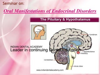
- 1. Oral Manifestations of Endocrinal Disorders Seminar on: www.indiandentalacademy.com INDIAN DENTAL ACADEMY Leader in continuing Dental Education
- 2. Contents Part-1 Introduction Classification Growth hormone Thyroid hormones Gonadal hormones Radiographic features Oral manifestation & dental consideration Part-2 Parathyroid hormones Hormones of Adrenal gland Oral manifestation & dental consideration Radiographic features Conclusion www.indiandentalacademy.com
- 3. What are hormones???? Hormones are the substances released from the cells that circulate and affect distant organs. The main physiologic function of hormones are growth, maintenance of homeostasis and reproduction. www.indiandentalacademy.com
- 4. Chemistry of hormones Hormones are classified into three types depending on their chemical nature. 1.Steroid derivative These are the hormones derived from cholesterol or its derivatives. Steroid hormones are corticosteroid and sex hormones. 2.Protein derivative These hormones are large or small peptides. Protein hormones are, the hormones secreted by pituitary gland, parathyroid gland, pancreas. 3.Derivative of amino acid- tyrosine There are two types of hormones which are derivatives of the amino acid called tyrosine. Thyroid hormones and adrenal medullary hormones are derived from tyrosine. www.indiandentalacademy.com
- 5. Hormones secreted by major endocrine glands Anterior pituitary 1.Growth hormone (GH) 2.Thyroid stimulating hormone (TSH) 3.Adrenocorticotropic hormone (ACTH) 4.Follicle stimulating hormone (FH) 5. Luteinizing hormone (LH) 6. Prolactin 7. Melanocyte stimulating hormone Posterior pituitary 1. Antidiuretic hormone (ADH) 2. Oxytocin Thyroid gland 1. Thyroxine 2. Triiodothyronine 3. Calcitonin www.indiandentalacademy.com
- 6. Parathyroid gland Parathormone Pancreas 1.Insulin 2.Glucagon 3.Somatostatin Adrenal cortex (i) Mineralcorticoids 1.Aldosterone 2.11 deoxycorticosterone (ii)Glucocorticoids 1.Cortisol 2.Corticosterone www.indiandentalacademy.com
- 7. (iii) Sex hormone 1. Androgen 2. Estrogen 3. Progesterone Adrenal medulla 1.Adrenaline (Epinephrine) 2.Noradrenaline (Norepinephrine) 3.Dopamine www.indiandentalacademy.com
- 8. Pituitary gland The pituitary gland is also known as hypophysis. It is small gland with diameter of 1cm & weighs about 0.5-1 gm. Physiologically gland is divided into portions namely, the anterior or adenohypophysis and posterior or neurohypophysis. www.indiandentalacademy.com
- 9. Actions: GH is responsible for growth of almost all parts of the body. 1) Metabolic effect: a)Effect of GH on protein metabolism- i)increases amino acid transport through cell membrane ii)increases translation of RNA. iii)increases the transcription of DNA to RNA. iv)decreases catabolism of proteins. b) Effect of GH on fat metabolism- GH cause mobilization of fats from the adipose tissues, and thus increase concentration of fatty acid in the body fluid. These fatty acid are available for the production of energy. Thus, GH spares proteins. Growth Hormone www.indiandentalacademy.com
- 10. c) Effect of GH on carbohydrate metabolism- The main action of GH on carbohydrate is conservation of glucose- i)Decrease the peripheral utilization of glucose ii)Increase in deposition of glycogen in the cells. iii)Decrease in the uptake of glucose by the cells. iv)Diabetogenic effect of GH 2) Effect of GH on Bones: GH is responsible for differentiation and development of bone. In later stages increases growth of skeleton. It increases length as well as thickness of the bone. www.indiandentalacademy.com
- 11. Antidiuretic hormone ADH is also called as vasopressin because it cause constriction of blood vessels. The major function of ADH is retention of water by acting on kidneys. Diabetes Insipidus is a syndrome of inability to hold on to free water by kidneys. It can be caused by inadequate pituitary production of ADH or resistance to ADH by the kidney. Excessive ADH- syndrome of inappropriate antidiuretic hormone (SIADH) is a syndrome of too much total body water and manifest as hyponatremia, that can lead to seizure and death if undetected. Vassopressor effect of ADH is very much more than the amount required to cause antidiuretic effect. www.indiandentalacademy.com
- 12. Disorders of Pituitary gland Gigantism Acromegaly Acromegalic gigantism Cushing disease Dwarfism Acromicria Simmond’s disease Diabetes Insipidus Syndrome of inappropriate hypersecretion of antidiuretic hormone (SIADH). Dystrophia Adiposogenitalis www.indiandentalacademy.com
- 13. Oral manifestation of Pituitary disorders Patients with GH excess have characteristic coarse facial appearance because of thick rubbery skin, enlarged nose and thick lips. They have macrocephaly, macrognathia, disproportionate mandibular growth and generalized diastema. Anterior open bite and malocclusion because of macrognathia and tooth migration. Intraorally excessive soft tissue growth usually presents as macroglossia and hypertrophy of pharyngeal and laryngeal tissues making the patient susceptible to sleep apnea. www.indiandentalacademy.com
- 15. GH deficiency presents with disproportionate growth of skull and facial skeleton giving them small facial appearance for their age. Tooth formation and growth of the alveolar regions of the jaws are abnormal and may be disproportionately smaller than adjacent anatomic structures, leading to crowding and malocclusion. Crowding and malocclusion cause high tendency for plaque accumulation and poor oral hygiene and hence may be prone for gingivitis and periodontal disease. Eruption of primary and secondary dentition and shedding of deciduous teeth is delayed. www.indiandentalacademy.com
- 16. Radiographic features Hyperpituitarism General radiographic features: -- The pituitary tumor responsible for hyperpituitarism often produces enlargement (balloning) of the sella turcica. --Skull radiograph may reveal enlargement of the paranasal sinuses(especially the frontal sinus). --Hyperpituitarism in adults also produces diffuse thickening of outer table of skull. Radiographic features of the jaws -- Enlargement of jaws most notably mandible. In acromegaly the angle between ramus and body of the mandible may increase. --Macroglossia may result in flaring of teeth and this feature is helpful in differentiation between acromegalic prognathism and inherited prognathism. www.indiandentalacademy.com
- 17. Radiographic features associated with the teeth: Tooth crowns are usually normal in size, although the roots of the posterior teeth often enlarge as a result of hypercementosis. Dental radiograph may demonstrate large pulp chambers (taurodontism). Supraerruption of posterior teeth may occur in an attempt to compensate for the growth of mandible. www.indiandentalacademy.com
- 18. Radiographic features of hypopituitarism: The crowns of the permanent form normally but their eruption is delayed several years. The third molar buds may be completely absent. Jaws are small especially mandible; this results in crowding and malocclusion www.indiandentalacademy.com
- 19. Dental management 50% of the GH excess patient develop hypertension and 10% develop cardiomegaly and over heart failure and 30% develop insulin resistance or DM type 2. Henceforth dental management of these patients must consider these complication and consultation with physician. Patients with GH deficiency require correction of dental and skeletal malocclussion. If a DI patient require any dental treatment, under general anesthesia, the anesthetist must monitor fluid and electrolyte intake because the urine of DI patient is solute-free water. The clinician should also avoid or reduce the use of glucocorticoids in DI patients, since glucocorticoids can increase renal loss of water and can further complicate DI. www.indiandentalacademy.com
- 20. Thyroid gland Thyroid is endocrine gland situated at the root of the neck on either side of the trachea. Normally it weighs about 20 to 40 gm. Thyroid has two lobes connected in the middle by an isthmus. It is larger in females than in males. Diseases of thyroid gland are more common in females than in males www.indiandentalacademy.com
- 21. Function of thyroid hormones 1) Effect on Basal Metabolic Rate: Thyroxine increases BMR of almost all the tissues of the body. 2) Effect on Protein Metabolism: Thyroxine increases the synthesis of protein by- i)By increasing translation of RNA. ii) By increasing the transcription of DNA to RNA. iii) By increasing the activity of mitochondria. iv)By increasing the activity of cellular enzyme. 3) Effect on fat metabolism: Increases the free fatty acid level in the blood.www.indiandentalacademy.com
- 22. 4) Effect on Carbohydrate metabolism: Thyroxine stimulates almost all the process involved in metabolism of glucose by- i) increasing absorption of glucose from GIT. ii) accelerates the transport of glucose through the cell membrane. iii) increases the breakdown of glycogen into glucose. iv) accelerates gluconeogenesis. 5) Effect on Plasma and Liver Fats: Thyroxine also increases deposition of fats in liver leading to fatty liver and decreases cholestrol level by increasing its excretion from liver cells into bile. www.indiandentalacademy.com
- 23. 6) Effect on Vitamin Metabolism: Thyroxine increases the formation of enzymes and vitamins may be utilized during the formation of the enzyme. 7) Effect on Body Temperature: It increases the heat production in the body by accelerating the various cellular metabolic processes and increasing BMR. 8) Effect on Growth: Lack of thyroxine can arrest growth and increase in thyroxine can accelerate the the growth of the body especially in children. 9) Effect on Body weight: Increase in thyroxine secretion decreases the body weight and decrease in thyroxine, increases the body weight. www.indiandentalacademy.com
- 24. 10) Effect on Blood: Thyroxine accelerates process of erythropoiesis. 11) Effect on Cardiovascular system: i) it increases heart rate ii) increases the force of contraction iii) increases the blood flow iv) increases the blood pressure 12) Effect on Respiration: Increases rate and force of respiration indirectly by increasing utilization of oxygen and formation of carbon dioxide. www.indiandentalacademy.com
- 25. 13) Effect on GIT: Increases the appetite & food intake. And also increases secretion and movements of GIT. 14) Effect on Central Nervous System: It is very essential for normal development and functioning CNS. 15) Effect on Skeletal Muscle: Thyroxine is very essential for normal activity of skeletal muscles. Slight increase in thyroxine can make muscle to work with rigor and can produce weakness in muscle due to catabolism of proteins, this condition is called as thyrotoxic myopathy. www.indiandentalacademy.com
- 26. 16) Effect on Sleep: Hypersecretion of thyroxine causes excessive stimulation of skeletal muscle and CNS. So the person feels tired, exhausted and feels like sleeping. 17) Effect on Sexual function: Hyposecretion of thyroid hormone in men cause libido and in women cause menorrhagia, polymenohorrhea & amenorrhoea and hypersecretion causes leads to impotence in men and oligomenorrhea. 18) Effect on other Endocrine gland: Because of it effect on metabolism, thyroxine increases the demand for secretion of other endocrine glands. www.indiandentalacademy.com
- 28. Basal metabolic rate = measure energy of cells for making its work = anabolism + catabolism. Its function determine by iodine level in blood: ↓ I- in blood: Regulate the gland to be functioning. Stimulate TRH Pituitary gland TSH stimulate thyroid gland thyroid hormones feed back inhibition. ↑ I- in the blood feed back mechanism no TRH no TSH no T3 and T4. Feed back mechanism works normally during the disease but the gland not responding to it. www.indiandentalacademy.com
- 30. Diagram showing the hypothalamic-pituitary-thyroid axis involved in the control of thyroid secretion. The secretion of thyroid-stimulating hormone (TSH) is regulated by the interaction of thyroid-releasing hormone (TRH) and an inhibitory factor (somatostatin). Thyroid hormones (T3 and T4) act directly on the pituitary to inhibit TSH secretion. Thyroid hormones also act at the hypothalamic level to stimulate somatostatin release. T4 is converted to T3 in the liver, kidney, and heart and in the pituitary and hypothalamus. T3 is more potent than T4 at all sites. www.indiandentalacademy.com
- 31. A, Thyroid enlargement in hyperthyroidism; B, exophthalmos. Disorders of thyroid gland 1.Hyperthyroidism Graves disease 2.Hypothyroidism Myxedema Cretinism 3. Goiter a. Toxic goiter b. Nontoxic goiter i) endemic colloid ii) idiopathic nontoxic www.indiandentalacademy.com
- 32. Signs & symptoms of hyperthyroidism & hypothyroidism www.indiandentalacademy.com
- 33. Diagnosis Diagnosis begins with history and physical examination. In patients with hyperthyroidism resulting from Grave’s disease the classical signs and symptoms are unusually characteristic enough to make diagnosis. In other patients laboratory studies become important. Serum levels of T3 and T4 are elevated, and the most useful tests are serum thyroxine or free thyroxine index, serum T3 and thyroidal radioactive iodine uptake (RAIU). Cretinism is often suspected at birth however the typical appearnce and behavioral changes may not become apparent until the third to sixth month of life. www.indiandentalacademy.com
- 34. It is important to determine whether the cause of hypothyroidism is primary (i.e associated with decreased function of the thyroid gland or secondary, involving of the hypothalamic-pituitary axis). Most sensitive test to is Radioimmunoassay for serum TSH. The diagnosis of initial primary hypothyroidism is made if the serum TSH levels are greater than two folds . When clinically hypothyroid patient presents with normal TSH and T3 –resin uptake but a low serum T4 further studies must be done to determine whether symptom are caused by pituitary or hypothalamic failure. TRH stimulation test shows no TSH elevation with TRH injection, whereas a definite rise is seen when cause is hypothalamic failure, which is indicative of secondary hypothyroidism.www.indiandentalacademy.com
- 35. As a part of a routine head & neck examination, the oral health practitioner should palpate the thyroid gland. Hyperthyroidism can exacerbate the patient’s response to dental pain and anxiety. Signs of thyroid disease include changes in occulomotor function, protrusion of eyes,excessive sweating, enlargement of thyroid, tounge or lingual thyroid tissue and difficulty in swalllowing. Patients may have increased susceptibility to dental caries and periodontal diseases. Accelerated dental eruption in children, maxillary or mandibular osteoporosis. Oral manifestation of thyroid gland disorder www.indiandentalacademy.com
- 36. In hypothyroidism, orofacial findings include facial myxedema, macroglossia, compromised periodontal health, delay tooth erruption, delayed wound healing, a hoarse voice. Salivary gland enlargement, changes in taste, and burning mouth syndrome and development of connective- tissue diseases such as Sjogren sydrome SLE& have also been reported. Hashimoto’s thyroditis have also been associated with xerostomia and impaired salivary secretion. www.indiandentalacademy.com
- 38. Radiographic features Hyperthyroidism It results in advanced in an advanced rate of dental development and early eruption, with premature loss of primary teeth. Adult may show generalized decrease in bone density or loss of some areas of edentulous alveolar bone. Hypothyroidism In children there is delayed closing of epiphyses and skull sutures with production numerous wormian bones(accessory bone suture). Effect on teeth include delay eruption, short roots and thinnning of the lamina dura. Maxilla and mandible are relatively small. www.indiandentalacademy.com
- 39. Dental considerations The most important concern in treating the patient with hyperthyroidism is the risk development of thyrotoxicosis or a “thyroid storm” which includes symptom of extreme irritability and delirium, hypotension, vomiting and diarrhea. It can be triggered by surgery, sepsis and trauma. Epinephrine is contraindicated and elective dental care should be differed in patient. Patient who have hyperthyroidism are susceptible to cardiovascular diseases, including atrial dysrhythmias, tachycardia and hypertension. www.indiandentalacademy.com
- 40. Certain analgesic must be used with caution in these patient. Aspirin and NSAID’s may cause increased levels of T4, leading to thyrotoxicosis. NSAID’s can also cause decrease effect of beta blocker. Patients with hyperthroidism have elevated blood pressure and may require increase attention and of longer duration to arrest the bleeding www.indiandentalacademy.com
- 41. Hypothyroidism Lethargy is common finding in patient with uncontrolled hypothyroidism and the oral health practitioner should be aware of lethargy which could indicate poorly controlled condition. Lethargy could become a concern due to diminished respiratory rate and increased risk of aspiration of dental material. These patients are susceptible to cardiovascular diseases, therefore required consultation with medical provider. Antibiotic prophylaxis is required for the patient with atrial fibrillation and cardiac valve pathology before invasive pathology. www.indiandentalacademy.com
- 42. Hypothyroidism patients are sensitive to Central nervous system depressants and barbiturates so these medications should be use cautiously. Patients with long-standing hypothyroidism may have increased subcutaneous mucopolysaccharides . The presence of excess subcutaneous mucopolysaccharides may decrease the ability of small vessels to constrict when cut and may result in increased bleeding from the infiltrated tissues, including mucosa and skin. Patients with hypothyroidism may have delayed wound healing due to decreased metabolic activity in fibroblasts. www.indiandentalacademy.com
- 43. Delayed wound healing may be associated with an increased risk for infection because of the longer exposure of the unhealed tissue to pathogenic organisms. Another cause of infection is as a result of drug side effect since one of the most commonly used drug (propylthiouracil) can cause agranulocytosis or leukopenia www.indiandentalacademy.com
- 44. Gonads and Gonadal dysfunction The gonads, like most other endocrine organs, are incorporated into endocrine axis; the hypothalamic-pituitary-gonadal (HPG) axis. The gonadal hormone released by pituitary are Follicle stimulating hormone and Luteinizing hormone. In male, LH stimulates testosterone production from leydig cells of testicles, and FSH stimulates sperm production by sertoli cells. In females, FSH stimulates maturation of the follicle and LH causes luteinization or maturation of follicle into corpeus luteum as well as production of ovarian estradiol. www.indiandentalacademy.com
- 45. Oral manifestation of Gonadal disorders Hypersecretion of female sex hormone commonly occurs in pregnancy. Hyposecretion of gonadal hormone occur during menopause. Pregnancy Pregnancy cause physiologic changes throughout the body that are relevance to dentist. In addition to hemodynamic and metabolic changes caused by pregnancy may require special dental treatment planning considerations for the protection of expectant mother and developing fetus. www.indiandentalacademy.com
- 46. Medical aspects- The normal pregnancy lasts for 40 weeks. During pregnancy there is a marked increase in cardiac output(30% to 50%), blood volume increases to help sustain fetus and the plasma volume may increase as much as 50% Other important physiologic changes of concern to dentist include mild hypotension, dyspnea on exertion, fatigability and an increase requirement for iron. The endocrine changes in pregnancy consits of increased production of female sex hormones and glucocorticoids . www.indiandentalacademy.com
- 47. Oral Changes Several unusual oral manifestation that may occur in pregnant woman such as melasma which disappears after delivery of new- born baby. High levels of female sex hormone cause increased capillary permeability, making susceptible to gingivitis (pregnancy gingivitis), gingival hyperplasia and pyogenic grannuloma (pregnancy tumor). www.indiandentalacademy.com
- 48. Increased angiogenesis, due to sex hormones coupled with gingival irritation by local factors such as plaque, is believed to cause pyogenic granuloma. It can happen at any time during pregnancy, but is reported to be most common in first pregnancies, during the first and the second trimesters. They are exuberant growths of grannulation tissue that develop in interdental region, these lesions may regress following birth, however surgical excison is usually warranted. The increased levels of female sex hormones during pregnancy are responsible for altered gingival response.This theory is supported by the fact that gingival inflammation tends to lessen in severity postpartum and that meticulous plaque control during pregnancy tends to minimize gingival inflammation during gestational period. www.indiandentalacademy.com
- 49. Salivary estrogen level has been suggested as a screening test to detect the risk potential for preterm labor. Salivary estrogen levels are higher in the women destined to have preterm babies than in women having normal term deliveries. Salivary estrogen also increases the proliferation and desquamation of the oral mucosa and an increase in subgingival crevicular fluid levels. The desquamating cells provide a suitable environment for bacterial growth by providing nutrition predisposing the pregnant woman to dental caries. www.indiandentalacademy.com
- 50. Treatment of the pregnant patient must be structured in manner that affords maximum protection to both mother and developing fetus. The following recommendations should be regarded as general guidelines that may modified based on patient’s medical or dental status or by communication with patient’s physician. The specific dental management concerns in patients with gonadal disorders are focused on the associated secondary disorders. The pregnant patient is susceptible to gestation diabetes since insulin action is antagonized by estrogen and progesterone. Patient education:- Explain need for good plaque control in view of relationship between local irritants, hormonal changes and gingival disease during pregnancy. Dental management www.indiandentalacademy.com
- 51. Elective and stressful procedure should be avoided during the first trimester and the last half of the third trimester. The second trimester is the safest period to provide dental care during pregnancy. Injudicious use of medications should be avoided during pregnancy. Radiographs are not contraindicated during pregnancy but the practioner should use good clinical judgement and limit the number of x-rays. Timing of dental treatment www.indiandentalacademy.com
- 52. First trimester (conception to 14th week) The most critical and rapid cell division and active organogenesis occur between the second and the eighth week of postconception. Therefore, the greater risk of susceptibility to stress and terratogens occurs during this time and 50% to 75% of all spontaneous abortions occur during this period. The recommendations are: 1. Educate the patient about maternal oral changes during pregnancy. 2. Emphasize strict oral hygiene instructions and thereby plaque control. 3. Limit dental treatment to periodontal prophylaxis and emergency treatments only. 4. Avoid routine radiographs. Use selectively and when needed. DENTAL MANAGEMENT GUIDELINES www.indiandentalacademy.com
- 53. Second trimester (14th to 28th week) Organogenesis is completed and therefore the risk to the fetus is low. This is the safest period for providing dental care during pregnancy. The recommendations are: 1. Oral hygiene, instruction, and plaque control. 2. Scaling, polishing, and curettage may be performed if necessary. 3. Control of active oral diseases, if any. 4. Elective dental care is safe. 5. Avoid routine radiographs. Use selectively and when needed. Third trimester (29th week until childbirth) Although there is no risk to the fetus during this trimester, the pregnant mother may experience an increasing level of discomfort. Short dental appointments should be scheduled with appropriate positioning while in the chair to prevent supine hypotension.www.indiandentalacademy.com
- 54. It is safe to perform routine dental treatment in the early part of the third trimester, but from the middle of the third trimester routine dental treatment should be avoided. The recommendations are: 1. Oral hygiene, instruction, and plaque control. 2. Scaling, polishing, and curettage may be performed if necessary. 3. Avoid elective dental care during the second half of the third trimester. 4. Avoid routine radiographs. Use selectively and when needed. www.indiandentalacademy.com
- 55. Radiographs in Pregnancy X-rays are a type of electromagnetic radiation that have the ability to ionize material through which it passes. Ionizing living matter results in damage to cells or DNA. Depending on the amount of radiation and the stage of pregnancy, damage to fetal cells may result in miscarriage, birth defects, or mental impairment. Dental radiographs may be prescribed during pregnancy, because radiation exposure to the fetus in utero is negligible. The dose to the fetus is about 1/50 000 of the direct exposure to the head. As a consequence, the effective body dose from the full mouth series of D speed films is less than 1x10⁻⁶ Gy . www.indiandentalacademy.com
- 56. Teratogenecity of radiation depends on fetal age and the dose of radiation. The greatest risk to the fetus for teratogenecity and death is during the first 10 days after conception. The most critical period of fetal development is between 4 and 18 weeks after conception. The chance of fetal teratogenecity with an exposure of 0.01Gy is about 0.1% and radiation doses of up to 0.05 Gy or less are not associated with significant increase in teratogenecity. Fetal exposure to radiation of more than 0.20 Gy will cause microcephaly and mental retardation. www.indiandentalacademy.com
- 58. Radiographs employed in dentistry such as the panoramic and full mouth intraoral series are generally safe during pregnancy. The average radiation doses absorbed by the fetus in panoramic and full mouth radiographs are 1.5x10⁻⁴ Gy and 10-5 Gy respectively. The dental radiation received is also 40-fold less than the naturally occurring background radiation. Although the risk of teratogenecity is exceedingly low with dental radiographs, the amount of radiation exposure to the pregnant mother and fetus can and must be minimized even further by using bitewing radiographs instead of panoramic radiographs, using high-speed films (E speed), the use of rectangular collimation instead of circular collimation, properly collimated beam, and lead aprons over the abdomen. www.indiandentalacademy.com
- 59. The ADA endorses US Food and Drug Administration (FDA) selection criteria for dental x-ray exposures, which states that ‘‘dental radiographs for pregnant patients may be prescribed according to the usual and customary selection criteria.’’ While considering potential risks of diagnostic imaging, the potential benefits must be considered. Radiography facilitates an accurate diagnosis. This in turn will lead to maintenance of health or timely initiation of corrective therapy when needed. Incomplete or inaccurate diagnosis delays appropriate therapy and may lead to inappropriate management, and further deterioration and complication become more likely. www.indiandentalacademy.com
- 60. Drugs Drugs are absorbed easily during pregnancy as the serum concentration for drug binding is lower than in the nonpregnant state. There is also a higher volume of drug distribution, lower maximum plasma concentration, lower plasma half-life, higher lipid solubility, and a higher clearance of the drugs. All these factors allow for easy transfer of an unbound drug across the placenta exposing the fetus to the drugs. Certain drugs are known to cause miscarriage, teratogenecity, and low birth weight of the fetus. Therefore, caution should be exercised when prescribing drugs to a pregnant women. Most drugs are excreted in breast milk, exposing the newborn to the drugs. Neonatal toxicity depends on the chemical properties, dose, frequency, duration of exposure to the drugs, and amount of milk consumed. www.indiandentalacademy.com
- 63. Menopause It represents decline of ovarian function and usually occurs between age of 40 and 55 year. Artificial menopause occur if the ovaries are removed or receive irradiation therapy. Signs and Symptom: • Symptom that develop are due to deficiency of estrogen. • Irritability, hot flashes, depression, paresthesias, insomnia, nervousness. • During advanced stages of menopause, osteoporosis resulting in back or joint pain may occur. www.indiandentalacademy.com
- 64. Oral Changes Complaints of burning mouth and tongue, taste abnormalities and dryness of the mucous membrane are not uncommon. Patients occasionally present with desquamative gingivitis. In this condition there is atrophy and ulceration of the gingival tissues that can produce extreme gingival pain and bleeding. Postmenopauasal women have increased susceptibility to osteporosis, so dental radiograph may demonstrate hypocalcified bone. These may predispose individual to dental caries, glossodynia, dysgeusia, unpleasant metallic taste and oral candiasis. There is also an increase in the incidence of Sjogren’s syndrome, pemphigus vulgaris, burning mouth syndrome and trigeminal neuralgia. www.indiandentalacademy.com
- 65. References Burket’s textbook of oral medicine, eleventh edition. Differential diagnosis of Oral and Maxillofacial lesions, Wood & Goaz, fifth edition White & Pharon, Textbook of Oral Radiology, fifth edition Essentials of Medical physiology, Sembulingam Dental management of patients with endocrine disorders-J Clin Exp Dent. 2010 Thyroid Disorders. Part I: Hyperthyroidism- J OOOE Vol. 101 No. 3 March 2006 Pregnancy and Lactation - J ORAL SURGERY ORAL MEDICINE ORAL PATHOLOGY ORAL RADIOL ENDOD June 2004, Vol. 97 No. 6 www.indiandentalacademy.com
- 67. Oral Manifestations of Endocrinal Disorders - II Guided by- Dr.S.S Degwekar Presented by- Neha Vyas Seminar on: www.indiandentalacademy.com
- 68. Contents Part-2 Parathyroid hormones Hormones of Adrenal gland Radiographic features Oral manifestation & dental consideration Conclusion Part-1 Introduction Classification Growth hormone Thyroid hormones Gonadal hormones Radiographic features Oral manifestation & dental consideration www.indiandentalacademy.com
- 69. Parathyroid gland Normally there are four parathyroid gland, which are located immediately behind thyroid gland at the upper and lower poles. Parathyroid glands are very small gland measuring about 6mm long, 3mm wide and 2mm thick with dark brown color. Parathormone secreted by the parathyroid gland is essential for the maintenance of blood calcium level within very narrow critical level. www.indiandentalacademy.com
- 70. Regulation of blood calcium level - Calcium taken through dietary substances is absorbed from GIT into blood and distributed to various parts of body. - While passing through kidney large quantity of calcium is filtered in the glomerulous. - From the filtrate 98-99% of calcium is reabsorbed through urine. - Depending upon the blood level the calcium may be deposited or reabsorbed from bone. - All these process are regulated by three hormone namely, Parathormone, 1,25 Dihydroxy Cholecalciferal, Calcitonin.www.indiandentalacademy.com
- 71. I. Parathormone - Secreted by chief cells of Parathyroid gland - Main function is to increase level of blood calcium by mobilizing calcium from bone. II. 1,25 Dihydroxy Cholecalciferal - Steroid hormone synthesized from vitamin D by means of series of hydroxylation reaction in liver and kidneys. - Action is to increase blood calcium by increasing calcium absorption from intestine. III. Calcitonin - Secreted by parafollicular cells of thyroid gland. - It is a calcium lowering hormone. It reduces blood calcium by decreasing bone resorption.www.indiandentalacademy.com
- 72. Regulation of Blood Calcium level Decreased blood calcium level Increased blood calcium level Parathyroid Kidneys Parathormone 1-25 Dihydroxy Cholecalciferal Bone Intestine Reabsorption and release of calcium Absorption of calcium thyroid Calcitonin Bone Deposition of calcium Normal blood calcium levelwww.indiandentalacademy.com
- 73. Parathormone Actions of PTH Maintain blood calcium level (9-11 mg %) by following mechanisms- 1) PTH is responsible for resorption of bone by following phase a) Rapid Phase Occurs within minutes of release of PTH. PTH gets attached with receptors of all membrane of osteoblast and osteocytes. b) Slow Phase Slow phase of bone resorption is by activation of osteoclasts. Effect on Bones www.indiandentalacademy.com
- 74. 2) PTH increases the reabsorption of calcium from the renal tubules. 3) PTH increases absorption of calcium ions from GIT because of formation of 1, 25 dihydroxy cholecalciferal from vitaminD. Effect on Kidneys Effect on GIT www.indiandentalacademy.com
- 75. Role of PTH in activation of vitamin D Vitamin D is essential for calcium absorption from GIT but it itself is not active substance, it has to be converted into 1,25 Dihydroxy cholecalciferal in liver and kidneys. Activation of vitamin D - There are various forms of vitamin D but important one is vitamin D3 (cholecalciferol) which is synthesized in the skin 7 dehydroxy cholestrol by action of UV rays from sunlight. - Activation of vitamin D3 occurs in following phase www.indiandentalacademy.com
- 76. Step 1. - Cholecalciferol is converted into 25 hydroxy cholecalciferol in liver, this process is limited and can be inhibited by 25, hydroxy cholecalciferol itself , this is known as feedback mechanism. - Inhibition is necessary for two reasons -- Regulation of amount of active vitamin D. -- Storage of vitamin D for months. -- 25, hydroxy cholecalciferol can be stored in the body for 2-5 days while vitamin D3 can be stored in liver for months. Step 2. 25 hydroxy cholecalciferol is converted into 1,25 dihydroxy cholecalciferol in kidney and it requires presence of parathormone. www.indiandentalacademy.com
- 77. Role of calcium in regulating 1,25 dihydroxy cholecalciferol - If more amount of calcium is present it then inhibits formation of 1,25 dihydroxy cholecalciferol in the following ways: 1) It directly supresses 25 hydroxy cholecalciferol conversion into 1,25 dihydroxy cholecalciferol , this effect is mild. 2)Increasing calcium ions causing decrease in PTH secretion hence supressing the conversion of 25 hydroxy cholecalciferol into 1, 25 dihydroxy cholecalciferol. -This makes calcium level in plasma to fall back to normal www.indiandentalacademy.com
- 78. Cholecalciferal (Vitamin D3) (inactive form of vitamin D) 25- hydroxy cholecalciferol 1,25 Dihydroxy cholecalciferol (active form of Vitamin D) 1,25 Dihydroxy cholecalciferol Calcium IntestineKidneyLiver Parathormone Inhibition Activation of Vitamin Dwww.indiandentalacademy.com
- 79. Actions of 1,25 Dihydroxy Cholecalciferol 1. It increases absorption of calcium from intestine by formation of calcium binding proteins, these proteins act as carrier protein for facilated diffusion by which calcium ions are transported and these proteins remain in the body even after removal of 1,25 dihydroxy cholecalciferol. 2. It increases the synthesis of calcium induced adenosine triphosphate in intestinal epithelium. 3. It increases the synthesis of alkaline phosphate in intestinal epithelium. www.indiandentalacademy.com
- 80. Regulation of Parathormone Secretion - Parathormone secretion is inversely proportional to blood calcium level. Increase in blood calcium level decreases PTH secretion. - Condition in which secretion of PTH is decreased 1. Excessive quantities of calcium in the diet. 2. Increased Vitamin D in diet. 3. Increase resorption of calcium from the bones various diseases of bone. - Decrease in calcium ion concentration causing increase Parathormone secretion as in case of rickets, pregnancy. www.indiandentalacademy.com
- 81. Calcitonin Secreted by parafollicular cells of thyroid gland. ActionsofCalcitonin - Maintenance of blood calcium level along with PTH. It reduces calcium level by acting on bones, kidney and intestine. 1. On Bones Facilitates deposition of calcium on bones. It also suppresses the activity of osteoclasts . 2. On Kidney Calcitonin increases excretion of calcium through urine by inhibiting the reabsoption of calcium from the renal tubules. 3. On Intestine It prevents absorption of calcium from intestine into the blood.www.indiandentalacademy.com
- 82. Hypoparathyroidism Hypoparathyroidism causes hypocalcemia by decreasing the resorption of calcium from bones. This causes neuromuscular hyperexcitability resulting in hypocalcemic tetany. Normally, tetany occurs if the plasma calcium level falls below 6 mg% from its normal value of 9.4mg%. The four types of hypoparathyroidism are- 1.DiGeorge’s Syndrome 2.Postoperative hypoparathyroidism 3. Idiopathic hypoparathyroidism 4.Pseudohypoparathyroidism Disorders of Parathyroid gland www.indiandentalacademy.com
- 83. Signs and symptoms of hypoparathyroidism Tetany is characteristic, as are paresthesias of the lips, tongue, fingers & feet and myalgia and spasm of facial musculature. The neuromuscular instability associated with hypocalcemia can be proved by tapping the facial muscles (Chvostek’s sign). Another useful test is Trousseau’s sign(carpodeal spasm) which occurs when blood supply to the hand is reduced by application of a blood pressure cuff above systolic pressure. Other symptoms include muscle weakness, cramps, heart palpitation, and bizzare behaviour patterns.Abnormalties of skin, nails, and teeth are common.The skin is coarse, scaly and dry and the nail beds are deformed; hair is thin and alopecia may be present. www.indiandentalacademy.com
- 85. Oral Manifestations Painful muscular spasms affect oral and laryngeal muscles. Despite low serum calcium levels, the maxilla and mandible are abnormally dense with well-calcified trabeculae. If the hypoparathyroidism is part of autoimmune polyendocrinopathy syndrome, oral mucocutaneous candiasis may be present in acute or chronic form. If the hypoparathyroidism occurs in still developing teeth there will be abnormalities in appearance and eruption pattern. www.indiandentalacademy.com
- 86. There may be enamel hypoplasia, single or parallel horizontal bands on enamel and poorly mineralized dentin. Other dental findings include malformed teeth, anodontia, short blunt root apices, elongated pulp chambers, impacted teeth and mandibular exostoses. If hypoparathyroidism occurs after dental development, there are no abnormalities seen in erupted teeth. www.indiandentalacademy.com
- 87. Hyperparathyroidism Hyperparathyroidism is a disease in which there may be a complex of biochemical, anatomic, and clinical abnormalities resulting from the increased secretion of PTH. The primary clinical orofacial signs and symptoms of hyperparathyroidism are reflections of the systemic effects of hypercalcemia. This entity is the most common cause of a generalized rarefaction of the jaws. Bones, stones abdominal groans, psychic moans with fatigue overtones. Patient complains of weakness, anorexia, nausea, vomiting, constipation, abdominal pains, muscular and joint pains, polyuria, polydipsia and emotional instability www.indiandentalacademy.com
- 88. Variety of osseous changes may be present these include metastatic calcification, subperiosteal erosion, ostetis fibrosa generalisata, disturbances in jaw bones, brown giant cell lesions and rarely osteoscelrosis. Metastatic calcification: ectopic calcification in soft tissue is most common cause of hyperparathyroidism. 45%-80% patient reports nephrolithiasis and nephrocalcinosis. Other soft tissues involved are subcutaneous tissues, walls of blood vessel, articular cartilages and joint capsules. Subperiosteal erosion: especially of middle phalanges, is considered hallmark of hyperparathyroid dysfunction. Loss of lamina dura has been considered a type of subperiosteal erosion, likening periodontal ligament to periosteum. www.indiandentalacademy.com
- 89. Osetitis fibrosa generalisata(cystica): The term osteitis fibrosa generalisata refers to pattern of generalized rarefaction seen in skeletal as a late change in primary, secondary, or tertiary hyperparathyroidism and occasionally in pseudohyperparathyroidism. Early symptoms include vague aches and pains, which may be uite disseminated later symptoms are severe bone pain and tenderness followed by fractures and development of deformities. On radiographs the bones may appear quite radiolucent with thin cortices and hazy, indistinct trabeculae. Some bones may be less homogenous, presenting a moth-eaten image. Regions in which trabaculae are completely missing have a cystlike appearance on radiographs. www.indiandentalacademy.com
- 90. Oral manifestation of hyperparathyroidism The primary clinical orofacial signs and symptoms of hyperparathyroidism are reflections of systemic hypercalcemia. The lytic jaw lesions or jaw tumors can increase in size, causing the bony cortex to expand, ultimately becoming destroyed. These tumors rarely expand into periosteum but can produce gingival swelling. Due to bony changes teeth become mobile, drift and cause malocclusion. With gradual loosening of the dentition periradicular radiolucencies develop, with increased periodontal pocketing, root resorption and dental pain. www.indiandentalacademy.com
- 91. Primary Hyperparathyroidism When excessive secretion arises from one or more parathyroid gland it is referred as primary hyperparathyroidism. It is result of a primary hyperplasia or a benign or malignant tumor of the parathyroid glands. Postmenopausal women are most commonly affected by hyperparathyroidism. Pathologic hyperparathyroidism may affect one or more members of the family, resulting in familial hyperparathyroidism. This type may also be associated with MEN syndrome types1, 2A & 2B or the hyperparathyroid jaw tumor syndrome(HPT-JT) In advanced cases the classic serum changes of increased levels of calcium and alkaline phosphatase and decreased levels of phosphorous are usually present. www.indiandentalacademy.com
- 92. Secondary Hyperparathyroidism Occurs when the parathyroid glands are stimulated to produce increased amount of PTH to correct abnormally low serum calcium levels. May also occur due to compensatory glandular enlargement in response to unusual hypocalcemia induced by metabolic disorders such as renal failure, deficiency of 1,25 dihydroxy vitamin D or malabsorption of calcium found in rickets, and some forms of osteomalacia. The low serum calcium levels stimulate increased production and secretion of PTH, which then induce bone resorption with liberation of calcium and phosphate ions. Contrary to the situation to primary hyperparathyroidism, in secondary hyperparathyroidism there is an inverse relationship between the levels of serum PTH and serum calcium.www.indiandentalacademy.com
- 93. Tertiary Hyperparathyroidism Parathyroid tumor develop due to long standing secondary hyperparathyroidism this is known as tertiary hyperparathyroidism. The increased PTH levels produced increased bone resorption and a resultant hypercalcemia. www.indiandentalacademy.com
- 94. Radiographic features General radiographic features: Subtle erosion of bone from the subperiosteal surfaces of phalanges of the hand. Demineralization of the skeleton results in an unusual radiolucent appearnce. Osteitis fibrosa cystica resulting in apparent bone structure. Brown tumor are peripheral or central tumors of bone are radiolucent. Pathologic calcifications of soft tissues is seen in kidneys and joint. In prominent hyperparathyroidism, the entire calvarium has a grannular appearance caused by the loss of central (diplopic) trabaculae and thinning of cortical tables. www.indiandentalacademy.com
- 95. Radiographic features of the jaws Demineralization and thinning of cortical boundaries often occur in the jaws in cortical boundaries such as inferior border, mandibular canal and the cortical outlines of maxillary sinuses. The density of the jaws is decreased. A change in the normal trabecular pattern may occur resulting in a ground-glass appearance of numerous, small, randomly oriented trabaculae. Brown tumors may appear in any bone but are more common in facial bones and jaws, lesions may be multiple within single bone. www.indiandentalacademy.com
- 97. Radiographic features of the teeth and associated structure Periapical radiographs reveal loss of lamina dura in patients (only about 10%) with hyperparathyroidism. The loss of lamina dura may either be complete or partial around a particular tooth. The result of loss of lamina dura may give the root a tapered appearance because decreased image contrast. Fully developed teeth are not affected except that they appear more radiopaque. www.indiandentalacademy.com
- 99. Fig 1. Photograph of the patient showing left palatal swelling and temporary fillings in teeth #14 and #15. Fig 2. A, Intraoral radiograph showing loss of lamina dura of mandibular anterior teeth. www.indiandentalacademy.com
- 100. Radiographic features of hypoparathyroidism The principal radiographic change is calcification of basal ganglia. On the skull radiographs this calcification appears flocculent and paired within the cerebral hemispheres on the posterioranterior view. Radiographic examination of the jaws may reveal enamel hypoplasia, external root resorption, delayed eruption, or root dilaceration. www.indiandentalacademy.com
- 103. Adrenal Gland Adrenal cortex Adrenal medulla Mineralcorticoid Glucocorticoid Sex steroid Cortiosterone 11 Deoxycorticosterone Cortisol Aldosterone Adrenaline Non Adrenaline Dopamine www.indiandentalacademy.com
- 105. Mineralcorticoids Secreted by Zona glomerulosa Aldosterone- life saving hormone Glucocorticoids Secreted by Zona fascilulata Cortisol- life protecting hormone Sex steroids Secreted by zona reticularis www.indiandentalacademy.com
- 106. Actions of Mineralcorticoids: 1) Effect on sodium reabsorbtion: Acts on distal convulated tubule and collecting duct and increases reabsortion of sodium . 2) Effect on extracellular fluid volume: Hypernatremia may develop Increased thurst Increased intake of water Increased ECF volume and blood pressurewww.indiandentalacademy.com
- 107. 3) Effect on blood pressure:- ECF volume blood volume Blood pressure Aldosterone escape or Escape phenomenon This cause two reactions:- a) Secretion of atrial natriuretic peptide(ANP) b) Pressure diuresis www.indiandentalacademy.com
- 108. 4) Effect on potassium ion: Increases excretion of potassium ion through renal tubule. decrease in aldosterone leads to hyperkalemia Decrease aldosterone Hyperkalemia Cardiac toxicity, weak contraction of heart Death Development of arrythmia Increase aldosterone Hypokalemia Muscular weakness www.indiandentalacademy.com
- 109. 5) Effect on hydrogen ion concentration: Aldosterone cause sodium reabsorption from renal tubule which causes excretion of hydrogen ion. Aldosterone is essential for acid base balance. 6) Effect on sweat and salivary gland: Sodium is reabsorbed from sweat glands and thus causes conservation of sodium. It has similar effect on salivary gland. 7) Effect on intestine: Aldsterone increases sodium reabsorption from intestine especially colon. www.indiandentalacademy.com
- 110. Glucocorticoids Actions:- 1) Effect on carbohydrate metabolism: Increases the blood sugar level by two ways- a) Promoting gluconeogenesis. b) Inhibiting glucose uptake & utilization by peripheral cells (anti-insulin action of glucocorticoid) 2) Effect on protein metabolism: Glucocorticoids cause catabolism of proteins. They decrease proteins in the cell and increase plasma amino acids and protein content in the liver www.indiandentalacademy.com
- 111. 4) Effect on mineral metabolism: Glucocorticoids causes increase in retention of sodium & calcium. And increases excretion of potassium. Hypersecretion oedema, hypertension, hypokalemia, muscular weakness 5) Effect on water metabolism: It increases quantity of water intake and causes increases excretion of water. 6) Effects on muscles: It increases release of amino acid from muscle by catabolism of proteins. Hypersecretion muscular weakness & hypokalemiawww.indiandentalacademy.com
- 112. 7) Effects on blood cells: Decreases eosinophils, basophils & lymphocytes. And increases neutrophils, RBC & platelets. 8) Effect on CNS: Decrease in the hormone causes irritability and lack of concentration. 9) Permissive action: eg- calorigenic effect of glucagon lypolytic pressor effects of catecholamines bronchodilator www.indiandentalacademy.com
- 113. 10) Effects on resistance to stress: Glucocorticoids provide increase in resistance to stress by: i) release of amino acid ii) release of fatty acid iii) enhances vascular reactivity to catecholamines & fatty acid iv) prevent severity of other changes in body due to stress. 11) Anti- inflammatory action: i) prevent release of histamine and proteolytic enzyme ii) decreases the permeability of capillaries iii) inhibit migration of leukocytes into affected area www.indiandentalacademy.com
- 114. 12) Anti-allergic action: Prevents various allergic reactions. 13)Immunosuppressive effect: Glucocorticoids suppress immune system by decreasing the number of T lymphocytes and lymphoid tissue. Regulation of secretion: FEEDBACK CONTROL: regulates its own secretion through negative feedback control by inhibiting release of CRF from hypothalamus & ACTH from anterior puititary. Cicardian Rhythm of ACTH: ACTH and cortisol levels are high in the morning and low in the evening. www.indiandentalacademy.com
- 115. Emotion , stress, trauma, and influence of cicardian rythm Hypothalamus Corticotropin Releasing harmone (CRF) Anterior Pituitary Adrenocorticotropic Hormone (ACTH) Adrenal Cortex cortisol Inhibitionfeedback Regulation of cortisol Secretionwww.indiandentalacademy.com
- 117. Disorders of adrenal cortex Hyperactivity of adrenal cortex: - Cushing syndrome - Hyperaldosteronism - Adrenogenital syndrome Hypoactivity of adrenal cortex: - Addison’s disease - Addison’s crisis or Adrenal crisis Congenital adrenal hyperplasia www.indiandentalacademy.com
- 118. Adrenal Medulla Medulla forms inner part of the gland and constitute 20% of adrenal gland. Made up of interlacing cords of cells which contain fine granules these are called as chromophil cells or chromaffin cells or pheochrom cells. Hormones of adrenal medulla are amines derived from catechol called catecholamines. Three catecholamines are secreted by adrenal medulla: 1. Adrenaline or Epinephrine 2. Noradrenaline or Norepinephrine 3. Dopamine www.indiandentalacademy.com
- 119. Actions : 1. Effect on Metabolism Adrenaline influences metabolic function more than non- adrenaline. a) General Metabolism Increases oxygen consumption, carbon dioxide removal and basal metabolic rate. Hence called as Calorigenic hormone. b) Carbohydrate Metabolism - Increased blood sugar level - Increased glycogenolysis in liver and muscle. c) Fat Metabolism - Adrenaline causes mobilization of free fatty acid from adipose tissue. www.indiandentalacademy.com
- 120. 2. Effect on Blood - Reduces blood coagulation time - Increases RBC and Hemoglobin content in blood by causing contraction of spleen and release of RBC into Circulation. 3. Effect on Heart - Adrenaline has stronger effect on heart than non-adrenaline. - Increases overall activity of heart i.e. Heart rate (Chronotropic effect), force of contraction (ionotropic effect) and excitability of heart muscle (bathomotropic effect). www.indiandentalacademy.com
- 121. 4.Effect on Blood vessel - Non-Adrenaline has stronger effects on blood vessels. - General Vasoconstrictors. - Total peripheral resistance increases by non-adrenaline. - Adrenaline also causes constriction of blood vessels it causes dilation of blood vessel in skeletal muscle, Liver and heart through beta-2 receptors. - Hence total peripheral resistance is reduced by adrenaline. 5.Effect on Blood Pressure -- Adrenaline - Increases systolic and decreases diastolic blood pressure. -- Non-Adrenaline - Increases diastolic pressure & slightly increases systolic blood pressure by action on heart. Hypertension develops in excessive secretion of catecholamines. www.indiandentalacademy.com
- 122. 6. Effect on Respiration - Adrenaline increases rate and force of respiration. 7. Effect on Skin - Increases secretion of sweat. 8. Effect on Skeletal Muscle - Adrenaline causes severe contraction and quick fatigue of skeletal muscle. - Vasodilatation of skeletal muscle. www.indiandentalacademy.com
- 123. 9. Effect on Smooth Muscle - Cathecolamines causes contraction of smooth muscles in : 1. Splenic capsule 2. Arrector pili 3. Spincters of GIT 4. Uterus 5. Gall bladder 6. Dilator pupillae of iris. - Cathecolamines causes relaxation of 1. Non spincteric part of GIT 2. Bronchioles 3. Urinary bladder. www.indiandentalacademy.com
- 124. 10. Effect on CNS - Increased activity of brain. - Release of adrenaline during fight or flight reaction after exposure to stress reaction . 11. Other Effects -- On salivary glands vasoconstriction in salivary gland causes mild increases in salivary secretion. -- Sweat Gland -Increased secretion of apocrine sweat gland. -- Lacrimal Gland - Increased secretory activity of lacrimal gland. www.indiandentalacademy.com
- 125. -- Spleen - causes contraction of smooth muscles of splenic capsule and release of RBC. -- ACTH - Adrenaline increases ACTH -- Nerve Fiber - Adrenaline decreases latency of action potential in nerve fiber. -- Renin Secretion - Catecholamines increases secretion of renin from juxtaglamerular apparatus of the kidney. www.indiandentalacademy.com
- 126. Pheochromocytoma (primary adrenal medullary tumor). Clinical presentation: - Episodic hypertension, headache, sweating, palpation and flushing. It is of interest to dentist because reports have appeared in literature describing patients with distinct symptom complex charecterized by endocrine neoplasms (pheochromocytoma & medullary carcinoma of thyroid gland) and multiple neuromas involving lips, tongue layrnx and eyelids. This condition is referred to as multiple mucosal nervous system or Sipple’s syndrome.www.indiandentalacademy.com
- 127. Oral manifestation of adrenal cortex Hyperadrenocorticism (Glucocorticoid excess or Cushing’s syndrome) Primary feature of Cushing’s syndrome is a round moon face due to muscle wasting and accumulation of fat. Surface capillaries in face and other skin regions become fragile, rendering them readily susceptible to haematomas after mild trauma. Facial skin has ruddy color that stimulate glowing health, acne and excessive facial hair are common. www.indiandentalacademy.com
- 128. Long standing Cushing’s syndrome produces delayed growth and development including skeletal and dental structures. Oral signs and symptoms of immunosuppression can be seen including oral candidiasis, herpes labialis, herpes zoster infections, gingival and periodontal diseases and impaired wound healing. www.indiandentalacademy.com
- 129. Hypoadrenocorticism (Glucocorticoid deficency, Addison’s disease) Primary orofacial feature is unusual skin pigmentation most intensely on sun exposed areas. On face freckle and mole become more intense as well as tan-like complexion. Mucocutaneous junction undergoes increased pigmentation including lips, and can also occur on intraoral mucosal surfaces. Oral pigmentation appear as irregular spots from pale brown to gray or black. www.indiandentalacademy.com
- 130. Dental management It is important to consider medical condition that include easy bruising, impaired wound healing, osteoporosis, hypertension, heart failure, DM, immunosuppression and depression and psychosis. Assessment of the ability to withstand stress is essential component. In adrenal insufficiency adrenal function is inadequate to produce adequate cortisol in response to stress and patient may experience severe hypotension, nausea, cardiovascular events, stroke, trauma and death. www.indiandentalacademy.com
- 131. Patients who are on chronic glucocorticoid therapy have decreases in subcutaneous collagen and the production of other extracellular proteins by fibroblast, so they may bleed and to bruise easily. There may also be related defects in the walls of small blood vessels, resulting in defective constriction of these vessels during bleeding. Wound healing is also impaired, and scar formation is less timely and less vigorous than in a normal subjects. Patients who are on chronic glucocorticoid therapy are considered to be immunocompromised and more than normally susceptible to infection. Antibiotic prophylaxis is decided on the basis of the underlying disease, however, and not on the basis of glucocorticoid therapy. www.indiandentalacademy.com
- 132. Hypoadrenocorticism Dental management is similar to that for patient who has taken long-term moderate to high doses of glucorticoids since Addison’s disease is frequently treated with exogenous glucocorticoids. The oral health must be recognize and provide initial management of an acute adrenal crisis (intramuscular or intravenous hydrocortisone) when treating these patients. www.indiandentalacademy.com
- 133. Radiographic feature of Cushing’s syndrome The primary radiographic feature of Cushing’s syndrome is generalized osteoporosis, which may have a granular bone pattern. This demineralization may result in pathologic fracture. The skull can cause diffuse thinning accompanied by a mottled appearance. The teeth may erupt prematurely, partial loss of lamina dura may occur. www.indiandentalacademy.com
- 134. Dental and oral findings associated with endocrine disorder Softtissueabnormalities:- Macroglossia Abnormalpigmentationofskinandmucosa Neckmasses(goiters,adenomas,carcinomas) Salivaryglanddysfunction(xerostomia) Ectopicglandulartissue(lingualthyroidgland) Radiographicabnormalities:- lossoflaminadura Delayedrootformation Enlargementofparanasalsinuses Abnormalcalcification Osteolyticlesions(giantcelltumors) Osteoporosis Alteredtrabaculaepattern(groundglass appearance) www.indiandentalacademy.com
- 135. Jaw and tooth abnormalities Disproportionate jaw growth(prognathism,micrognathia) Malocclusion Tooth spacing and flaring Retarded or accelerated eruption pattern Defects in enamel and dentin formation www.indiandentalacademy.com
- 136. Conclusion It is important for dentist to have sufficient information with which to suspect endocrinopathy, to describe the clinical and radiographic oral manifestation of various endocrine disorder and to understand dental management considerations in patients with suspected or proven endocrine dysfunction. www.indiandentalacademy.com
- 137. References Burket’s textbook of oral medicine, eleventh edition. Differential diagnosis of Oral and Maxillofacial lesions, Wood & Goaz, fifth edition White & Pharon, Textbook of Oral Radiology, fifth edition Essentials of medical physiology, Sembulingam Oral manifestations established the diagnosis of hyperparathyroidism: a rare case report- journal of indian academy of oral madicine and radiology 2011 Dental management of patients with endocrine disorders-J Clin Exp Dent. 2010 www.indiandentalacademy.com