SlideShare a Scribd company logo
1 of 58
Dual Energy CT
SOMATOM Definition


Answers for life.
3
4
Contents

Dual Source CT with SOMATOM Definition                    6

Dual Energy CT                                            9

How Dual Energy CT Works                                 10

Direct Angiography and Bone Removal                      13
with Plaque Highlighting
Case 1 – Carotid Angiography                             14
Case 2 – Aortic Angiography                              16

Plaque Characterization                                  19
Case 1 – Carotid Plaques                                 20
Case 2 – Aortic Stent                                    22

Assessment of Lung Perfusion                             25
Case 1 – Detection of Pulmonary Embolism                 26
Case 2 – Assessment of Perfusion Defects                 28

Virtual Non-Contrast Images                              31
Case 1 – Differentiation of Renal Masses                 32
Case 2 – Identification of Stones in Contrast Material   34

Characterization of Renal Calculi                        37
Cases 1 and 2 –                                          38
Differentiation of Uric Acid and Calcified Stones

Visualization of Tendons and Ligaments                   41
Case 1 – Tendons of Foot and Ankle                       42
Case 2 – Tendons of Hand and Wrist                       44

New Applications                                         46
Optimum Contrast Blending                                47
Assessment of Myocardial Perfusion                       48
Differentiation of Brain Hemorrhage and                  50
Contrast Enhancement
Detection of Pulmonary Embolism in Lung Vessels          52
Characterization of Gout Tophi                           54


                                                         5
Dual Source CT with
    SOMATOM Definition

         The idea behind Dual Source CT is as simple as it is
         ingenious: It is merely using two X-ray sources and
         two detectors at the same time. The result? You get
         double temporal resolution, double speed, and
         twice the power, while lowering dose even further.
         It provides images of exceptional quality and is an
         amazing tool to explore new clinical opportunities.

         The benefits that Dual Source CT holds for you and
         your patients are astounding. SOMATOM® Definition
         allows you to scan any heart at any heart rate
         without the need of beta-blockers – at the lowest
         radiation dose ever achieved in Siemens CT.

         Moreover, it provides one-stop diagnoses, regardless
         of size*, condition, and heart rate of the patient,
         saving precious time and money in acute care.
         And imagine all the additional clinical opportunities
         Spiral Dual Energy scanning offers in CT by
         characterizing materials in a single scan.

         Reaching excellence in CT is not only about having
         the most innovative scanner: It is also about
         pushing clinical boundaries to a higher level,
         providing advantages nobody wants to miss.
         We make a difference by offering a complete and
         comprehensive solution dedicated to all clinical
         needs, by turning complex examinations into easy
         CT routine.


        * Up to 300 kg/200 cm (660 lbs/79‘‘).
6
7
8
Dual Energy CT


   It has always been an aim to collect as much          Clinical Benefits
   information as possible for differentiation of
   tissues. Providing Spiral Dual Energy scanning,       • Direct subtraction of bone even
   SOMATOM Definition opens the door to a new              in complicated anatomical
   world of characterization, visualizing the chemical
                                                           regions.
   composition of material.

   The idea of Dual Energy is not new to the CT          • Display of atherosclerotic
   community. Earlier approaches, including two            plaques.
   subsequent scans at different tube voltages or
   two subsequent scans at the same position, failed     • Virtual unenhanced images.
   to seamlessly align the imaged anatomy and to
   capture the same phase of contrast enhancement.       • Evaluation of lung perfusion.
   SOMATOM Definition overcomes this limitation by
   permitting the use of two X-ray sources at two
   different kV levels simultaneously. The result is     • Display of lung vessels affected
   two spiral data sets acquired simultaneously in a       by pulmonary emboli.
   single scan providing diverse information, which
   allows you to differentiate, characterize, isolate,   • Visualization of tendons and
   and distinguish the imaged tissue and material.         ligaments.
   Many applications are already available for daily     • Kidney stone characterization.
   clinical use, such as an accurate subtraction of
   bone in CTAs, assessment of pulmonary perfusion,
   characterization of kidney stones or iodine removal   • Visualization of iodine
   from liver scans to generate a virtual unenhanced       concentration in the
   image. What’s more, new applications continuously       myocardium.
   increase the clinical value of Dual Energy CT, for
   example characterization of atherosclerotic plaques   • Differentiation between old and
   or assessment of myocardial perfusion.                  fresh intracranial bleedings.
   By enabling not only faster and more reliable
                                                         • Visualization of uric acid crystals
   diagnoses, but also by further broadening the
   application spectrum of CT, Spiral Dual Energy          in peripheral extremities.
   makes a difference for everybody’s daily work.


                                                                                              9
How Dual Energy CT Works


        The X-ray tube’s kilo voltage (kV) determines the
        average energy of the photons in the X-ray beam.
        Changing the tube potential results in an alteration
        of photon energy and a corresponding modification
        of the attenuation of the X-ray beam in the materials
        scanned. In other words, X-ray absorption is energy-
        dependent, for example, scanning an object with
        80 kV results in a different attenuation than with
        140 kV. In addition, this attenuation also depends
        on the type of material or tissue scanned. Iodine,
        for instance, has its maximum attenuation at low
        energy, while its CT-density is only about half in
        high-energy scans. The attenuation of bones, on
        the other hand, changes much less when scanned
        with low photon energies compared to high-voltage
        examinations.
                                                                      80 kV      140 kV
                                                                 Attenuation B   Attenuation A
        Spiral Dual Energy CT exploits this effect: Two X-ray
        sources running simultaneously at different voltages
        acquire two data sets showing different attenuation
        levels. In the resulting images, the material-specific
        difference in attenuation makes a classification
        of the elementary chemical composition of the
        scanned tissue feasible. Depending on the clinical
        question, the images obtained at 140 and 80 kV
        potential are further processed with specific
        software algorithms implemented in the syngo
        Dual Energy software. In addition, fused images are
        provided for initial diagnosis. These are created as
        weighted average which have a low image noise and
        a normal attenuation like images scanned at 120 kV
        tube potential.


10
Energy 1:                                         Energy 2:


       Bone 670 HU                                         Bone 450 HU

                                           Iodine 296 HU                              Iodine 144 HU


                             80 kV                                           140 kV


Because X-ray absorption is energy-dependent,
changing the tube’s kilo voltage results in
a material-specific change of attenuation.


                                                                                                 11
12
Direct Angiography and Bone
Removal with Plaque Highlighting
syngo Dual Energy Direct Angio with Bone Removal


As a minimally invasive procedure in comparison to DSA, multislice
spiral CT (MSCT) has become the first-line modality for many angiographic
applications. For routine practice and especially in the emergency room,
CTA is the standard of care because it is less time consuming, less
susceptible to motion artifacts, and much less prone to complications.
Today, MSCT is the standard modality in the detection of aortic aneurysms,
especially in emergencies, when pulmonary embolism, acute heart
attack, and aortic dissection need to be ruled out by CT simultaneously.
Visualization of the aorta is possible beyond the vessel wall, also showing,
for example, intramural thrombi or periaortic infections. The latest
technical evolution of MSCT to DSCT makes direct bone removal possible
based on a spectral differentiation between iodine and bone in Dual
Energy CT. Thus, a direct visualization of the aorta and branching vessels is
feasible. Moreover, MSCT is the perfect tool for pre- and postoperative
assessment and evaluation with endovascular aortic repair (EVAR), and
Dual Energy CT can help to differentiate endoleaks from calcifications in
the thrombosed lumen.

In neurovascular angiography, the immediate availability and fast exam in
MSCT have also proven highly beneficial in the non-invasive investigation
of supra-aortic extracranial and intracranial vessels for the assessment of
acute stroke. Recent studies show that supraaortic CT angiography (CTA) is
also ideally suited for the detection of intracranial aneurysms of 3 mm and
larger in cases of subarachnoidal hemorrhage. With dual energy material
differentiation, it has also become possible to perform reliable bone and
calcified plaque subtraction out of CTA volume data, so that aneurysms in
close vicinity to the skull base are more easily identified.

CT angiography is also gaining importance in the assessment of peripheral
arterial occlusive disease. Dual Energy CT can help to cope with those huge
datasets exceeding 1,000 images by generating a single maximum intensity
projection without the superimposition of bones. Additionally, plaques can
be identified and removed or highlighted in this image to support the grading
of stenoses and to assist therapeutic planning.


                                                                                13
Case 1 – Carotid Angiography


     W. Eicher, MD                                             Examination Protocol
     Landeskrankenhaus Klagenfurt,
     Klagenfurt, Austria                                       Scanner               SOMATOM Definition
                                                               Scan area             supraaortic vessels from
     History                                                                         aortic arch to top of head
                                                               Scan length           278 mm
     A 79-year-old male patient was referred to CT for         Scan time             8s
     suspected occlusion of the right carotid artery. His      Scan direction        caudocranial
     past medical history included second stage PAOD           kV                    140 kV and 80 kV
     (Peripheral Arterial Obstructive Disease).                Effective mAs         51 mAs and 186 mAs
                                                               Rotation time         0.33 s
                                                               Slice collimation     64 x 0.6 mm
     Diagnosis                                                 CTDIvol               9.8 mGy
                                                               Sex                   M
     CT angiography revealed an occlusion of the right         Contrast
     common carotid artery close to its origin. There was
     a moderate stenosis of the proximal right vertebral       Contrast material     Ultravist 370
     artery. Additionally, a high-grade stenosis of the left   Volume                80 ml
     carotid bulb involving the common, internal, and          Flow rate             6 ml/s
     external carotid artery was found. The left vertebral     Start delay           4 s after trigger
     artery was occluded as well. Intracranially, the          Saline chaser bolus   50 ml, 6 ml/s
     distal right internal carotid artery was supplied by      Postprocessing application
     collateral flow. The anterior, middle, and posterior
     cerebral arteries were all perfused.                      syngo Dual Energy Direct Angio with Bone Removal


     Comments

     With Dual Energy CT, maximum intensity projections
     of the extra- and intracranial arteries can be
     generated without superimposing bones, similar
     to MRA. These images make it possible to screen
     datasets very quickly for pathology. In complex
     datasets, the technique provides an excellent
     overview of the vasculature including occlusions,
     stenoses, plaques, and collateral vessels.
14
[A] VRT   [B] MIP

A         B


          C


          [C] VRT


                    15
Case 2 – Aortic Angiography


     P. Rossi, MD; F. Civaia, MD                              Examination Protocol
     Centre Cardio-Thoracique de Monaco
     Monte Carlo, Monaco                                      Scanner               SOMATOM Definition
                                                              Scan area             thorax and abdomen
     History                                                  Scan length           541 mm
                                                              Scan time             13 s
     A 70-year-old woman with chronic hypertension            Scan direction        craniocaudal
     was referred for follow-up aortic angiography after      kV                    140 kV and 80 kV
     endovascular repair of a dissecting aortic aneurysm.     Effective mAs         42 mAs and 180 mAs
     She had a type B dissection of the thoracic aorta        Rotation time         0.5 s
     extending into the abdominal aorta and iliac             Slice collimation     14 x 1.2 mm
     arteries.                                                CTDIvol               10.2 mGy
                                                              Sex                   F
                                                              Contrast
     Diagnosis
                                                              Contrast material     Ultravist 370
     CTA showed the stent in the true lumen of the type       Volume                120 ml
     B dissection in segment III of the thoracic aorta.       Flow rate             4 ml/s
     There was a persistent entry and perfusion of the        Start delay           7 s after trigger
     false lumen. The dissection membrane extended            Saline chaser bolus   50 ml, 4 ml/s
     into the left iliac artery. The left renal artery was    Postprocessing application
     occluded.
                                                              syngo Dual Energy Direct Angio with Bone Removal

     Comments

     Dual Energy CTA provides an excellent overview of
     pathology. A stent, a perfused false lumen, a missing
     renal artery, and a dissection of the iliac artery are
     all evident in a single MIP image.


16
A         B                  C


[A] VRT   [B] Inverted MIP   [C] MIP


                                       17
18
Plaque Characterization
syngo Dual Energy Hardplaque Display


With simultaneous dual energy scanning, automated plaque detection and
characterization has become viable. The first step is to reliably differentiate
calcified plaques from the contrast-opacified lumen of the vessel. The new
syngo Dual Energy Hardplaques algorithm helps to differentiate plaque
from iodine by color-coding both differently. This aim is accomplished
better than with the bone removal algorithm, because this algorithm
is designed to differentiate and mark calcium and iodine rather than
completely removing calcium-containing structures from the dataset.
Thus, the plaque load becomes obvious and it is possible to quantify
atherosclerotic lesions, for example, to monitor treatment response to
statin therapy. Also, the visual grading of calcified stenoses can be
improved, because the algorithm helps to delineate plaque and lumen
more exactly. Additionally, this data can be used to improve the automatic
grading of stenoses by postprocessing software.

The algorithm can also be used to differentiate calcification from contrast
enhancement or to visualize an additional contrast enhancement in
calcified lesions. This can be helpful, for example, to identify endoleaks
 in a partially calcified thrombosed false lumen after endovascular repair
of aneurysms or dissections. In calcified lesions of liver or spleen, for
instance, metastases or parasitic cysts, the algorithm can be used to assess
contrast enhancement even without an additional unenhanced scan.


                                                                                  19
Case 1 – Carotid Plaques


     Centre Cardio-Thoracique                               Examination Protocol
     de Monaco, Monaco
                                                            Scanner               SOMATOM Definition

     History                                                Scan area             aortic arch to circle of Willis
                                                            Scan length           250 mm
     A 63-year-old hypertensive patient presented with      Scan time             4s
     transient amaurosis fugax and aphasia for eight        Scan direction        caudocranial
     hours. MRI angiography was not possible due to         kV                    140 kV and 80 kV
     a cardiac pacemaker.                                   Effective mAs         55 mAs and 234 mAs
                                                            Rotation time         0.33 s
                                                            Slice collimation     0.6 mm
     Diagnosis                                              CTDIvol               9.1 mGy
                                                            Pitch                 0.7
     Abnormal anatomic findings of the carotid arteries     Sex                   F
     were excluded. Fused dual energy images showed         Contrast
     calcified plaques at both carotid bifurcations. A
     severe vascular stenosis due to calcified or soft      Contrast material     Ultravist 370
     plaque was ruled out.                                  Volume                80 ml
                                                            Flow rate             5 ml/s
                                                            Start delay           2 s after trigger
     Comments                                               Saline chaser bolus   50 ml, 5 ml/s
                                                            Postprocessing application
     Dual Energy CTA makes a differentiation of iodine-
     filled vessel lumina from calcified vessel plaques     syngo Dual Energy Hardplaque Display
     feasible, making a more accurate quantification of
     carotid stenosis possible. Color-coded visualization
     further simplifies diagnosis and presentation.


20
A                                      B                                      C


[A] Axial dual energy image of a       [B] Coronary dual energy image (MPR)   [C] Sagittal dual energy image (MPR)
calcified plaque at the bifurcation    of the same plaque.                    of the same plaque.
of the left carotid artery. Note the
differentiation of iodine (blue) and
calcium (red).


                                                                                                                21
Case 2 – Aortic Stent


     T. R. C. Johnson, MD                              Examination Protocol
     Grosshadern University Hospital
     Munich, Germany                                   Scanner               SOMATOM Definition
                                                       Scan area             thorax
     History                                           Scan length           350 mm
                                                       Scan time             8s
     A 45-year-old male patient was referred for       Scan direction        craniocaudal
     follow-up one year after endovascular repair      kV                    140 kV and 80 kV
     of an aortic aneurysm.                            Effective mAs         50 mAs and 214 mAs
                                                       Rotation time         0.5 s
                                                       Slice collimation     14 x 1.2 mm
     Diagnosis                                         CTDIvol               11.1 mGy
                                                       Sex                   M
     Aortic angiography showed a stent in the          Contrast
     descending thoracic aorta. Hyperdense material
     was noted in the thrombosed part of the lumen     Contrast material     Ultravist 370
     outside the stent. There was no connection        Volume                120 ml
     to the proximal or distal end of the stent. The   Flow rate             4 ml/s
     hardplaques algorithm color-coded these areas     Start delay           5 s after trigger
     as iodine, confirming an endoleak type III.       Saline chaser bolus   50 ml, 4 ml/s
                                                       Postprocessing application

     Comments                                          syngo Dual Energy Hardplaque Display

     The hardplaques algorithm is helpful to clearly
     differentiate calcifications from contrast
     enhancement. The single phase acquisition in
     dual energy technique was sufficient to make
     the diagnosis in this case.


22
[A] Volume-rendered image showing                                               [E + F] Axial and coronal color-
the contrast-opacified aorta with the                                           coded result images of the
stent.                                  [C + D] Axial and coronal images        hardplaques algorithm. The
                                        showing some hyperdense material        hyperdense material is identified
[B] Maximum intensity projection        on the right side of the stent in the   as iodine, confirming an
visualizing the struts of the stent.    thrombosed lumen.                       endoleak.

A                                       C                                       E


B                                       D                                       F


                                                                                                              23
24
Assessment of Lung Perfusion
syngo Dual Energy Lung PBV


CT angiography has recently become the diagnostic standard for non-
invasive detection of pulmonary embolism. However, it seems that there
is some discrepancy between the size or location of emboli and their
clinical relevance. Therefore, additional perfusion imaging may add
relevant information.

With Dual Energy CT it is possible to color-code the iodine distribution
in the lung parenchyma, and thus to visualize perfusion defects caused
by small emboli as areas with reduced color density. At the same time,
the hemodynamic significance of larger emboli can be assessed. This
method was evaluated in two clinical studies at the University of Munich.
Perfusion defects were observed in the dual energy perfusion analysis in
all patients with occlusive pulmonary embolism. There was also a good
agreement between dual energy perfusion assessment and pulmonary
perfusion scintigraphy. Hence, Dual Energy CT of pulmonary perfusion is
a feasible method for detecting and assessing the functional relevance of
pulmonary embolism without additional dose, contrast application, or
examination time.


                                                                            25
Case 1 – Detection of
     Pulmonary Embolism
     J. Ferda, PhD                                           Examination Protocol
     Fakultni Nemocnice
     Pilsen, Czech Republic                                  Scanner               SOMATOM Definition
                                                             Scan area             thorax
     History                                                 Scan length           271 mm
                                                             Scan time             9s
     A 19-year-old woman presented with sudden breath        Scan direction        craniocaudal
     shortness and chest pain.                               kV                    140 kV and 80 kV
                                                             Effective mAs         22 mAs and 155 mAs
                                                             Rotation time         0.5 s
     Diagnosis                                               Slice collimation     14 x 1.2 mm
                                                             CTDIvol               5.2 mGy
     The primarily reconstructed angiographic images         Contrast
     showed subtle hypodense material in the lateral
     segment of the right middle lobe. With the dual-        Contrast material     Ultravist 370
     energy-based perfusion analysis, two small              Volume                80 ml
     peripheral perfusion defects were visualized,           Flow rate             4 ml/s
     one in the lateral and one in the medial segment        Start delay           7 s after trigger
     of the middle lobe.                                     Saline chaser bolus   50 ml, 4 ml/s
                                                             Postprocessing application

     Comments                                                syngo Dual Energy Lung PBV

     The perfusion information was helpful in confirming
     pulmonary embolism in the presence of the subtle
     angiographic finding. Additionally, a small perfusion
     defect was diagnosed on the basis of the perfusion
     information which might have been missed in
     normal angiography. Thus, Dual Energy CT can
     improve the sensitivity for pulmonary embolism
     and may be able to replace perfusion scintigraphy
     in some instances.


26
[A] Axial reconstruction with color-   [B] Volume-rendered image showing that        [D] Coronal reconstruction showing
coded perfusion information.           most of the lung parenchyma is perfused       the embolic material in the right
Note the hypodense areas in the        normally.                                     middle lobe artery. Note that the lung
segmental lateral middle lobe artery                                                 parenchyma has a normal density in
and the corresponding wedge-shaped     [C] Color-coded axial image showing both      lung window.
subpleural perfusion defect.           perfusion defects in the right middle lobe.

A                                                                                                 B


                                                                                                  C


                                                                                                  D


                                                                                                                        27
Case 2 – Assessment of Perfusion Defects


     B. Ghaye, MD; J.-F. Monville, MD                      Examination Protocol
     University Hospital of Liège
     Liège, Belgium                                        Scanner               SOMATOM Definition
                                                           Scan area             thorax
     History                                               Scan length           342 mm
                                                           Scan time             8s
     A 76-year-old male presented in the emergency         Scan direction        caudocranial
     department with sudden onset of dyspnea.              kV                    140 kV and 80 kV
     Pulmonary embolism was suspected and the patient      Effective mAs         51 mAs and 213 mAs
     was referred for CT pulmonary angiography.            Rotation time         0.5 s
                                                           Slice collimation     1.2 mm
                                                           CTDIvol               6.96 mGy
     Diagnosis                                             Sex                   M
                                                           Contrast
     The angiographic reconstruction as weighted
     average images revealed massive central pulmonary     Contrast material     Ultravist 370
     embolism. The additional perfusion assessment         Volume                100 ml
     showed large perfusion defects in the segments        Flow rate             5 ml/s
     corresponding to the emboli. Based on the perfusion   Start delay           6 s after trigger
     assessment, it was estimated that only about half     Saline chaser bolus   50 ml, 5 ml/s
     of the lung parenchyma remained perfused and          Postprocessing application
     that blood oxygenization was severely impaired.
                                                           syngo Dual Energy Lung PBV

     Comments

     With Dual Energy CT, both morphological and
     functional on lung perfusion are obtained with a
     single exam. This can contribute to a comprehensive
     workup of acute conditions such as pulmonary
     embolism or chronic pulmonary diseases such as
     pulmonary hypertension, emphysematic, or fibrotic
     diseases. The dual energy perfusion information
     may even render additional perfusion scintigraphy
     unnecessary in some instances, thus saving time,
     money, and radiation exposure.
28
A                                            B                                       C


[A] Axial reconstruction with color-coded    [B] Coronal reconstruction. Only the    [C] Volume-rendered image with
dual energy perfusion information. Note      apical parts show a normal perfusion.   color-coded perfusion information.
the large perfusion defects in both lungs.                                           There are large perfusion defects
                                                                                     in the right middle and lower lobe
                                                                                     and in the lingual and lower lobe
                                                                                     on the left.


                                                                                                                      29
30
Virtual Non-Contrast Images
syngo Dual Energy Virtual Unenhanced


Multi-detector row CT scanners have substantially broadened the
spectrum of clinical applications and examination techniques, but the
basic principle of image acquisition with contrast media being used
for better delineation of organs and specific structures has remained
the same since the introduction of CT in the 1970s.

However, if pathology is only very subtly delineated, contrast media
may mask the sought-after pathology. Such is the case with intramural
hematomas in the aorta or small kidney stones. With Dual Energy CT, this
clinical problem can be overcome: Thanks to the particularly stronger
enhancement rate of iodine at low tube voltage, it is possible to remove
the iodine content of any object in the image mathematically without
removing the object itself. This results in “virtual non-contrast” images.
Hence, it is possible to scan at any phase of contrast enhancement
and non-contrast images can be generated afterwards. The non-contrast
phase can be omitted for all imaging protocols in clinical routine, which
can help to reduce dose significantly. Whenever needed for diagnosis,
non-enhanced images can be generated without any further effort.


                                                                             31
Case 1 – Differentiation
     of Renal Masses
     R. P. Hartmann, MD                                     Examination Protocol
     Mayo Clinic College of Medicine
     Rochester, MN, USA                                     Scanner             SOMATOM Definition
                                                            Scan area           abdomen
                                                            Scan length         427 mm
     History
                                                            Scan time           15 s
                                                            Scan direction      craniocaudal
     A 70-year-old female patient presented for follow-up
                                                            kV                  140 kV and 80 kV
     of a suspected solid mass in the lower pole of the
                                                            Eff. mAs            50 mAs and 190 mAs
     left kidney.
                                                            Rotation time       0.5 s
                                                            Slice collimation   14 x 1.2 mm
                                                            CTDIvol             10.2 mGy
     Diagnosis
                                                            Sex                 F
     There was a 1.2 cm mass in the lower pole of the       Contrast
     left kidney. Based on the presence of iodine signal
                                                            Volume              80 ml
     centrally, it had to be considered a solid mass.
                                                            Flow rate           4 ml/s
     This was also established based on an increase
                                                            Start delay         70 s
     in attenuation from true non-contrast to post-
     contrast images of 70 HU. This was considered          Postprocessing application
     worrisome for a renal cell carcinoma. A second
                                                            syngo Dual Energy Virtual Unenhanced
     lesion in the mid portion of the right kidney did
     not have iodine signal and was considered to be
     a benign cyst.


     Comment

     The use of the iodine overlay image that can be
     produced from a dual energy acquisition allows
     for the differentiation of a renal cyst from a solid
     mass. It is not necessary to acquire a non-contrast
     scan prior to dual energy scanning, because the
     iodine enhancement can be identified in the color-
     coded overlay or in virtual non-contrast images
     generated with the dual energy information.


32
[A] Non-contrast coronal MPR.       A   B
    No stones were identified in
                  either kidney.


  [B] Coronal image of the right
  kidney with a low attenuation
 mass in the mid portion (arrow)
   that has a typical appearance
                  of a renal cyst.


    [C] Coronal image of the left
   kidney with an indeterminate
   lesion along the medial lower
                    pole (arrow).


[D] Coronal iodine overlay image      C   D
    depicting both lesions (cyst in
 the right kidney with signal void
       and mass in the left kidney
  identified on average weighted
                  series (arrows).


                                              33
Case 2 – Identification of Stones
     in Contrast Material
     R. P. Hartmann, MD                                     Examination Protocol
     Mayo Clinic College of Medicine
     Rochester, MN, USA                                     Scanner               SOMATOM Definition
                                                            Scan area             upper abdomen
     History                                                Scan length           274 mm
                                                            Scan time             10 s
     A 66-year-old male patient presented for follow-up     Scan direction        craniocaudal
     exam of known nephrolithiasis. His prior history       kV                    140 kV and 80 kV
     included bladder transitional cell carcinoma.          Effective mAs         40 mAs and 200 mAs
                                                            Rotation time         0.5 s
                                                            Slice collimation     14 x 1.2 mm
     Diagnosis                                              CTDIvol               10.1 mGy
                                                            Sex                   M
     In the weighted average reconstructions, the           Contrast
     contrast material in the renal collecting system
     made it impossible to identify calculi. In the         Volume                80 ml
     calculated virtual non-contrast images, two renal      Flow rate             4 ml/s
     calculi were identified. No other abnormalities        Start delay           300 s
     of the kidneys, ureters, or bladder were observed.     Saline chaser bolus   50 ml, 4 ml/s
     The stones were lodged within the intrarenal           Postprocessing application
     collecting system and were slightly larger than
     on previous exams.                                     syngo Dual Energy Virtual Unenhanced


     Comments

     The virtual non-contrast images produced from the
     excretory phase of a CT urogram are capable of
     detecting the stones that are evident on a true non-
     contrast acquisition. Given this capability of Dual
     Energy CT, a separate non-contrast acquisition is
     not needed.


34
[A] Coronal MPR image from a       A   B
   non-contrast CT acquisition
   demonstrates stones in the
 upper and lower poles of the
         left kidney (arrows).


   [B] Coronal MPR image after
    iodine subtraction from the
    excretory phase. The stones
     previously obscured by the
surrounding iodinated contrast
are now easily visible (arrows).


    [C] Coronal MPR from dual
   energy data acquired in the
    excretory phase. The areas
   determined to be iodine are     C   D
depicted with color. The stones
       are not visible (arrows).


[D] Coronal MPR from the dual
energy mixed images acquired
   during the excretory phase.
     The stones shown on the
 non-contrast acquisitions are
                now obscured.


                                           35
36
Characterization of Renal Calculi
syngo Dual Energy Calculi Characterization


In recent years, the ability of MDCT to provide rapid contiguous thin-slice
imaging through the abdomen has led to the supersession of traditional
techniques in the detection of nephrolithiasis, urolithiasis, renal masses,
and urothelial neoplasms such as transitional cell carcinoma. Improved CT
acquisition techniques have led to increased sensitivity and speed. They
also require less radiation dose than traditional X-ray urograms.

To date, a contrast and a non-contrast scan were required in multislice
CT Urography (CTU) for the safe detection of stones and calcifications,
and the characterization of renal masses. Spiral Dual Energy CT, with
its potential for delivering a virtual non-contrast image set, makes the
acquisition of an initial non-contrast scan obsolete. Saving dose and time,
a virtual non-contrast image set can be produced from a CT acquisition
performed during an excretory phase, when the intrarenal collecting
systems and ureters are opacified with iodinated contrast.

Studies at the the Mayo Clinic College of Medicine have shown that
Spiral Dual Energy CT also provides a highly accurate method of predicting
stone compositions on the basis of the various materials’ specific
X-ray attenuation characteristics. After the Spiral Dual Energy CT data
acquisition, detailed stone characterization was performed using micro CT.
For all stones, micro CT confirmed the major mineral compositions as
determined by infrared spectroscopy. This opens up an interesting
perspective in the therapy of patients with uric acid stones. The expensive
and somewhat risky shock wave lithotripsy procedure can be avoided.
Instead, such patients may be treated through urinary alkalization, which
is likely to dissolve uric acid stones. Studies at the University of Munich
using the SOMATOM Definition have confirmed that Dual Energy CT is able
to reliably differentiate uric acid calculi from other types of renal stones,
both in vitro and in vivo.


                                                                                37
Cases 1 and 2 – Differentiation
     of Uric Acid and Calcified Stones
     A. Graser, MD; T. R. C. Johnson, MD;                Diagnosis
     C. R. Becker, MD
     Grosshadern University Hospital                     The red color-code of the kidney stone shown
     Munich, Germany                                     in patient 1 indicated a uric acid calculus.
                                                         Approximately 12 hours after the Dual Energy CT
                                                         scan, the stone passed spontaneously and was
     History                                             analyzed. The chemical analysis confirmed that
                                                         the calculus consisted entirely of uric acid. The
     A 34-year-old male (1) and a 55-year-old male (2)   blue color-code shown in patient 2 characterized
     patient were both referred for MDCT evaluation of   a calcified stone. Based on this dual energy study,
     known urolithiasis.                                 the stone was removed by ureterorenoscopy.
                                                         The lab analysis of the removed stone confirmed
                                                         that the stone consisted of calcium oxalate.


                                                         Comment

                                                         The dual-energy-based differentiation of renal
                                                         calculi makes it possible to treat patients with uric
                                                         acid stones medically and to initiate mechanical
     Examination Protocol                                removal in patients with other types of calculi.

     Scanner                           SOMATOM Definition              SOMATOM Definition
     Patient                           Patient 1                       Patient 2
     Scan area                         abdomen                         lower abdomen
     Scan length                       377 mm                          215 mm
     Scan time                         14 s                            8s
     Scan direction                    craniocaudal                    craniocaudal
     kV                                140 kV and 80 kV                140 kV and 80 kV
     Effective mAs                     64 mAs and 352 mAs              69 mAs and 351 mAs
     Rotation time                     0.5 s                           0.5 s
     Slice collimation                 1.2 mm                          1.2 mm
     CTDIvol                           11.7 mGy                        11.7 mGy
     Sex                               M                               M
     Postprocessing application
     syngo Dual Energy Calculi Characterization
38
[A] Using conventional     A   B
     CT imaging the kidney
stones (arrows) can clearly
 be visualized; however, its
    composition cannot be
             characterized.


  [B] + [D] In patient 1 the
       kidney stone can be
 characterized as uric acid
 stone, color-coded in red.


       [C] The dual energy
   characterization shows
        that the calculus of
    patient 2 is a calcified
stone, color-coded in blue
   (as cortical bone in the    C   D
                    image).


                                       39
40
Visualization of Tendons
and Ligaments
syngo Dual Energy Musculoskeletal


Dual energy techniques based on the recently introduced Dual Source CT
with Spiral Dual Energy capabilities promise to offer additional diagnostic
information regarding the integrity of ligaments, tendons, and potentially
cartilage. However, MRI remains the imaging modality of choice in many
cases.

Dual Energy CT makes it possible to differentiate and visualize tendons
and ligaments. This can be of great value in trauma patients where CT is
performed to evaluate fractures. If the CT scan is performed in dual energy
technique, bones can be assessed as well as tendons and ligaments in the
same dataset. Although MRI is necessary to detect minor abnormalities,
such as edema in a tendon, dual energy evaluation is available with the
dataset obtained for the initial evaluation, and it is generally sufficient to
exclude tendon ruptures or to display dislocations. Also, the differentiation
of cartilage may be of interest, for example, to weigh the possibility of
joint reconstruction against the necessity of replacement in elderly trauma
patients.


                                                                                 41
Case 1 – Tendons of Foot and Ankle


     C. Sun, MD                                               Examination Protocol
     ASAN Medical Center
     Seoul, Korea                                             Scanner             SOMATOM Definition
                                                              Scan area           foot and ankle
     History                                                  Scan length         274 mm
                                                              Scan time           55 s
     After a bicycle accident, a 27-year-old man was          Scan direction      craniocaudal
     referred for CT to rule out bony extensor hallucis       kV                  140 kV and 80 kV
     longus tendon avulsion.                                  Effective mAs       50 mAs and 90 mAs
                                                              Rotation time       1.0 s
                                                              Slice collimation   20 x 0.6 mm
     Diagnosis                                                CTDIvol             9.2 mGy
                                                              Sex                 M
     On the anterior aspect of the right foot there was       Postprocessing application
     a defect and proximal retraction of the extensor
     hallucis longus tendon. There was no bony avulsion       syngo Dual Energy Musculoskeletal
     and no evidence of fractures or other injuries.


     Comments

     With Dual Energy CT it is possible not only to exclude
     a fracture or bony avulsion but also to evaluate the
     tendons and ligaments directly. Without Dual Energy
     CT, the relevant diagnosis would not have been
     made in this patient.


42
[A] Sagittal MPR image with      A   B
color-coded tendons. Note the
  interruption of the extensor
       hallucis longus tendon.


[B] Axial MPR image. Note the
        color-coded flexor and
     extensor tendons and the
          absent first extensor.


  [C] Volume-rendered image
from a superior view showing
the extensor tendons and the
   interrupted hallux tendon.


     [D] VRT in a plantar view     C   D
  showing the flexor tendons.


                                           43
Case 2 – Tendons of Hand and Wrist


     T. R. C. Johnson, MD                                    Examination Protocol
     Grosshadern University Hospital
     Munich, Germany                                         Scanner             SOMATOM Definition
                                                             Scan area           wrist
     History                                                 Scan length         133 mm
                                                             Scan time           4s
     A 45-year-old female patient was referred for           kV                  140 kV and 80 kV
     diagnostic workup of chronic pain in the wrist          Effective mAs       38 mAs and 100 mAs
     six years after an intraarticular fracture of           Rotation time       1.0 s
     the radius.                                             Slice collimation   64 x 0.6 mm
                                                             CTDIvol             9.1 mGy
                                                             Sex                 F
     Diagnosis                                               Postprocessing application

     The SL joint space showed a triangular configuration,   syngo Dual Energy Musculoskeletal
     and the color-coding of the ligaments and tendons
     showed no signal in the area of the scapholunate
     interosseous ligaments. Rupture of the ligaments
     with scapholunar dissociation was suspected.
     Additionally, there was severe radiocarpal arthrosis
     with an uneven articular surface as remnant of the
     previous fracture. Joint space to os lunatum was
     noticeably narrowed.


     Comments

     Dual Energy makes it possible to visualize both
     bones and ligaments in a single exam. Although
     Dual Energy CT cannot replace MRI for a detailed
     evaluation, it can be useful to assess the presence
     and continuity of ligaments and tendons without
     additional radiation exposure. Considering that
     CT is mostly performed as primary modality to rule
     out fractures, this technique can help to route the
     further diagnostic workup.
44
[A] + [B] Volume-rendered       A   B
images showing the continuity
        of the flexor tendons.


         [C] + [D] Color-coded
reconstructions in coronal and
           sagittal orientation.


                                   C   D


                                           45
New Applications


     Dual Energy CT is very young. As early as two years
     after the first dual energy applications were
     invented, they have been fully integrated in the
     DSCT system and provide clinically useful additional
     information without additional effort in daily
     clinical routine at many hospitals.

     Additionally, Dual Energy CT has opened up a whole
     new field of research and science. Many academic
     centers worldwide work with this new technology
     and identify further clinical applications. As a
     manufacturer Siemens has taken this opportunity to
     integrate these new applications into the syngo Dual
     Energy software as fast as possible. Therefore,
     the group of postprocessing applications
     selectable in the menu of the software
     is growing continuously, and Siemens is
     proud to present several new and already
     FDA cleared application classes.


46
Optimum Contrast
Blending
syngo Dual Energy Optimum Contrast


Dual Energy datasets usually consist of images
obtained at 80 and 140 kVp. The different
postprocessing algorithms exploit the spectral
behavior of certain materials or tissues to identify
or quantify them. Additionally, average images can
be calculated to provide a ”normal” CT image with
a low noise and normal attenuation properties,
because the 140 kVp images lack contrast, while
the 80 kVp images are very noisy. However, special,
non-linear blending algorithms can provide a better
image than mere averaging.

By adding more of the contrast information from
the 80 kVp images in areas of homogeneous soft
tissue, and more of the detail from the 140 kVp
images in high-contrast areas such as lung or bone,
an optimized image can be calculated. This image
provides less noise and more contrast than linear
average images.


                                                       47
Assessment of
     Myocardial Perfusion
     syngo Dual Energy Heart PBV


     With its high temporal resolution, Dual Source CT
     has greatly improved coronary CT angiography,
     because it is less sensitive to high heart rates or
     arrhythmia than other types of scanners. With Dual
     Energy CT, it is now possible to color-code iodine
     content to visualize organ perfusion.

     A new protocol now offers an ECG-gated dual
     energy scan to assess myocardial perfusion.
     The corresponding syngo Dual Energy Heart PBV
     software color-codes myocardial perfusion,
     so that both coronary artery morphology and
     myocardial perfusion can be assessed in a single
     CT scan.


48
[A] Volume-rendered       A   B   C
     image of the heart
 scanned in dual energy
             technique.


     [B] – [D] MPRs with
  color-coded perfusion
    information in short
 axis, two chamber, and
    coronal orientation.
      Note the perfusion
      defect of the basal    D   E   F
posterior left ventricular
                     wall.


  [E] + [F] Cut volume-
        rendered images
 showing the perfusion
 defect of the posterior
  wall in short and long
                    axis.


      [G] – [I] Short axis   G   H   I

     SPECT images in an
 apical, mid-ventricular,
         and basal plane
confirming the posterior
       perfusion defect.


                                         49
Differentiation of
     Brain Hemorrhage and
     Contrast Enhancement
     syngo Dual Energy Brain Hemorrhage


     A primary task of cranial CT is to rule out
     hemorrhage, and this is why an unenhanced
     scan is usually obtained even in other clinical
     questions in which a contrast-enhanced scan
     is necessary, for example to rule out neoplasms.

     With syngo Dual Energy Brain Hemorrhage, a
     virtual non-contrast image can be generated
     from a contrast-enhanced dual energy scan.
     This application may make it possible to discard
     the pre-contrast scan in some instances.


50
[A] The normal reconstruction of     A   B
     the contrast-enhanced Dual
         Energy CT scan shows a
      hyperdense area at the left
      parietal lobe, suggestive of
    subarachnoidal hemorrhage.


 [B] The syngo Dual Energy Brain
   Hemorrhage application color-
 codes the center of the lesion to
   indicate iodine enhancement.


[C] The color-coding alone shows
  a distinct spot of enhancement.


       [D] T1-weighted MRI after     C   D
    administration of gadolinium
    contrast confirms the central
     enhancement, either due to
       neoplastic tissue or active
                        bleeding.


                                             51
Detection of Pulmonary
     Embolism in Lung
     Vessels
     syngo Dual Energy Lung Vessels


     syngo Dual Energy Lung Vessels color-codes the
     iodine content of lung vessels. Vessels that are
     affected by pulmonary embolism and, therefore,
     show a significantly lower iodine concentration
     than non-affected vessels are assigned a different
     color than those with a high iodine content. With
     the color-coding, the affected vessels are much
     easier to identify. In this manner, Dual Energy CT can
     improve the sensitivity for pulmonary embolism by
     demonstrating perfusion defects in the parenchyma
     and additionally by highlighting affected arteries.


52
[B] Corresponding angiographic
                                      [A] Color-coded sagittal MPR of the    image reconstructed from the
                                      right lung showing normally perfused   same scan. There is embolic
                                      vessels in turquoise and embolized     material in the lower and middle
                                      vessels in red.                        lobe arteries.

                                      A                                      B


C                                     D                                      E


[C] Color-coded coronal MPR showing   [D] Angulated coronal angiographic     [E] Color-coded volume-rendered
embolism to both lower lobes.         image showing the emboli in the        image of the pulmonary vessels
                                      lower lobe arteries.                   demonstrating the emboli in the
                                                                             arteries.

                                                                                                           53
Characterization
     of Gout Tophi
     syngo Dual Energy Gout


     The syngo Dual Energy Gout application visualizes
     deposits of uric acid crystals which are characteristic
     for gout tophi. Additionally, the application color-
     codes iodine enhancement so that tophi with active
     inflammatory changes can be differentiated from
     stable ones. This way, both the molecular cause
     and the activity of the disease can be shown in a
     single scan.


54
A                                              B


[A] Axial image of both feet showing massive   [B] The red color-coding confirms uric acid in
deformities and calcified masses.              the masses, identifying them as gout tophi.


                                                                                                55
Courtesy List
• ASAN Medical Center, Seoul, Korea
• Centre Cardio-Thoracique de Monaco,
  Monte Carlo, Monaco
• CHU De Charleroi, Charleroi, Belgium
• Fakultni Nemocnice/Pilsen, Czech Republic
• Grosshadern University Hospital, Munich, Germany
• Landeskrankenhaus Klagefurt/Klagenfurt, Austria
• Mayo Clinic College of Medicine
  Rochester, MN, USA
• Medical University of South Carolina,
  Charleston, SC, USA
• University Hospital of Liège, Liège, Belgium
• University of British Columbia, Vancouver
  General Hospital/Vancouver Canada


56
57
On account of certain regional limitations of sales rights
                                                             Local Contact Information
and service availability, we cannot guarantee that all
products included in this brochure are available through
the Siemens sales organization worldwide. Availability       In the USA:
and packaging may vary by country and is subject             Siemens Medical Solutions USA, Inc.
to change without prior notice. Some/All of the features     51 Valley Stream Parkway
and products described herein may not be available in        Malvern, PA 19355
the United States.
                                                             Phone: +1 888 826 9702
The information in this document contains general            Phone: +1 610 448 4500
technical descriptions of specifications and options as      Fax: +1 610 448 2554
well as standard and optional features which do not
always have to be present in individual cases.               In China:                             In Asia:
                                                             China Medical Solutions               Siemens Medical Solutions
Siemens reserves the right to modify the design,
                                                             7, Wangjing Zhonghuan Nanlu           Asia Pacific Headquarters
packaging, specifications, and options described herein
without prior notice. Please contact your local Siemens      Chaoyang District                     The Siemens Center
sales representative for the most current information.       P.O.Box 8543                          60 MacPherson Road
                                                             Beijing 100102, P.R.China             Singapore 348615
Note: Any technical data contained in this document          Phone: +86 10 6476 8888               Phone: +65 6490 8182
may vary within defined tolerances. Original images          Fax: +86 10 6476 4706                 Fax: +65 6490 8183
always lose a certain amount of detail when reproduced.

The statements contained herein are based on the             In Japan:                             In Germany:
actual experience of Siemens customers. Siemens              Siemens-Asahi                         Siemens AG
maintains data on file to support these claims. However,     Medical Technologies Ltd.             Healthcare Sector
these statements do not suggest or constitute a              Takanawa Park Tower 14 F              Computed Tomography
warranty that all product experience will yield similar      20-14, Higashi-Gotanda 3-chome        Siemensstr. 1
results. Results may vary, based on the particular
                                                             Shinagawa-ku                          DE-91301 Forchheim
circumstances of individual sites and users.
                                                             Tokyo 141-8644                        Germany
Please find fitting accessories:                             Phone: +81 35423 8510                 Phone: +49 9191 18 0
www.siemens.com/medical-accessories                          Fax: +81 35423 8740                   Fax: +49 9191 18 9998


Global Siemens Headquarters                                  Global Siemens                        Legal Manufacturer
                                                             Healthcare Headquarters
Siemens AG                                                                                         Siemens AG
Wittelsbacherplatz 2                                         Siemens AG                            Wittelsbacherplatz 2
80333 Muenchen                                               Healthcare Sector                     DE-80333 Muenchen
Germany                                                      Henkestr. 127                         Germany
                                                             91052 Erlangen
                                                             Germany
                                                             Phone: +49 9131 84-0
                                                             www.siemens.com/healthcare


www.siemens.com/healthcare


Order No. A91CT-00615-04C1-7600 | Printed in Germany | CC CT WS 08085. | © 11.2008, Siemens AG

More Related Content

What's hot

Dose reduction technique in ct scan
Dose reduction technique in ct scanDose reduction technique in ct scan
Dose reduction technique in ct scanMohd Aiman Azmardi
 
Basic principle of ct and ct generations
Basic principle of ct and ct generationsBasic principle of ct and ct generations
Basic principle of ct and ct generationsTarunGoyal66
 
CT Generation (Generation of CT)
CT Generation (Generation of CT)CT Generation (Generation of CT)
CT Generation (Generation of CT)Upakar Paudel
 
Ct tube and detectors
Ct tube and detectorsCt tube and detectors
Ct tube and detectorssandip suman
 
Computed Tomography Dose Index
Computed Tomography Dose IndexComputed Tomography Dose Index
Computed Tomography Dose IndexAnjan Dangal
 
Quality Assurance Programme in Computed Tomography
Quality Assurance Programme in Computed TomographyQuality Assurance Programme in Computed Tomography
Quality Assurance Programme in Computed TomographyRamzee Small
 
Digital breast tomosynthesis
Digital breast tomosynthesisDigital breast tomosynthesis
Digital breast tomosynthesisYashawant Yadav
 
CT Angiography presentation
CT Angiography presentation CT Angiography presentation
CT Angiography presentation Shatha M
 
Post processing of computed tomography
Post processing of computed tomographyPost processing of computed tomography
Post processing of computed tomographyBeuniquewithNehaSing
 
Multi slice ct ppt
Multi slice ct pptMulti slice ct ppt
Multi slice ct pptAnand Rk
 
Dual energy CT in radiotherapy: Current applications and future outlook
Dual energy CT in radiotherapy: Current applications and future outlookDual energy CT in radiotherapy: Current applications and future outlook
Dual energy CT in radiotherapy: Current applications and future outlookWookjin Choi
 
Physics of Multidetector CT Scan
Physics of Multidetector CT ScanPhysics of Multidetector CT Scan
Physics of Multidetector CT ScanDr Varun Bansal
 
Ct radiation dose & safety lecture 1
Ct radiation dose & safety lecture 1Ct radiation dose & safety lecture 1
Ct radiation dose & safety lecture 1Gamal Mahdaly
 
Chapter 6 image quality in ct
Chapter 6 image quality in ct Chapter 6 image quality in ct
Chapter 6 image quality in ct Muntaser S.Ahmad
 
Difference Between Single Slice and Multi Slice CT Scanner
Difference Between Single Slice and Multi Slice CT ScannerDifference Between Single Slice and Multi Slice CT Scanner
Difference Between Single Slice and Multi Slice CT ScannerAbdul Rehman (R)(CT)(MR)
 

What's hot (20)

Dose reduction technique in ct scan
Dose reduction technique in ct scanDose reduction technique in ct scan
Dose reduction technique in ct scan
 
Basic principle of ct and ct generations
Basic principle of ct and ct generationsBasic principle of ct and ct generations
Basic principle of ct and ct generations
 
CT Generation (Generation of CT)
CT Generation (Generation of CT)CT Generation (Generation of CT)
CT Generation (Generation of CT)
 
Ct tube and detectors
Ct tube and detectorsCt tube and detectors
Ct tube and detectors
 
Computed Tomography Dose Index
Computed Tomography Dose IndexComputed Tomography Dose Index
Computed Tomography Dose Index
 
Perfusion ct
Perfusion ctPerfusion ct
Perfusion ct
 
Quality Assurance Programme in Computed Tomography
Quality Assurance Programme in Computed TomographyQuality Assurance Programme in Computed Tomography
Quality Assurance Programme in Computed Tomography
 
Digital breast tomosynthesis
Digital breast tomosynthesisDigital breast tomosynthesis
Digital breast tomosynthesis
 
CT Angiography presentation
CT Angiography presentation CT Angiography presentation
CT Angiography presentation
 
Nuclear imaging
Nuclear imagingNuclear imaging
Nuclear imaging
 
Post Processing of CT Thorax
Post Processing of CT ThoraxPost Processing of CT Thorax
Post Processing of CT Thorax
 
Post processing of computed tomography
Post processing of computed tomographyPost processing of computed tomography
Post processing of computed tomography
 
Multi slice ct ppt
Multi slice ct pptMulti slice ct ppt
Multi slice ct ppt
 
Computed tomography
Computed tomography Computed tomography
Computed tomography
 
Dual energy CT in radiotherapy: Current applications and future outlook
Dual energy CT in radiotherapy: Current applications and future outlookDual energy CT in radiotherapy: Current applications and future outlook
Dual energy CT in radiotherapy: Current applications and future outlook
 
Physics of Multidetector CT Scan
Physics of Multidetector CT ScanPhysics of Multidetector CT Scan
Physics of Multidetector CT Scan
 
Ct protocols of thorax
Ct protocols of thoraxCt protocols of thorax
Ct protocols of thorax
 
Ct radiation dose & safety lecture 1
Ct radiation dose & safety lecture 1Ct radiation dose & safety lecture 1
Ct radiation dose & safety lecture 1
 
Chapter 6 image quality in ct
Chapter 6 image quality in ct Chapter 6 image quality in ct
Chapter 6 image quality in ct
 
Difference Between Single Slice and Multi Slice CT Scanner
Difference Between Single Slice and Multi Slice CT ScannerDifference Between Single Slice and Multi Slice CT Scanner
Difference Between Single Slice and Multi Slice CT Scanner
 

Viewers also liked

CT Somatom Definition - Dual Source - SIEMENS
CT Somatom Definition - Dual Source - SIEMENSCT Somatom Definition - Dual Source - SIEMENS
CT Somatom Definition - Dual Source - SIEMENSJhon Arriaga Cordova
 
CT perfusion physics and its application in Neuroimaging
CT perfusion physics and its application in NeuroimagingCT perfusion physics and its application in Neuroimaging
CT perfusion physics and its application in NeuroimagingDr.Suhas Basavaiah
 
Robust breathing signal extraction from cone beam CT projections based on ada...
Robust breathing signal extraction from cone beam CT projections based on ada...Robust breathing signal extraction from cone beam CT projections based on ada...
Robust breathing signal extraction from cone beam CT projections based on ada...Wookjin Choi
 
Artefacts mr & ct
Artefacts   mr & ctArtefacts   mr & ct
Artefacts mr & ctRakesh Ca
 
COMPARISON BETWEEN 128 TO 328 COMPUTED TOMOGRAPHY SLICE SCANNERS
COMPARISON BETWEEN 128 TO 328  COMPUTED TOMOGRAPHY  SLICE  SCANNERS COMPARISON BETWEEN 128 TO 328  COMPUTED TOMOGRAPHY  SLICE  SCANNERS
COMPARISON BETWEEN 128 TO 328 COMPUTED TOMOGRAPHY SLICE SCANNERS BISHAL KHANAL
 
Spectral x-ray photon counting
Spectral x-ray photon countingSpectral x-ray photon counting
Spectral x-ray photon countingGunnar Maehlum
 
Integrated Detector Electronics (IDEAS) ASIC product update
Integrated Detector Electronics (IDEAS) ASIC product updateIntegrated Detector Electronics (IDEAS) ASIC product update
Integrated Detector Electronics (IDEAS) ASIC product updateGunnar Maehlum
 
iMinds The Conference: Johan de Mey
iMinds The Conference: Johan de MeyiMinds The Conference: Johan de Mey
iMinds The Conference: Johan de Meyimec
 
principals of computed tomography / dental implant courses
principals of computed tomography / dental implant coursesprincipals of computed tomography / dental implant courses
principals of computed tomography / dental implant coursesIndian dental academy
 

Viewers also liked (20)

Dual energy CT
Dual energy CTDual energy CT
Dual energy CT
 
CT definition flash - SIEMENS
CT definition flash - SIEMENSCT definition flash - SIEMENS
CT definition flash - SIEMENS
 
CT Somatom Definition - Dual Source - SIEMENS
CT Somatom Definition - Dual Source - SIEMENSCT Somatom Definition - Dual Source - SIEMENS
CT Somatom Definition - Dual Source - SIEMENS
 
CT perfusion physics and its application in Neuroimaging
CT perfusion physics and its application in NeuroimagingCT perfusion physics and its application in Neuroimaging
CT perfusion physics and its application in Neuroimaging
 
Dual energy case study
Dual energy case studyDual energy case study
Dual energy case study
 
Robust breathing signal extraction from cone beam CT projections based on ada...
Robust breathing signal extraction from cone beam CT projections based on ada...Robust breathing signal extraction from cone beam CT projections based on ada...
Robust breathing signal extraction from cone beam CT projections based on ada...
 
L 5 ct physics
L 5 ct physicsL 5 ct physics
L 5 ct physics
 
CT Artifacts
CT ArtifactsCT Artifacts
CT Artifacts
 
Artefacts mr & ct
Artefacts   mr & ctArtefacts   mr & ct
Artefacts mr & ct
 
Somatom session 29
Somatom session 29Somatom session 29
Somatom session 29
 
FieldStrength 50 advanced neuro
FieldStrength 50 advanced neuroFieldStrength 50 advanced neuro
FieldStrength 50 advanced neuro
 
Somatom Definition Edge -SIEMENS
Somatom Definition Edge -SIEMENSSomatom Definition Edge -SIEMENS
Somatom Definition Edge -SIEMENS
 
FieldStrength 49
FieldStrength 49FieldStrength 49
FieldStrength 49
 
COMPARISON BETWEEN 128 TO 328 COMPUTED TOMOGRAPHY SLICE SCANNERS
COMPARISON BETWEEN 128 TO 328  COMPUTED TOMOGRAPHY  SLICE  SCANNERS COMPARISON BETWEEN 128 TO 328  COMPUTED TOMOGRAPHY  SLICE  SCANNERS
COMPARISON BETWEEN 128 TO 328 COMPUTED TOMOGRAPHY SLICE SCANNERS
 
Spectral x-ray photon counting
Spectral x-ray photon countingSpectral x-ray photon counting
Spectral x-ray photon counting
 
Integrated Detector Electronics (IDEAS) ASIC product update
Integrated Detector Electronics (IDEAS) ASIC product updateIntegrated Detector Electronics (IDEAS) ASIC product update
Integrated Detector Electronics (IDEAS) ASIC product update
 
iMinds The Conference: Johan de Mey
iMinds The Conference: Johan de MeyiMinds The Conference: Johan de Mey
iMinds The Conference: Johan de Mey
 
Somatom sessions 31
Somatom sessions 31Somatom sessions 31
Somatom sessions 31
 
DTI lecture 100710
DTI lecture 100710DTI lecture 100710
DTI lecture 100710
 
principals of computed tomography / dental implant courses
principals of computed tomography / dental implant coursesprincipals of computed tomography / dental implant courses
principals of computed tomography / dental implant courses
 

Similar to Dual Energy CT - SIEMENS

COMPUTED TOMOGRAPHY SCAN
COMPUTED TOMOGRAPHY SCANCOMPUTED TOMOGRAPHY SCAN
COMPUTED TOMOGRAPHY SCANShounak Nandi
 
MDCT Principles and Applications- Avinesh Shrestha
MDCT Principles and Applications- Avinesh ShresthaMDCT Principles and Applications- Avinesh Shrestha
MDCT Principles and Applications- Avinesh ShresthaAvinesh Shrestha
 
2007 roma, campus biomedico, università di ingegnieria. quale raporto tra bi...
2007 roma, campus biomedico, università di ingegnieria. quale raporto tra bi...2007 roma, campus biomedico, università di ingegnieria. quale raporto tra bi...
2007 roma, campus biomedico, università di ingegnieria. quale raporto tra bi...Centro Diagnostico Nardi
 
DUAL ENERGY CT-1.pptx
DUAL ENERGY CT-1.pptxDUAL ENERGY CT-1.pptx
DUAL ENERGY CT-1.pptxJyotiDalal22
 
J0362069073
J0362069073J0362069073
J0362069073theijes
 
Basic principle of ct and ct generations
Basic principle of ct and ct generationsBasic principle of ct and ct generations
Basic principle of ct and ct generationsTarun Goyal
 
Recent Avancement of CT Scan
Recent Avancement of CT ScanRecent Avancement of CT Scan
Recent Avancement of CT ScanAmit Dutta Roy
 
A presentation on X-Ray applications in various fields .
A presentation on X-Ray applications in various fields .A presentation on X-Ray applications in various fields .
A presentation on X-Ray applications in various fields .Himanshu Dixit
 
Imaging physics and limitations
Imaging physics and limitationsImaging physics and limitations
Imaging physics and limitationsRad Tech
 
FINAL CTCA CAMRT presentation - Yili
FINAL CTCA CAMRT presentation - YiliFINAL CTCA CAMRT presentation - Yili
FINAL CTCA CAMRT presentation - YiliYili Wang
 
Technical aspects of dental CBCT - Dr. Kavan Gandhi
Technical aspects of dental CBCT - Dr. Kavan GandhiTechnical aspects of dental CBCT - Dr. Kavan Gandhi
Technical aspects of dental CBCT - Dr. Kavan GandhiKAVAN GANDHI
 
MRI PROCEDURE OF BRAIN
MRI PROCEDURE OF BRAIN MRI PROCEDURE OF BRAIN
MRI PROCEDURE OF BRAIN SUJAN KARKI
 

Similar to Dual Energy CT - SIEMENS (20)

COMPUTED TOMOGRAPHY SCAN
COMPUTED TOMOGRAPHY SCANCOMPUTED TOMOGRAPHY SCAN
COMPUTED TOMOGRAPHY SCAN
 
MDCT Principles and Applications- Avinesh Shrestha
MDCT Principles and Applications- Avinesh ShresthaMDCT Principles and Applications- Avinesh Shrestha
MDCT Principles and Applications- Avinesh Shrestha
 
2007 roma, campus biomedico, università di ingegnieria. quale raporto tra bi...
2007 roma, campus biomedico, università di ingegnieria. quale raporto tra bi...2007 roma, campus biomedico, università di ingegnieria. quale raporto tra bi...
2007 roma, campus biomedico, università di ingegnieria. quale raporto tra bi...
 
DUAL ENERGY CT-1.pptx
DUAL ENERGY CT-1.pptxDUAL ENERGY CT-1.pptx
DUAL ENERGY CT-1.pptx
 
J0362069073
J0362069073J0362069073
J0362069073
 
Ct brain presentation
Ct brain presentationCt brain presentation
Ct brain presentation
 
Basic principle of ct and ct generations
Basic principle of ct and ct generationsBasic principle of ct and ct generations
Basic principle of ct and ct generations
 
Cone beam CT: Concepts and applicationss.pdf
Cone beam CT: Concepts and applicationss.pdfCone beam CT: Concepts and applicationss.pdf
Cone beam CT: Concepts and applicationss.pdf
 
Cone beam CT: concepts and applications.pptx
Cone beam CT: concepts and applications.pptxCone beam CT: concepts and applications.pptx
Cone beam CT: concepts and applications.pptx
 
MSCT & DECT.pptx
MSCT & DECT.pptxMSCT & DECT.pptx
MSCT & DECT.pptx
 
Recent Avancement of CT Scan
Recent Avancement of CT ScanRecent Avancement of CT Scan
Recent Avancement of CT Scan
 
A presentation on X-Ray applications in various fields .
A presentation on X-Ray applications in various fields .A presentation on X-Ray applications in various fields .
A presentation on X-Ray applications in various fields .
 
Imaging physics and limitations
Imaging physics and limitationsImaging physics and limitations
Imaging physics and limitations
 
FINAL CTCA CAMRT presentation - Yili
FINAL CTCA CAMRT presentation - YiliFINAL CTCA CAMRT presentation - Yili
FINAL CTCA CAMRT presentation - Yili
 
Technical aspects of dental CBCT - Dr. Kavan Gandhi
Technical aspects of dental CBCT - Dr. Kavan GandhiTechnical aspects of dental CBCT - Dr. Kavan Gandhi
Technical aspects of dental CBCT - Dr. Kavan Gandhi
 
CT scan
CT scanCT scan
CT scan
 
OCT Machines
OCT Machines OCT Machines
OCT Machines
 
Medical-Imaging.pdf
Medical-Imaging.pdfMedical-Imaging.pdf
Medical-Imaging.pdf
 
MRI PROCEDURE OF BRAIN
MRI PROCEDURE OF BRAIN MRI PROCEDURE OF BRAIN
MRI PROCEDURE OF BRAIN
 
CT SCAN
CT SCANCT SCAN
CT SCAN
 

More from Jhon Arriaga Cordova (20)

Resonancia no ecoplanar en colesteatoma.pptx
Resonancia no ecoplanar en colesteatoma.pptxResonancia no ecoplanar en colesteatoma.pptx
Resonancia no ecoplanar en colesteatoma.pptx
 
Oído.pptx
Oído.pptxOído.pptx
Oído.pptx
 
Toshiba Visions 23
Toshiba Visions 23Toshiba Visions 23
Toshiba Visions 23
 
Ct somatom sessions-33
Ct somatom sessions-33Ct somatom sessions-33
Ct somatom sessions-33
 
Somatom sessions 32
Somatom sessions 32Somatom sessions 32
Somatom sessions 32
 
Somatom sessions 30
Somatom sessions 30Somatom sessions 30
Somatom sessions 30
 
Somatom sessions 26
Somatom sessions 26Somatom sessions 26
Somatom sessions 26
 
Somatom sessions 25
Somatom sessions 25Somatom sessions 25
Somatom sessions 25
 
Somatom sessions 24
Somatom sessions 24Somatom sessions 24
Somatom sessions 24
 
Somatom session 29
Somatom session 29Somatom session 29
Somatom session 29
 
Somatom session 28
Somatom session 28Somatom session 28
Somatom session 28
 
Somatom session 27
Somatom session 27Somatom session 27
Somatom session 27
 
Somatom session 27
Somatom session 27Somatom session 27
Somatom session 27
 
Neurography Supplement to Issue 50
Neurography Supplement to Issue 50Neurography Supplement to Issue 50
Neurography Supplement to Issue 50
 
ISMRM Edition - Issue 49
ISMRM Edition - Issue 49ISMRM Edition - Issue 49
ISMRM Edition - Issue 49
 
Pediatric Imaging - Issue 47
Pediatric Imaging - Issue 47Pediatric Imaging - Issue 47
Pediatric Imaging - Issue 47
 
ISMRM Edition – Issue 58
ISMRM Edition – Issue 58ISMRM Edition – Issue 58
ISMRM Edition – Issue 58
 
Pediatric SPR Edition – Issue 57
Pediatric SPR Edition – Issue 57Pediatric SPR Edition – Issue 57
Pediatric SPR Edition – Issue 57
 
SCMR Edition – Issue 56
SCMR Edition – Issue 56SCMR Edition – Issue 56
SCMR Edition – Issue 56
 
Magnetom flash 55
Magnetom flash 55Magnetom flash 55
Magnetom flash 55
 

Recently uploaded

Are Multi-Cloud and Serverless Good or Bad?
Are Multi-Cloud and Serverless Good or Bad?Are Multi-Cloud and Serverless Good or Bad?
Are Multi-Cloud and Serverless Good or Bad?Mattias Andersson
 
DevEX - reference for building teams, processes, and platforms
DevEX - reference for building teams, processes, and platformsDevEX - reference for building teams, processes, and platforms
DevEX - reference for building teams, processes, and platformsSergiu Bodiu
 
WordPress Websites for Engineers: Elevate Your Brand
WordPress Websites for Engineers: Elevate Your BrandWordPress Websites for Engineers: Elevate Your Brand
WordPress Websites for Engineers: Elevate Your Brandgvaughan
 
Digital Identity is Under Attack: FIDO Paris Seminar.pptx
Digital Identity is Under Attack: FIDO Paris Seminar.pptxDigital Identity is Under Attack: FIDO Paris Seminar.pptx
Digital Identity is Under Attack: FIDO Paris Seminar.pptxLoriGlavin3
 
What is DBT - The Ultimate Data Build Tool.pdf
What is DBT - The Ultimate Data Build Tool.pdfWhat is DBT - The Ultimate Data Build Tool.pdf
What is DBT - The Ultimate Data Build Tool.pdfMounikaPolabathina
 
A Deep Dive on Passkeys: FIDO Paris Seminar.pptx
A Deep Dive on Passkeys: FIDO Paris Seminar.pptxA Deep Dive on Passkeys: FIDO Paris Seminar.pptx
A Deep Dive on Passkeys: FIDO Paris Seminar.pptxLoriGlavin3
 
Transcript: New from BookNet Canada for 2024: BNC CataList - Tech Forum 2024
Transcript: New from BookNet Canada for 2024: BNC CataList - Tech Forum 2024Transcript: New from BookNet Canada for 2024: BNC CataList - Tech Forum 2024
Transcript: New from BookNet Canada for 2024: BNC CataList - Tech Forum 2024BookNet Canada
 
"Subclassing and Composition – A Pythonic Tour of Trade-Offs", Hynek Schlawack
"Subclassing and Composition – A Pythonic Tour of Trade-Offs", Hynek Schlawack"Subclassing and Composition – A Pythonic Tour of Trade-Offs", Hynek Schlawack
"Subclassing and Composition – A Pythonic Tour of Trade-Offs", Hynek SchlawackFwdays
 
Passkey Providers and Enabling Portability: FIDO Paris Seminar.pptx
Passkey Providers and Enabling Portability: FIDO Paris Seminar.pptxPasskey Providers and Enabling Portability: FIDO Paris Seminar.pptx
Passkey Providers and Enabling Portability: FIDO Paris Seminar.pptxLoriGlavin3
 
The Ultimate Guide to Choosing WordPress Pros and Cons
The Ultimate Guide to Choosing WordPress Pros and ConsThe Ultimate Guide to Choosing WordPress Pros and Cons
The Ultimate Guide to Choosing WordPress Pros and ConsPixlogix Infotech
 
Tampa BSides - Chef's Tour of Microsoft Security Adoption Framework (SAF)
Tampa BSides - Chef's Tour of Microsoft Security Adoption Framework (SAF)Tampa BSides - Chef's Tour of Microsoft Security Adoption Framework (SAF)
Tampa BSides - Chef's Tour of Microsoft Security Adoption Framework (SAF)Mark Simos
 
"Debugging python applications inside k8s environment", Andrii Soldatenko
"Debugging python applications inside k8s environment", Andrii Soldatenko"Debugging python applications inside k8s environment", Andrii Soldatenko
"Debugging python applications inside k8s environment", Andrii SoldatenkoFwdays
 
Dev Dives: Streamline document processing with UiPath Studio Web
Dev Dives: Streamline document processing with UiPath Studio WebDev Dives: Streamline document processing with UiPath Studio Web
Dev Dives: Streamline document processing with UiPath Studio WebUiPathCommunity
 
Connect Wave/ connectwave Pitch Deck Presentation
Connect Wave/ connectwave Pitch Deck PresentationConnect Wave/ connectwave Pitch Deck Presentation
Connect Wave/ connectwave Pitch Deck PresentationSlibray Presentation
 
Streamlining Python Development: A Guide to a Modern Project Setup
Streamlining Python Development: A Guide to a Modern Project SetupStreamlining Python Development: A Guide to a Modern Project Setup
Streamlining Python Development: A Guide to a Modern Project SetupFlorian Wilhelm
 
Gen AI in Business - Global Trends Report 2024.pdf
Gen AI in Business - Global Trends Report 2024.pdfGen AI in Business - Global Trends Report 2024.pdf
Gen AI in Business - Global Trends Report 2024.pdfAddepto
 
New from BookNet Canada for 2024: Loan Stars - Tech Forum 2024
New from BookNet Canada for 2024: Loan Stars - Tech Forum 2024New from BookNet Canada for 2024: Loan Stars - Tech Forum 2024
New from BookNet Canada for 2024: Loan Stars - Tech Forum 2024BookNet Canada
 
The State of Passkeys with FIDO Alliance.pptx
The State of Passkeys with FIDO Alliance.pptxThe State of Passkeys with FIDO Alliance.pptx
The State of Passkeys with FIDO Alliance.pptxLoriGlavin3
 
SIP trunking in Janus @ Kamailio World 2024
SIP trunking in Janus @ Kamailio World 2024SIP trunking in Janus @ Kamailio World 2024
SIP trunking in Janus @ Kamailio World 2024Lorenzo Miniero
 
Merck Moving Beyond Passwords: FIDO Paris Seminar.pptx
Merck Moving Beyond Passwords: FIDO Paris Seminar.pptxMerck Moving Beyond Passwords: FIDO Paris Seminar.pptx
Merck Moving Beyond Passwords: FIDO Paris Seminar.pptxLoriGlavin3
 

Recently uploaded (20)

Are Multi-Cloud and Serverless Good or Bad?
Are Multi-Cloud and Serverless Good or Bad?Are Multi-Cloud and Serverless Good or Bad?
Are Multi-Cloud and Serverless Good or Bad?
 
DevEX - reference for building teams, processes, and platforms
DevEX - reference for building teams, processes, and platformsDevEX - reference for building teams, processes, and platforms
DevEX - reference for building teams, processes, and platforms
 
WordPress Websites for Engineers: Elevate Your Brand
WordPress Websites for Engineers: Elevate Your BrandWordPress Websites for Engineers: Elevate Your Brand
WordPress Websites for Engineers: Elevate Your Brand
 
Digital Identity is Under Attack: FIDO Paris Seminar.pptx
Digital Identity is Under Attack: FIDO Paris Seminar.pptxDigital Identity is Under Attack: FIDO Paris Seminar.pptx
Digital Identity is Under Attack: FIDO Paris Seminar.pptx
 
What is DBT - The Ultimate Data Build Tool.pdf
What is DBT - The Ultimate Data Build Tool.pdfWhat is DBT - The Ultimate Data Build Tool.pdf
What is DBT - The Ultimate Data Build Tool.pdf
 
A Deep Dive on Passkeys: FIDO Paris Seminar.pptx
A Deep Dive on Passkeys: FIDO Paris Seminar.pptxA Deep Dive on Passkeys: FIDO Paris Seminar.pptx
A Deep Dive on Passkeys: FIDO Paris Seminar.pptx
 
Transcript: New from BookNet Canada for 2024: BNC CataList - Tech Forum 2024
Transcript: New from BookNet Canada for 2024: BNC CataList - Tech Forum 2024Transcript: New from BookNet Canada for 2024: BNC CataList - Tech Forum 2024
Transcript: New from BookNet Canada for 2024: BNC CataList - Tech Forum 2024
 
"Subclassing and Composition – A Pythonic Tour of Trade-Offs", Hynek Schlawack
"Subclassing and Composition – A Pythonic Tour of Trade-Offs", Hynek Schlawack"Subclassing and Composition – A Pythonic Tour of Trade-Offs", Hynek Schlawack
"Subclassing and Composition – A Pythonic Tour of Trade-Offs", Hynek Schlawack
 
Passkey Providers and Enabling Portability: FIDO Paris Seminar.pptx
Passkey Providers and Enabling Portability: FIDO Paris Seminar.pptxPasskey Providers and Enabling Portability: FIDO Paris Seminar.pptx
Passkey Providers and Enabling Portability: FIDO Paris Seminar.pptx
 
The Ultimate Guide to Choosing WordPress Pros and Cons
The Ultimate Guide to Choosing WordPress Pros and ConsThe Ultimate Guide to Choosing WordPress Pros and Cons
The Ultimate Guide to Choosing WordPress Pros and Cons
 
Tampa BSides - Chef's Tour of Microsoft Security Adoption Framework (SAF)
Tampa BSides - Chef's Tour of Microsoft Security Adoption Framework (SAF)Tampa BSides - Chef's Tour of Microsoft Security Adoption Framework (SAF)
Tampa BSides - Chef's Tour of Microsoft Security Adoption Framework (SAF)
 
"Debugging python applications inside k8s environment", Andrii Soldatenko
"Debugging python applications inside k8s environment", Andrii Soldatenko"Debugging python applications inside k8s environment", Andrii Soldatenko
"Debugging python applications inside k8s environment", Andrii Soldatenko
 
Dev Dives: Streamline document processing with UiPath Studio Web
Dev Dives: Streamline document processing with UiPath Studio WebDev Dives: Streamline document processing with UiPath Studio Web
Dev Dives: Streamline document processing with UiPath Studio Web
 
Connect Wave/ connectwave Pitch Deck Presentation
Connect Wave/ connectwave Pitch Deck PresentationConnect Wave/ connectwave Pitch Deck Presentation
Connect Wave/ connectwave Pitch Deck Presentation
 
Streamlining Python Development: A Guide to a Modern Project Setup
Streamlining Python Development: A Guide to a Modern Project SetupStreamlining Python Development: A Guide to a Modern Project Setup
Streamlining Python Development: A Guide to a Modern Project Setup
 
Gen AI in Business - Global Trends Report 2024.pdf
Gen AI in Business - Global Trends Report 2024.pdfGen AI in Business - Global Trends Report 2024.pdf
Gen AI in Business - Global Trends Report 2024.pdf
 
New from BookNet Canada for 2024: Loan Stars - Tech Forum 2024
New from BookNet Canada for 2024: Loan Stars - Tech Forum 2024New from BookNet Canada for 2024: Loan Stars - Tech Forum 2024
New from BookNet Canada for 2024: Loan Stars - Tech Forum 2024
 
The State of Passkeys with FIDO Alliance.pptx
The State of Passkeys with FIDO Alliance.pptxThe State of Passkeys with FIDO Alliance.pptx
The State of Passkeys with FIDO Alliance.pptx
 
SIP trunking in Janus @ Kamailio World 2024
SIP trunking in Janus @ Kamailio World 2024SIP trunking in Janus @ Kamailio World 2024
SIP trunking in Janus @ Kamailio World 2024
 
Merck Moving Beyond Passwords: FIDO Paris Seminar.pptx
Merck Moving Beyond Passwords: FIDO Paris Seminar.pptxMerck Moving Beyond Passwords: FIDO Paris Seminar.pptx
Merck Moving Beyond Passwords: FIDO Paris Seminar.pptx
 

Dual Energy CT - SIEMENS

  • 1. Dual Energy CT SOMATOM Definition Answers for life.
  • 2.
  • 3. 3
  • 4. 4
  • 5. Contents Dual Source CT with SOMATOM Definition 6 Dual Energy CT 9 How Dual Energy CT Works 10 Direct Angiography and Bone Removal 13 with Plaque Highlighting Case 1 – Carotid Angiography 14 Case 2 – Aortic Angiography 16 Plaque Characterization 19 Case 1 – Carotid Plaques 20 Case 2 – Aortic Stent 22 Assessment of Lung Perfusion 25 Case 1 – Detection of Pulmonary Embolism 26 Case 2 – Assessment of Perfusion Defects 28 Virtual Non-Contrast Images 31 Case 1 – Differentiation of Renal Masses 32 Case 2 – Identification of Stones in Contrast Material 34 Characterization of Renal Calculi 37 Cases 1 and 2 – 38 Differentiation of Uric Acid and Calcified Stones Visualization of Tendons and Ligaments 41 Case 1 – Tendons of Foot and Ankle 42 Case 2 – Tendons of Hand and Wrist 44 New Applications 46 Optimum Contrast Blending 47 Assessment of Myocardial Perfusion 48 Differentiation of Brain Hemorrhage and 50 Contrast Enhancement Detection of Pulmonary Embolism in Lung Vessels 52 Characterization of Gout Tophi 54 5
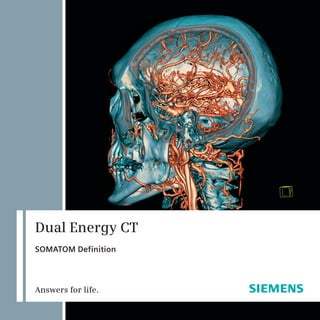
  • 6. Dual Source CT with SOMATOM Definition The idea behind Dual Source CT is as simple as it is ingenious: It is merely using two X-ray sources and two detectors at the same time. The result? You get double temporal resolution, double speed, and twice the power, while lowering dose even further. It provides images of exceptional quality and is an amazing tool to explore new clinical opportunities. The benefits that Dual Source CT holds for you and your patients are astounding. SOMATOM® Definition allows you to scan any heart at any heart rate without the need of beta-blockers – at the lowest radiation dose ever achieved in Siemens CT. Moreover, it provides one-stop diagnoses, regardless of size*, condition, and heart rate of the patient, saving precious time and money in acute care. And imagine all the additional clinical opportunities Spiral Dual Energy scanning offers in CT by characterizing materials in a single scan. Reaching excellence in CT is not only about having the most innovative scanner: It is also about pushing clinical boundaries to a higher level, providing advantages nobody wants to miss. We make a difference by offering a complete and comprehensive solution dedicated to all clinical needs, by turning complex examinations into easy CT routine. * Up to 300 kg/200 cm (660 lbs/79‘‘). 6
  • 7. 7
  • 8. 8
  • 9. Dual Energy CT It has always been an aim to collect as much Clinical Benefits information as possible for differentiation of tissues. Providing Spiral Dual Energy scanning, • Direct subtraction of bone even SOMATOM Definition opens the door to a new in complicated anatomical world of characterization, visualizing the chemical regions. composition of material. The idea of Dual Energy is not new to the CT • Display of atherosclerotic community. Earlier approaches, including two plaques. subsequent scans at different tube voltages or two subsequent scans at the same position, failed • Virtual unenhanced images. to seamlessly align the imaged anatomy and to capture the same phase of contrast enhancement. • Evaluation of lung perfusion. SOMATOM Definition overcomes this limitation by permitting the use of two X-ray sources at two different kV levels simultaneously. The result is • Display of lung vessels affected two spiral data sets acquired simultaneously in a by pulmonary emboli. single scan providing diverse information, which allows you to differentiate, characterize, isolate, • Visualization of tendons and and distinguish the imaged tissue and material. ligaments. Many applications are already available for daily • Kidney stone characterization. clinical use, such as an accurate subtraction of bone in CTAs, assessment of pulmonary perfusion, characterization of kidney stones or iodine removal • Visualization of iodine from liver scans to generate a virtual unenhanced concentration in the image. What’s more, new applications continuously myocardium. increase the clinical value of Dual Energy CT, for example characterization of atherosclerotic plaques • Differentiation between old and or assessment of myocardial perfusion. fresh intracranial bleedings. By enabling not only faster and more reliable • Visualization of uric acid crystals diagnoses, but also by further broadening the application spectrum of CT, Spiral Dual Energy in peripheral extremities. makes a difference for everybody’s daily work. 9
  • 10. How Dual Energy CT Works The X-ray tube’s kilo voltage (kV) determines the average energy of the photons in the X-ray beam. Changing the tube potential results in an alteration of photon energy and a corresponding modification of the attenuation of the X-ray beam in the materials scanned. In other words, X-ray absorption is energy- dependent, for example, scanning an object with 80 kV results in a different attenuation than with 140 kV. In addition, this attenuation also depends on the type of material or tissue scanned. Iodine, for instance, has its maximum attenuation at low energy, while its CT-density is only about half in high-energy scans. The attenuation of bones, on the other hand, changes much less when scanned with low photon energies compared to high-voltage examinations. 80 kV 140 kV Attenuation B Attenuation A Spiral Dual Energy CT exploits this effect: Two X-ray sources running simultaneously at different voltages acquire two data sets showing different attenuation levels. In the resulting images, the material-specific difference in attenuation makes a classification of the elementary chemical composition of the scanned tissue feasible. Depending on the clinical question, the images obtained at 140 and 80 kV potential are further processed with specific software algorithms implemented in the syngo Dual Energy software. In addition, fused images are provided for initial diagnosis. These are created as weighted average which have a low image noise and a normal attenuation like images scanned at 120 kV tube potential. 10
  • 11. Energy 1: Energy 2: Bone 670 HU Bone 450 HU Iodine 296 HU Iodine 144 HU 80 kV 140 kV Because X-ray absorption is energy-dependent, changing the tube’s kilo voltage results in a material-specific change of attenuation. 11
  • 12. 12
  • 13. Direct Angiography and Bone Removal with Plaque Highlighting syngo Dual Energy Direct Angio with Bone Removal As a minimally invasive procedure in comparison to DSA, multislice spiral CT (MSCT) has become the first-line modality for many angiographic applications. For routine practice and especially in the emergency room, CTA is the standard of care because it is less time consuming, less susceptible to motion artifacts, and much less prone to complications. Today, MSCT is the standard modality in the detection of aortic aneurysms, especially in emergencies, when pulmonary embolism, acute heart attack, and aortic dissection need to be ruled out by CT simultaneously. Visualization of the aorta is possible beyond the vessel wall, also showing, for example, intramural thrombi or periaortic infections. The latest technical evolution of MSCT to DSCT makes direct bone removal possible based on a spectral differentiation between iodine and bone in Dual Energy CT. Thus, a direct visualization of the aorta and branching vessels is feasible. Moreover, MSCT is the perfect tool for pre- and postoperative assessment and evaluation with endovascular aortic repair (EVAR), and Dual Energy CT can help to differentiate endoleaks from calcifications in the thrombosed lumen. In neurovascular angiography, the immediate availability and fast exam in MSCT have also proven highly beneficial in the non-invasive investigation of supra-aortic extracranial and intracranial vessels for the assessment of acute stroke. Recent studies show that supraaortic CT angiography (CTA) is also ideally suited for the detection of intracranial aneurysms of 3 mm and larger in cases of subarachnoidal hemorrhage. With dual energy material differentiation, it has also become possible to perform reliable bone and calcified plaque subtraction out of CTA volume data, so that aneurysms in close vicinity to the skull base are more easily identified. CT angiography is also gaining importance in the assessment of peripheral arterial occlusive disease. Dual Energy CT can help to cope with those huge datasets exceeding 1,000 images by generating a single maximum intensity projection without the superimposition of bones. Additionally, plaques can be identified and removed or highlighted in this image to support the grading of stenoses and to assist therapeutic planning. 13
  • 14. Case 1 – Carotid Angiography W. Eicher, MD Examination Protocol Landeskrankenhaus Klagenfurt, Klagenfurt, Austria Scanner SOMATOM Definition Scan area supraaortic vessels from History aortic arch to top of head Scan length 278 mm A 79-year-old male patient was referred to CT for Scan time 8s suspected occlusion of the right carotid artery. His Scan direction caudocranial past medical history included second stage PAOD kV 140 kV and 80 kV (Peripheral Arterial Obstructive Disease). Effective mAs 51 mAs and 186 mAs Rotation time 0.33 s Slice collimation 64 x 0.6 mm Diagnosis CTDIvol 9.8 mGy Sex M CT angiography revealed an occlusion of the right Contrast common carotid artery close to its origin. There was a moderate stenosis of the proximal right vertebral Contrast material Ultravist 370 artery. Additionally, a high-grade stenosis of the left Volume 80 ml carotid bulb involving the common, internal, and Flow rate 6 ml/s external carotid artery was found. The left vertebral Start delay 4 s after trigger artery was occluded as well. Intracranially, the Saline chaser bolus 50 ml, 6 ml/s distal right internal carotid artery was supplied by Postprocessing application collateral flow. The anterior, middle, and posterior cerebral arteries were all perfused. syngo Dual Energy Direct Angio with Bone Removal Comments With Dual Energy CT, maximum intensity projections of the extra- and intracranial arteries can be generated without superimposing bones, similar to MRA. These images make it possible to screen datasets very quickly for pathology. In complex datasets, the technique provides an excellent overview of the vasculature including occlusions, stenoses, plaques, and collateral vessels. 14
  • 15. [A] VRT [B] MIP A B C [C] VRT 15
  • 16. Case 2 – Aortic Angiography P. Rossi, MD; F. Civaia, MD Examination Protocol Centre Cardio-Thoracique de Monaco Monte Carlo, Monaco Scanner SOMATOM Definition Scan area thorax and abdomen History Scan length 541 mm Scan time 13 s A 70-year-old woman with chronic hypertension Scan direction craniocaudal was referred for follow-up aortic angiography after kV 140 kV and 80 kV endovascular repair of a dissecting aortic aneurysm. Effective mAs 42 mAs and 180 mAs She had a type B dissection of the thoracic aorta Rotation time 0.5 s extending into the abdominal aorta and iliac Slice collimation 14 x 1.2 mm arteries. CTDIvol 10.2 mGy Sex F Contrast Diagnosis Contrast material Ultravist 370 CTA showed the stent in the true lumen of the type Volume 120 ml B dissection in segment III of the thoracic aorta. Flow rate 4 ml/s There was a persistent entry and perfusion of the Start delay 7 s after trigger false lumen. The dissection membrane extended Saline chaser bolus 50 ml, 4 ml/s into the left iliac artery. The left renal artery was Postprocessing application occluded. syngo Dual Energy Direct Angio with Bone Removal Comments Dual Energy CTA provides an excellent overview of pathology. A stent, a perfused false lumen, a missing renal artery, and a dissection of the iliac artery are all evident in a single MIP image. 16
  • 17. A B C [A] VRT [B] Inverted MIP [C] MIP 17
  • 18. 18
  • 19. Plaque Characterization syngo Dual Energy Hardplaque Display With simultaneous dual energy scanning, automated plaque detection and characterization has become viable. The first step is to reliably differentiate calcified plaques from the contrast-opacified lumen of the vessel. The new syngo Dual Energy Hardplaques algorithm helps to differentiate plaque from iodine by color-coding both differently. This aim is accomplished better than with the bone removal algorithm, because this algorithm is designed to differentiate and mark calcium and iodine rather than completely removing calcium-containing structures from the dataset. Thus, the plaque load becomes obvious and it is possible to quantify atherosclerotic lesions, for example, to monitor treatment response to statin therapy. Also, the visual grading of calcified stenoses can be improved, because the algorithm helps to delineate plaque and lumen more exactly. Additionally, this data can be used to improve the automatic grading of stenoses by postprocessing software. The algorithm can also be used to differentiate calcification from contrast enhancement or to visualize an additional contrast enhancement in calcified lesions. This can be helpful, for example, to identify endoleaks in a partially calcified thrombosed false lumen after endovascular repair of aneurysms or dissections. In calcified lesions of liver or spleen, for instance, metastases or parasitic cysts, the algorithm can be used to assess contrast enhancement even without an additional unenhanced scan. 19
  • 20. Case 1 – Carotid Plaques Centre Cardio-Thoracique Examination Protocol de Monaco, Monaco Scanner SOMATOM Definition History Scan area aortic arch to circle of Willis Scan length 250 mm A 63-year-old hypertensive patient presented with Scan time 4s transient amaurosis fugax and aphasia for eight Scan direction caudocranial hours. MRI angiography was not possible due to kV 140 kV and 80 kV a cardiac pacemaker. Effective mAs 55 mAs and 234 mAs Rotation time 0.33 s Slice collimation 0.6 mm Diagnosis CTDIvol 9.1 mGy Pitch 0.7 Abnormal anatomic findings of the carotid arteries Sex F were excluded. Fused dual energy images showed Contrast calcified plaques at both carotid bifurcations. A severe vascular stenosis due to calcified or soft Contrast material Ultravist 370 plaque was ruled out. Volume 80 ml Flow rate 5 ml/s Start delay 2 s after trigger Comments Saline chaser bolus 50 ml, 5 ml/s Postprocessing application Dual Energy CTA makes a differentiation of iodine- filled vessel lumina from calcified vessel plaques syngo Dual Energy Hardplaque Display feasible, making a more accurate quantification of carotid stenosis possible. Color-coded visualization further simplifies diagnosis and presentation. 20
  • 21. A B C [A] Axial dual energy image of a [B] Coronary dual energy image (MPR) [C] Sagittal dual energy image (MPR) calcified plaque at the bifurcation of the same plaque. of the same plaque. of the left carotid artery. Note the differentiation of iodine (blue) and calcium (red). 21
  • 22. Case 2 – Aortic Stent T. R. C. Johnson, MD Examination Protocol Grosshadern University Hospital Munich, Germany Scanner SOMATOM Definition Scan area thorax History Scan length 350 mm Scan time 8s A 45-year-old male patient was referred for Scan direction craniocaudal follow-up one year after endovascular repair kV 140 kV and 80 kV of an aortic aneurysm. Effective mAs 50 mAs and 214 mAs Rotation time 0.5 s Slice collimation 14 x 1.2 mm Diagnosis CTDIvol 11.1 mGy Sex M Aortic angiography showed a stent in the Contrast descending thoracic aorta. Hyperdense material was noted in the thrombosed part of the lumen Contrast material Ultravist 370 outside the stent. There was no connection Volume 120 ml to the proximal or distal end of the stent. The Flow rate 4 ml/s hardplaques algorithm color-coded these areas Start delay 5 s after trigger as iodine, confirming an endoleak type III. Saline chaser bolus 50 ml, 4 ml/s Postprocessing application Comments syngo Dual Energy Hardplaque Display The hardplaques algorithm is helpful to clearly differentiate calcifications from contrast enhancement. The single phase acquisition in dual energy technique was sufficient to make the diagnosis in this case. 22
  • 23. [A] Volume-rendered image showing [E + F] Axial and coronal color- the contrast-opacified aorta with the coded result images of the stent. [C + D] Axial and coronal images hardplaques algorithm. The showing some hyperdense material hyperdense material is identified [B] Maximum intensity projection on the right side of the stent in the as iodine, confirming an visualizing the struts of the stent. thrombosed lumen. endoleak. A C E B D F 23
  • 24. 24
  • 25. Assessment of Lung Perfusion syngo Dual Energy Lung PBV CT angiography has recently become the diagnostic standard for non- invasive detection of pulmonary embolism. However, it seems that there is some discrepancy between the size or location of emboli and their clinical relevance. Therefore, additional perfusion imaging may add relevant information. With Dual Energy CT it is possible to color-code the iodine distribution in the lung parenchyma, and thus to visualize perfusion defects caused by small emboli as areas with reduced color density. At the same time, the hemodynamic significance of larger emboli can be assessed. This method was evaluated in two clinical studies at the University of Munich. Perfusion defects were observed in the dual energy perfusion analysis in all patients with occlusive pulmonary embolism. There was also a good agreement between dual energy perfusion assessment and pulmonary perfusion scintigraphy. Hence, Dual Energy CT of pulmonary perfusion is a feasible method for detecting and assessing the functional relevance of pulmonary embolism without additional dose, contrast application, or examination time. 25
  • 26. Case 1 – Detection of Pulmonary Embolism J. Ferda, PhD Examination Protocol Fakultni Nemocnice Pilsen, Czech Republic Scanner SOMATOM Definition Scan area thorax History Scan length 271 mm Scan time 9s A 19-year-old woman presented with sudden breath Scan direction craniocaudal shortness and chest pain. kV 140 kV and 80 kV Effective mAs 22 mAs and 155 mAs Rotation time 0.5 s Diagnosis Slice collimation 14 x 1.2 mm CTDIvol 5.2 mGy The primarily reconstructed angiographic images Contrast showed subtle hypodense material in the lateral segment of the right middle lobe. With the dual- Contrast material Ultravist 370 energy-based perfusion analysis, two small Volume 80 ml peripheral perfusion defects were visualized, Flow rate 4 ml/s one in the lateral and one in the medial segment Start delay 7 s after trigger of the middle lobe. Saline chaser bolus 50 ml, 4 ml/s Postprocessing application Comments syngo Dual Energy Lung PBV The perfusion information was helpful in confirming pulmonary embolism in the presence of the subtle angiographic finding. Additionally, a small perfusion defect was diagnosed on the basis of the perfusion information which might have been missed in normal angiography. Thus, Dual Energy CT can improve the sensitivity for pulmonary embolism and may be able to replace perfusion scintigraphy in some instances. 26
  • 27. [A] Axial reconstruction with color- [B] Volume-rendered image showing that [D] Coronal reconstruction showing coded perfusion information. most of the lung parenchyma is perfused the embolic material in the right Note the hypodense areas in the normally. middle lobe artery. Note that the lung segmental lateral middle lobe artery parenchyma has a normal density in and the corresponding wedge-shaped [C] Color-coded axial image showing both lung window. subpleural perfusion defect. perfusion defects in the right middle lobe. A B C D 27
  • 28. Case 2 – Assessment of Perfusion Defects B. Ghaye, MD; J.-F. Monville, MD Examination Protocol University Hospital of Liège Liège, Belgium Scanner SOMATOM Definition Scan area thorax History Scan length 342 mm Scan time 8s A 76-year-old male presented in the emergency Scan direction caudocranial department with sudden onset of dyspnea. kV 140 kV and 80 kV Pulmonary embolism was suspected and the patient Effective mAs 51 mAs and 213 mAs was referred for CT pulmonary angiography. Rotation time 0.5 s Slice collimation 1.2 mm CTDIvol 6.96 mGy Diagnosis Sex M Contrast The angiographic reconstruction as weighted average images revealed massive central pulmonary Contrast material Ultravist 370 embolism. The additional perfusion assessment Volume 100 ml showed large perfusion defects in the segments Flow rate 5 ml/s corresponding to the emboli. Based on the perfusion Start delay 6 s after trigger assessment, it was estimated that only about half Saline chaser bolus 50 ml, 5 ml/s of the lung parenchyma remained perfused and Postprocessing application that blood oxygenization was severely impaired. syngo Dual Energy Lung PBV Comments With Dual Energy CT, both morphological and functional on lung perfusion are obtained with a single exam. This can contribute to a comprehensive workup of acute conditions such as pulmonary embolism or chronic pulmonary diseases such as pulmonary hypertension, emphysematic, or fibrotic diseases. The dual energy perfusion information may even render additional perfusion scintigraphy unnecessary in some instances, thus saving time, money, and radiation exposure. 28
  • 29. A B C [A] Axial reconstruction with color-coded [B] Coronal reconstruction. Only the [C] Volume-rendered image with dual energy perfusion information. Note apical parts show a normal perfusion. color-coded perfusion information. the large perfusion defects in both lungs. There are large perfusion defects in the right middle and lower lobe and in the lingual and lower lobe on the left. 29
  • 30. 30
  • 31. Virtual Non-Contrast Images syngo Dual Energy Virtual Unenhanced Multi-detector row CT scanners have substantially broadened the spectrum of clinical applications and examination techniques, but the basic principle of image acquisition with contrast media being used for better delineation of organs and specific structures has remained the same since the introduction of CT in the 1970s. However, if pathology is only very subtly delineated, contrast media may mask the sought-after pathology. Such is the case with intramural hematomas in the aorta or small kidney stones. With Dual Energy CT, this clinical problem can be overcome: Thanks to the particularly stronger enhancement rate of iodine at low tube voltage, it is possible to remove the iodine content of any object in the image mathematically without removing the object itself. This results in “virtual non-contrast” images. Hence, it is possible to scan at any phase of contrast enhancement and non-contrast images can be generated afterwards. The non-contrast phase can be omitted for all imaging protocols in clinical routine, which can help to reduce dose significantly. Whenever needed for diagnosis, non-enhanced images can be generated without any further effort. 31
  • 32. Case 1 – Differentiation of Renal Masses R. P. Hartmann, MD Examination Protocol Mayo Clinic College of Medicine Rochester, MN, USA Scanner SOMATOM Definition Scan area abdomen Scan length 427 mm History Scan time 15 s Scan direction craniocaudal A 70-year-old female patient presented for follow-up kV 140 kV and 80 kV of a suspected solid mass in the lower pole of the Eff. mAs 50 mAs and 190 mAs left kidney. Rotation time 0.5 s Slice collimation 14 x 1.2 mm CTDIvol 10.2 mGy Diagnosis Sex F There was a 1.2 cm mass in the lower pole of the Contrast left kidney. Based on the presence of iodine signal Volume 80 ml centrally, it had to be considered a solid mass. Flow rate 4 ml/s This was also established based on an increase Start delay 70 s in attenuation from true non-contrast to post- contrast images of 70 HU. This was considered Postprocessing application worrisome for a renal cell carcinoma. A second syngo Dual Energy Virtual Unenhanced lesion in the mid portion of the right kidney did not have iodine signal and was considered to be a benign cyst. Comment The use of the iodine overlay image that can be produced from a dual energy acquisition allows for the differentiation of a renal cyst from a solid mass. It is not necessary to acquire a non-contrast scan prior to dual energy scanning, because the iodine enhancement can be identified in the color- coded overlay or in virtual non-contrast images generated with the dual energy information. 32
  • 33. [A] Non-contrast coronal MPR. A B No stones were identified in either kidney. [B] Coronal image of the right kidney with a low attenuation mass in the mid portion (arrow) that has a typical appearance of a renal cyst. [C] Coronal image of the left kidney with an indeterminate lesion along the medial lower pole (arrow). [D] Coronal iodine overlay image C D depicting both lesions (cyst in the right kidney with signal void and mass in the left kidney identified on average weighted series (arrows). 33
  • 34. Case 2 – Identification of Stones in Contrast Material R. P. Hartmann, MD Examination Protocol Mayo Clinic College of Medicine Rochester, MN, USA Scanner SOMATOM Definition Scan area upper abdomen History Scan length 274 mm Scan time 10 s A 66-year-old male patient presented for follow-up Scan direction craniocaudal exam of known nephrolithiasis. His prior history kV 140 kV and 80 kV included bladder transitional cell carcinoma. Effective mAs 40 mAs and 200 mAs Rotation time 0.5 s Slice collimation 14 x 1.2 mm Diagnosis CTDIvol 10.1 mGy Sex M In the weighted average reconstructions, the Contrast contrast material in the renal collecting system made it impossible to identify calculi. In the Volume 80 ml calculated virtual non-contrast images, two renal Flow rate 4 ml/s calculi were identified. No other abnormalities Start delay 300 s of the kidneys, ureters, or bladder were observed. Saline chaser bolus 50 ml, 4 ml/s The stones were lodged within the intrarenal Postprocessing application collecting system and were slightly larger than on previous exams. syngo Dual Energy Virtual Unenhanced Comments The virtual non-contrast images produced from the excretory phase of a CT urogram are capable of detecting the stones that are evident on a true non- contrast acquisition. Given this capability of Dual Energy CT, a separate non-contrast acquisition is not needed. 34
  • 35. [A] Coronal MPR image from a A B non-contrast CT acquisition demonstrates stones in the upper and lower poles of the left kidney (arrows). [B] Coronal MPR image after iodine subtraction from the excretory phase. The stones previously obscured by the surrounding iodinated contrast are now easily visible (arrows). [C] Coronal MPR from dual energy data acquired in the excretory phase. The areas determined to be iodine are C D depicted with color. The stones are not visible (arrows). [D] Coronal MPR from the dual energy mixed images acquired during the excretory phase. The stones shown on the non-contrast acquisitions are now obscured. 35
  • 36. 36
  • 37. Characterization of Renal Calculi syngo Dual Energy Calculi Characterization In recent years, the ability of MDCT to provide rapid contiguous thin-slice imaging through the abdomen has led to the supersession of traditional techniques in the detection of nephrolithiasis, urolithiasis, renal masses, and urothelial neoplasms such as transitional cell carcinoma. Improved CT acquisition techniques have led to increased sensitivity and speed. They also require less radiation dose than traditional X-ray urograms. To date, a contrast and a non-contrast scan were required in multislice CT Urography (CTU) for the safe detection of stones and calcifications, and the characterization of renal masses. Spiral Dual Energy CT, with its potential for delivering a virtual non-contrast image set, makes the acquisition of an initial non-contrast scan obsolete. Saving dose and time, a virtual non-contrast image set can be produced from a CT acquisition performed during an excretory phase, when the intrarenal collecting systems and ureters are opacified with iodinated contrast. Studies at the the Mayo Clinic College of Medicine have shown that Spiral Dual Energy CT also provides a highly accurate method of predicting stone compositions on the basis of the various materials’ specific X-ray attenuation characteristics. After the Spiral Dual Energy CT data acquisition, detailed stone characterization was performed using micro CT. For all stones, micro CT confirmed the major mineral compositions as determined by infrared spectroscopy. This opens up an interesting perspective in the therapy of patients with uric acid stones. The expensive and somewhat risky shock wave lithotripsy procedure can be avoided. Instead, such patients may be treated through urinary alkalization, which is likely to dissolve uric acid stones. Studies at the University of Munich using the SOMATOM Definition have confirmed that Dual Energy CT is able to reliably differentiate uric acid calculi from other types of renal stones, both in vitro and in vivo. 37
  • 38. Cases 1 and 2 – Differentiation of Uric Acid and Calcified Stones A. Graser, MD; T. R. C. Johnson, MD; Diagnosis C. R. Becker, MD Grosshadern University Hospital The red color-code of the kidney stone shown Munich, Germany in patient 1 indicated a uric acid calculus. Approximately 12 hours after the Dual Energy CT scan, the stone passed spontaneously and was History analyzed. The chemical analysis confirmed that the calculus consisted entirely of uric acid. The A 34-year-old male (1) and a 55-year-old male (2) blue color-code shown in patient 2 characterized patient were both referred for MDCT evaluation of a calcified stone. Based on this dual energy study, known urolithiasis. the stone was removed by ureterorenoscopy. The lab analysis of the removed stone confirmed that the stone consisted of calcium oxalate. Comment The dual-energy-based differentiation of renal calculi makes it possible to treat patients with uric acid stones medically and to initiate mechanical Examination Protocol removal in patients with other types of calculi. Scanner SOMATOM Definition SOMATOM Definition Patient Patient 1 Patient 2 Scan area abdomen lower abdomen Scan length 377 mm 215 mm Scan time 14 s 8s Scan direction craniocaudal craniocaudal kV 140 kV and 80 kV 140 kV and 80 kV Effective mAs 64 mAs and 352 mAs 69 mAs and 351 mAs Rotation time 0.5 s 0.5 s Slice collimation 1.2 mm 1.2 mm CTDIvol 11.7 mGy 11.7 mGy Sex M M Postprocessing application syngo Dual Energy Calculi Characterization 38
  • 39. [A] Using conventional A B CT imaging the kidney stones (arrows) can clearly be visualized; however, its composition cannot be characterized. [B] + [D] In patient 1 the kidney stone can be characterized as uric acid stone, color-coded in red. [C] The dual energy characterization shows that the calculus of patient 2 is a calcified stone, color-coded in blue (as cortical bone in the C D image). 39
  • 40. 40
  • 41. Visualization of Tendons and Ligaments syngo Dual Energy Musculoskeletal Dual energy techniques based on the recently introduced Dual Source CT with Spiral Dual Energy capabilities promise to offer additional diagnostic information regarding the integrity of ligaments, tendons, and potentially cartilage. However, MRI remains the imaging modality of choice in many cases. Dual Energy CT makes it possible to differentiate and visualize tendons and ligaments. This can be of great value in trauma patients where CT is performed to evaluate fractures. If the CT scan is performed in dual energy technique, bones can be assessed as well as tendons and ligaments in the same dataset. Although MRI is necessary to detect minor abnormalities, such as edema in a tendon, dual energy evaluation is available with the dataset obtained for the initial evaluation, and it is generally sufficient to exclude tendon ruptures or to display dislocations. Also, the differentiation of cartilage may be of interest, for example, to weigh the possibility of joint reconstruction against the necessity of replacement in elderly trauma patients. 41
  • 42. Case 1 – Tendons of Foot and Ankle C. Sun, MD Examination Protocol ASAN Medical Center Seoul, Korea Scanner SOMATOM Definition Scan area foot and ankle History Scan length 274 mm Scan time 55 s After a bicycle accident, a 27-year-old man was Scan direction craniocaudal referred for CT to rule out bony extensor hallucis kV 140 kV and 80 kV longus tendon avulsion. Effective mAs 50 mAs and 90 mAs Rotation time 1.0 s Slice collimation 20 x 0.6 mm Diagnosis CTDIvol 9.2 mGy Sex M On the anterior aspect of the right foot there was Postprocessing application a defect and proximal retraction of the extensor hallucis longus tendon. There was no bony avulsion syngo Dual Energy Musculoskeletal and no evidence of fractures or other injuries. Comments With Dual Energy CT it is possible not only to exclude a fracture or bony avulsion but also to evaluate the tendons and ligaments directly. Without Dual Energy CT, the relevant diagnosis would not have been made in this patient. 42
  • 43. [A] Sagittal MPR image with A B color-coded tendons. Note the interruption of the extensor hallucis longus tendon. [B] Axial MPR image. Note the color-coded flexor and extensor tendons and the absent first extensor. [C] Volume-rendered image from a superior view showing the extensor tendons and the interrupted hallux tendon. [D] VRT in a plantar view C D showing the flexor tendons. 43
  • 44. Case 2 – Tendons of Hand and Wrist T. R. C. Johnson, MD Examination Protocol Grosshadern University Hospital Munich, Germany Scanner SOMATOM Definition Scan area wrist History Scan length 133 mm Scan time 4s A 45-year-old female patient was referred for kV 140 kV and 80 kV diagnostic workup of chronic pain in the wrist Effective mAs 38 mAs and 100 mAs six years after an intraarticular fracture of Rotation time 1.0 s the radius. Slice collimation 64 x 0.6 mm CTDIvol 9.1 mGy Sex F Diagnosis Postprocessing application The SL joint space showed a triangular configuration, syngo Dual Energy Musculoskeletal and the color-coding of the ligaments and tendons showed no signal in the area of the scapholunate interosseous ligaments. Rupture of the ligaments with scapholunar dissociation was suspected. Additionally, there was severe radiocarpal arthrosis with an uneven articular surface as remnant of the previous fracture. Joint space to os lunatum was noticeably narrowed. Comments Dual Energy makes it possible to visualize both bones and ligaments in a single exam. Although Dual Energy CT cannot replace MRI for a detailed evaluation, it can be useful to assess the presence and continuity of ligaments and tendons without additional radiation exposure. Considering that CT is mostly performed as primary modality to rule out fractures, this technique can help to route the further diagnostic workup. 44
  • 45. [A] + [B] Volume-rendered A B images showing the continuity of the flexor tendons. [C] + [D] Color-coded reconstructions in coronal and sagittal orientation. C D 45
  • 46. New Applications Dual Energy CT is very young. As early as two years after the first dual energy applications were invented, they have been fully integrated in the DSCT system and provide clinically useful additional information without additional effort in daily clinical routine at many hospitals. Additionally, Dual Energy CT has opened up a whole new field of research and science. Many academic centers worldwide work with this new technology and identify further clinical applications. As a manufacturer Siemens has taken this opportunity to integrate these new applications into the syngo Dual Energy software as fast as possible. Therefore, the group of postprocessing applications selectable in the menu of the software is growing continuously, and Siemens is proud to present several new and already FDA cleared application classes. 46
  • 47. Optimum Contrast Blending syngo Dual Energy Optimum Contrast Dual Energy datasets usually consist of images obtained at 80 and 140 kVp. The different postprocessing algorithms exploit the spectral behavior of certain materials or tissues to identify or quantify them. Additionally, average images can be calculated to provide a ”normal” CT image with a low noise and normal attenuation properties, because the 140 kVp images lack contrast, while the 80 kVp images are very noisy. However, special, non-linear blending algorithms can provide a better image than mere averaging. By adding more of the contrast information from the 80 kVp images in areas of homogeneous soft tissue, and more of the detail from the 140 kVp images in high-contrast areas such as lung or bone, an optimized image can be calculated. This image provides less noise and more contrast than linear average images. 47
  • 48. Assessment of Myocardial Perfusion syngo Dual Energy Heart PBV With its high temporal resolution, Dual Source CT has greatly improved coronary CT angiography, because it is less sensitive to high heart rates or arrhythmia than other types of scanners. With Dual Energy CT, it is now possible to color-code iodine content to visualize organ perfusion. A new protocol now offers an ECG-gated dual energy scan to assess myocardial perfusion. The corresponding syngo Dual Energy Heart PBV software color-codes myocardial perfusion, so that both coronary artery morphology and myocardial perfusion can be assessed in a single CT scan. 48
  • 49. [A] Volume-rendered A B C image of the heart scanned in dual energy technique. [B] – [D] MPRs with color-coded perfusion information in short axis, two chamber, and coronal orientation. Note the perfusion defect of the basal D E F posterior left ventricular wall. [E] + [F] Cut volume- rendered images showing the perfusion defect of the posterior wall in short and long axis. [G] – [I] Short axis G H I SPECT images in an apical, mid-ventricular, and basal plane confirming the posterior perfusion defect. 49
  • 50. Differentiation of Brain Hemorrhage and Contrast Enhancement syngo Dual Energy Brain Hemorrhage A primary task of cranial CT is to rule out hemorrhage, and this is why an unenhanced scan is usually obtained even in other clinical questions in which a contrast-enhanced scan is necessary, for example to rule out neoplasms. With syngo Dual Energy Brain Hemorrhage, a virtual non-contrast image can be generated from a contrast-enhanced dual energy scan. This application may make it possible to discard the pre-contrast scan in some instances. 50
  • 51. [A] The normal reconstruction of A B the contrast-enhanced Dual Energy CT scan shows a hyperdense area at the left parietal lobe, suggestive of subarachnoidal hemorrhage. [B] The syngo Dual Energy Brain Hemorrhage application color- codes the center of the lesion to indicate iodine enhancement. [C] The color-coding alone shows a distinct spot of enhancement. [D] T1-weighted MRI after C D administration of gadolinium contrast confirms the central enhancement, either due to neoplastic tissue or active bleeding. 51
  • 52. Detection of Pulmonary Embolism in Lung Vessels syngo Dual Energy Lung Vessels syngo Dual Energy Lung Vessels color-codes the iodine content of lung vessels. Vessels that are affected by pulmonary embolism and, therefore, show a significantly lower iodine concentration than non-affected vessels are assigned a different color than those with a high iodine content. With the color-coding, the affected vessels are much easier to identify. In this manner, Dual Energy CT can improve the sensitivity for pulmonary embolism by demonstrating perfusion defects in the parenchyma and additionally by highlighting affected arteries. 52
  • 53. [B] Corresponding angiographic [A] Color-coded sagittal MPR of the image reconstructed from the right lung showing normally perfused same scan. There is embolic vessels in turquoise and embolized material in the lower and middle vessels in red. lobe arteries. A B C D E [C] Color-coded coronal MPR showing [D] Angulated coronal angiographic [E] Color-coded volume-rendered embolism to both lower lobes. image showing the emboli in the image of the pulmonary vessels lower lobe arteries. demonstrating the emboli in the arteries. 53
  • 54. Characterization of Gout Tophi syngo Dual Energy Gout The syngo Dual Energy Gout application visualizes deposits of uric acid crystals which are characteristic for gout tophi. Additionally, the application color- codes iodine enhancement so that tophi with active inflammatory changes can be differentiated from stable ones. This way, both the molecular cause and the activity of the disease can be shown in a single scan. 54
  • 55. A B [A] Axial image of both feet showing massive [B] The red color-coding confirms uric acid in deformities and calcified masses. the masses, identifying them as gout tophi. 55
  • 56. Courtesy List • ASAN Medical Center, Seoul, Korea • Centre Cardio-Thoracique de Monaco, Monte Carlo, Monaco • CHU De Charleroi, Charleroi, Belgium • Fakultni Nemocnice/Pilsen, Czech Republic • Grosshadern University Hospital, Munich, Germany • Landeskrankenhaus Klagefurt/Klagenfurt, Austria • Mayo Clinic College of Medicine Rochester, MN, USA • Medical University of South Carolina, Charleston, SC, USA • University Hospital of Liège, Liège, Belgium • University of British Columbia, Vancouver General Hospital/Vancouver Canada 56
  • 57. 57
  • 58. On account of certain regional limitations of sales rights Local Contact Information and service availability, we cannot guarantee that all products included in this brochure are available through the Siemens sales organization worldwide. Availability In the USA: and packaging may vary by country and is subject Siemens Medical Solutions USA, Inc. to change without prior notice. Some/All of the features 51 Valley Stream Parkway and products described herein may not be available in Malvern, PA 19355 the United States. Phone: +1 888 826 9702 The information in this document contains general Phone: +1 610 448 4500 technical descriptions of specifications and options as Fax: +1 610 448 2554 well as standard and optional features which do not always have to be present in individual cases. In China: In Asia: China Medical Solutions Siemens Medical Solutions Siemens reserves the right to modify the design, 7, Wangjing Zhonghuan Nanlu Asia Pacific Headquarters packaging, specifications, and options described herein without prior notice. Please contact your local Siemens Chaoyang District The Siemens Center sales representative for the most current information. P.O.Box 8543 60 MacPherson Road Beijing 100102, P.R.China Singapore 348615 Note: Any technical data contained in this document Phone: +86 10 6476 8888 Phone: +65 6490 8182 may vary within defined tolerances. Original images Fax: +86 10 6476 4706 Fax: +65 6490 8183 always lose a certain amount of detail when reproduced. The statements contained herein are based on the In Japan: In Germany: actual experience of Siemens customers. Siemens Siemens-Asahi Siemens AG maintains data on file to support these claims. However, Medical Technologies Ltd. Healthcare Sector these statements do not suggest or constitute a Takanawa Park Tower 14 F Computed Tomography warranty that all product experience will yield similar 20-14, Higashi-Gotanda 3-chome Siemensstr. 1 results. Results may vary, based on the particular Shinagawa-ku DE-91301 Forchheim circumstances of individual sites and users. Tokyo 141-8644 Germany Please find fitting accessories: Phone: +81 35423 8510 Phone: +49 9191 18 0 www.siemens.com/medical-accessories Fax: +81 35423 8740 Fax: +49 9191 18 9998 Global Siemens Headquarters Global Siemens Legal Manufacturer Healthcare Headquarters Siemens AG Siemens AG Wittelsbacherplatz 2 Siemens AG Wittelsbacherplatz 2 80333 Muenchen Healthcare Sector DE-80333 Muenchen Germany Henkestr. 127 Germany 91052 Erlangen Germany Phone: +49 9131 84-0 www.siemens.com/healthcare www.siemens.com/healthcare Order No. A91CT-00615-04C1-7600 | Printed in Germany | CC CT WS 08085. | © 11.2008, Siemens AG