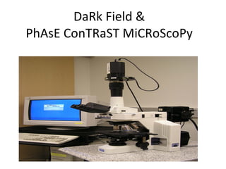
3. I. Dark field contrast microscopy takes advantage of objects that “scatter” light - this requires a special condenser that can “angle” the incident light II. Phase contrast microscopy takes advantage of objects that alter the phase of incident light - This requires “phase rings” in the condenser and in the objective lens III. Fluorescence microscopy take advantage of inherently fluorescent Material of biological objected that can be fluorescenlty labeled.
4. Single Cell Organism ( Tetrahymena ) observed with: “ Advanced” Light Microscopic Methods Bright Field Dark Field Phase Contrast Fluorescence (Dr. Gorovksy)
5. Recall that a specimen (e.g a live cell) with few structures that strongly absorb light provides little contrast with bright field microscopy e.g., the single celled protist Tetrahymena condenser lens objective lens
6. Dark Field Microscopy : Some objects can alter the light path by diffraction & or light scattering condenser lens objective lens
7. If the light pathway is angled without a specimen the field is dark... objective lens … but any object that diffracts light will be detected! Dark Field seen condenser lens objective lens condenser lens
8.
9.
10.
11.
12.
13.
14.
15.
16.
17.
18.
19.
20.
21.
22. Phase contrast is obtained with the help of the annular diaphragm by separating the central & direct ray from the diffracted rays
![[object Object],[object Object],[object Object],[object Object],[object Object]](data:image/gif;base64,R0lGODlhAQABAIAAAAAAAP///yH5BAEAAAAALAAAAAABAAEAAAIBRAA7)