Muscle tissue 2
•Download as PPTX, PDF•
17 likes•2,088 views

Muscle physiology, mechanism of muscle contraction.

Recommended
More Related Content
What's hot
What's hot (20)
Viewers also liked
Viewers also liked (18)
Similar to Muscle tissue 2
Similar to Muscle tissue 2 (20)
More from James H. Workman
More from James H. Workman (20)
Recently uploaded
Recently uploaded (20)
Muscle tissue 2
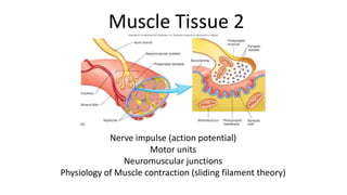
- 1. Muscle Tissue 2 Nerve impulse (action potential) Motor units Neuromuscular junctions Physiology of Muscle contraction (sliding filament theory)
- 2. Nerve impulse Necessary for muscle contraction Also known as action potential Momentary change in electrical potential due to rapid changes in ion concentration
- 3. Motor neuron Neuron with cell body located in the brain or spinal cord Ends at neuromuscular junction / synaptic end bulb Motor unit The functional unit of a skeletal muscle Consists of a motor neuron and the muscle fibers it stimulates muscles with large ratio motor units (1 neuron: many muscle fibers) provide powerful contractions but cannot provide delicate control muscles with small ratio motor units (1 neuron: few muscle fibers) provide delicate control for very precise movements
- 7. Resting potential Ion pumps actively maintain concentration gradients [Na + ] much higher outside cell [K + ] much higher inside cell 3 Na + transported out for every 2 K + transported in Results in net – 70 mV charge inside nerve cell; cell is polarized
- 8. Action potential Nerve impulse Na + channels open, Na + rush in to nerve axon Internal cell environment no longer polarized K + channels open; K + rush out of nerve Repolarizes cell Process travels down length of cell axon
- 13. Neuromuscular junction Meeting point at which motor neuron meets muscle fiber Synapse Region of communication between two neurons or a neuron and a target cell Neurotransmitter Chemical that crosses synaptic cleft (gap) allowing for communication between two cells Acetylcholine (ACh) is the neurotransmitter released from synaptic end bulb of motor neurons
- 16. Nerve impulse initiates contraction Nerve impulse arrives at NMJ Acetylcholine (Ach) released via exocytosis across synaptic cleft ACh stimulates Na + / K+ ion channels to open, muscle cell depolarizes Depolarization of sarcolemma causes Ca2+ release Ca2+ allows myosin / actin crossbridge and ultimately, sarcomere shortening
- 23. Key players of muscle contraction Myosin ~300 molecules comprise each thick filament Imagine two golf club twisted together Actin Thin filament molecules extending from anchoring points in the Z disc Titin Extends from Z disc to M line One of largest molecules known to exist; molar mass = 3million grams
- 24. Key players of muscle contraction Tropomyosin Part of thin filament; blocks sites where actin / myosin bind Troponin Holds tropomyosin in position. When Ca2+ is present, troponin changes shape (conformational change) Linked to tropomyosin, actin/mysoin binding site is uncovered when Ca2+ is present in sarcoplasm. Myomesin Forms M line of sarcomere
- 30. Sarcomere regions Z line (disc) Lateral terminus of sarcomere; separate adjacent sarcomeres A band Region where thick filaments present; darker appearance some overlap of thin filament in relaxed sarcomere more overlap of thin filament in contracted sarcomere
- 31. Sarcomere regions I band Region where thin filaments (no thick filaments) are present Lighter in appearance Narrowed in contracted sarcomere H zone Center of A band, thick filaments only Disappears during sarcomere contraction M line Central to sarcomere, myomesin proteins anchor myosin and titin proteins