Understanding the Craniovertebral Junction: Anatomy, Embryology, Radiology and Common Anomalies
•Download as PPTX, PDF•
58 likes•10,117 views

The craniovertebral junction consists of the occiput, atlas, axis, and connecting ligaments. It is a transition zone between the mobile cranium and spinal column. Common anomalies include occipitalization of the atlas, basilar invagination, atlantoaxial dislocation, and platybasia. Radiological assessment involves measurements of angles and distances on plain radiographs and CT/MRI to evaluate for abnormalities. Key measurements include the Chamberlain's line, McGregor's line, and posterior atlantodental interval.

Recommended
More Related Content
What's hot
What's hot (20)
Similar to Understanding the Craniovertebral Junction: Anatomy, Embryology, Radiology and Common Anomalies
Similar to Understanding the Craniovertebral Junction: Anatomy, Embryology, Radiology and Common Anomalies (20)
Recently uploaded
Recently uploaded (20)
Understanding the Craniovertebral Junction: Anatomy, Embryology, Radiology and Common Anomalies
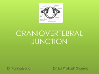
- 1. CRANIOVERTEBRAL JUNCTION Dr Kanhaiya lal Dr Jai Prakash Sharma
- 2. The craniovertebral (CVJ) is a collective term that refers to the occiput (posterior skull base), atlas, axis, and supporting ligaments. It is a transition zone b/w a mobile cranium & spinal column. It encloses the soft tissue structures of the cervico medullary junction (medulla, spinal cord, and lower cranial nerves
- 3. Contents Anatomy of CV junction Embryology Classification of CV Anomalies Clinical features Anatomical and radiological aspects Specific anomalies – AA dislocation, Dens dysplasia, KFS,ACM,
- 4. Parts of CV Junction include:- The Occiput First Cervical Vertebra (Atlas) Second Cervical Vertebra (Axis) Their articulations and Connecting ligaments
- 5. ANATOMY OF CVJ (ARTICULAR) b/w occiput & atlas Upper surfaces of C1 lateral masses is cup-like or concave which fit into the ball & socket configuration with occipital condyle, united by articular capsules surr. the AO joint & by the ant. & post. AO membranes. ( flexion 10*, extension 25*). 4 synovial joints b/w atlas & axis – 2 median –front & back of dens (Pivot variety) 2 lateral –b/w opposing articular facets (Plane variety) Each joint has its own capsule & synovial cavity. rotation is upto90* & approx ½ occurs at the A-A joint.
- 6. ANATOMY OF CVJ(LIGAMENTOUS) Principal stabilizing ligaments of C1 - -Transverse atlantal ligament -Alar ligaments Secondary stabilizing ligaments of CVJ are more elastic & weaker than the primary ligaments. -Apical ligament -Anterior & posterior A-O membranes -Tectorial membrane -Ligamentum flavum -Capsular ligaments
- 10. NEURAL structures related are – Caudal brainstem (Medulla) Fourth ventricle Rostral part of spinal cord Lower cranial (9,10,11 {only N that passes through the FM},12) & upper cervical nerves (The C1(SUBOCCIPITAL NERVE) , C2, and C3 nerves with both rami). In cerebellum, only the tonsils, biventral lobules & the lower part of the vermis (nodule, uvula & pyramid) ,
- 11. LYMPHATICS primarily into the retropharyngeal LN & then into the deep cervical chain. These LN’s also drain the nasopharynx & hence retrograde infection may affect the synovial lining of the CVJ complex with resultant neck stiffness & instability.
- 12. ARTERIAL The major arteries related to CVJ are :- Vertebral arteries (m/c) Postero-inferior cerebellar arteries (PICA) Meningeal branches of the vertebral External and internal carotid arteries.
- 13. EMBRYOLOGY & DEVELOPMENT Between 3rd& 5thweek: -The first four sclerotomes (which forms the vertebral bodies ) do not forms the vertebral bodies and fuse to form the occipital bone. CV junction is developed from 4 these and first 2 cervical sclerotome.
- 14. occipital sclerotomes 1, 2 – basiocciput 3- Jugular tubercles 4 (Proatlas) -ant. tubercle of clivus, apical cap of dens, ant.margin of FM, occipital condyles, lateral atlantal mass, sup.portion of post.arch of atlas. cervical sclerotome. 1- ant.arch of atlas post.inf,portion of c1, odontoid process. 2-rest of axis vertebra
- 15. APPLICATIONS Dysgenesis of the occipital sclerotome-may flatten the clivus-platybasia. basiocciput and rim of foramen magnum are underdeveloped, the odontoid and arch of atlas may invaginate - Basilar invagination. The proatlas may develop into separate vertebrae -Occipital vertebra proatlas fails to fuse with the atlas (cervical sclerotome), a rare anomaly termed bipartite articular facets. dens body may fail to fuse with apical part (occipital sclerotome)- Bicornuate dens Failure of segmentation b/w the axis & the 3rd cervical vertebra-Klippel–Feil syndrome.
- 16. Classification of CV Anomalies Congenital Malformation of occipital sclerotomes Clivus segmentation anomalies Condylar hypoplasia Assimilation of atlas Malformation of atlas Assimilation of atlas Atlantoaxial fusion. Aplasia of atlas arches. Malformation of axis. Irregular Atlantoaxial segmentations. Dens dysplasia Segmentations failure of c2-c3
- 17. Developmental and acquired At foraman magnum-stenosis Sec.basilar invagination-OI,Pagets ds,o malacia,rickets,RA. Atlantoaxial instability 1. down syndrome 2. Ehlers danlos syndrome 3. MPS 4. Trauma 5. Infection –TB 6. Tumors- Mets,FD,EG,chordom,osteoblastoma,NF Spontaneous rotatory subluxation-Grisel syndrome
- 18. Other classification Bony Anomalies Major Anomalies 1 Occipitalization 2 Basilar Invagination 3 Dens Dysplasia 4 Atlanto- axial dis. 5 Platybasia Minor Anomalies Dysplasia of Atlas Dysplasia of occipital condyles, clivus, etc. Soft Tissue anomalies Arnold-Chiari Malformation Syringomyelia/ Syringobulbia
- 19. Clinical features A. Cervical symptoms and signs- pain suboccipital region rediating vertex,stifness in 85% B. Myelopathic Features- long tract involvement and wasting C. CN involvement- IX, X,XI,XI inI 20% D. Vascular in 15% Transient Attack of V-B insufficiency E. Sensory symptom of post.column involvement. F. Cerebellar symptoms/signs- Nystagmus, Ataxia, intention tremor, dysarthria G. Features of Raised ICT- usually seen in Pts. Having basilar impresssion and/or ACM ATAXIA,ATROPHY,SRYNX –POOR PROGNOSIS
- 21. INVESTIGATIONS 1) X Rays Antero-posterior view Lateral view Open mouth view for dens Stress X-Rays (neutral,flexion,extention) Tomogram (rare) hypoplastic odontoid, occipitalisation of atlas are clearly visualised. CT Scan and 3D recon MRI conventional and dynamic Myelogram & Ventriculogram (used before the CT) Angiography
- 22. X Ray
- 24. CRANIOMETRY: Craniometry of the CVJ uses a series of lines, planes & angles to define the normal anatomic relationships of the CVJ. These measurements can be taken on plain X rays, 3D CT or on MRI. No single measurement is helpful. disadvantage --anatomic structures and planes vary within a normal range.
- 25. The important lines are 1. Chamberlain’s line 2. Wackenheim’s clivus canal line 3. Mc Gregor’s line (basal line) 4. Height index of Klaus 5. Mc rae’s line ( foramen magnum line) 6. Boogard,s line 7. FISHGOLD’S DIGASTRIC LINE 8. FISHGOLD’S BIMASTOID LINE The important angles are 1. Basal angle 2. Bull’s angle 3. Boogard,s angle
- 26. Normal landmark
- 27. Chamberlain’s line From tip of hard palate to posterior tip of Foramen Magnum (opisthion). If Tip of dens below this line ±3 mm Or <1/3 of odontoid above the line If not defined basilar Invagination
- 28. Mc Gregor’s line (basal line) Line drawn from posterior tip of Hard palate to lowest part of Occiput Odontoid tip >5mm above = Basilar Invagination Position changed with flexion and extension so not used. Should be used when lowest part of occipital bone is not Foramen Magnum.
- 29. Wackenheim’s clivus canal line Line drawn along clivus into cervical spinal canal Odontoid is ventral and tangential to this line If not –suggest AAD or BI
- 30. Welcher’s Basal Angle Nasion to tuberculum sella Tuberculum sellae to the basion along plane of the clivus Normal – 1240 - 142 > 1400 = platybasia < 1300 is seen in achondroplasia
- 31. Schmidt – Fischer angle Angle of axis of Atlanto-Occipital joint 125 +/- 2 degrees Angle is wider in condylar hypoplasia O C2 AA JT AO JT C1 C1
- 32. Mc rae’s line ( foramen magnum line) Joins anterior and posterior edges of Foramen magnum Tip of odontoid is below this line. When sagittal diameter of canal <20mm, in patient of >8 yr of age neurological symptoms occur – Foramen Magnum Stenosis
- 33. Boogard ‘s Line Nasion to Opisthion Basion should lie below this line Altered in basilar impression
- 34. FISHGOLD’S DIGASTRIC LINE Biventer line Connects the digastric grooves ( fossae for digastric muscles on undersurface of skull just medial to mastoid process) Line is normally 10mm above the atlanto-occipital junction. Upper limit of dens. Central axis of dens should perpendicular to this line. Corresponds to McRae’s line on lateral view If not suggest unilateral condylar hypoplasia.
- 35. FISHGOLD’S BIMASTOID LINE Line connecting tip of mastoid process. At level of atlanto-occipital junction Odontoid process should be less than 10 mm above this line
- 36. HEIGHT INDEX OF KLAUS Distance between tip of dens and tuberculum cruciate line( line drawn from tuberculum sella to internal occipital protruberence) 40-41mm normal In basilar invagination <30 mm
- 38. Boogard ‘s Angle 1st line between Dorsum sellae to Basion & Mc Rae’s line. Average - 1220 > 1350 Basillar impression
- 39. BULL’S ANGLE Line representing prolongation of hard palate and line joining the midpoints of the ant & post arches of C1. Normal : <100 Basilar invagination - >130
- 40. Lines and angles used in radiologic diagnosis of C.V anomalies. Parameter Normal range limits • Basal angle < 140 degree • Boogard’s angle < 135 degree • Bull’s angle < 13 degree • Chamberlain’s line < one third of odontoid above this line • Mcgregor’s line < 5 mm • Mcrae line odontoid lies below this • Klaus height index =40 mm • Atlanto-odontoid space upto 3 mm in adults upto 5 mm in children • EDFM = 19mm *
- 43. Ranawat method Line joining center of the anterior arch of C1 to post ring & another line along the axis of the odontoid from the centre of the pedicle of C2 to 1st line Normal distance between C-1 and C-2 in M 17 mm (±2 SD) F 15 mm (± 2 SD). A decrease in this distance indicates cephalad migration of C-2. C2 C1
- 44. Anomalies
- 45. Occipitalization of atlas/assimilation 50% of all cvj anomaly Failure of segmentation btw last occipital and first spinal sclerotome. S/S gradual or sudden onset by trauma No movement btw OA –leads increases stress at AA joint –get instability Associated –with basilar invagination,occipital vertebra,KF syndrome When incompletely assimilated, the atlas arches appear too high on the lateral plain radiograph or, when completely assimilated, are not visible at all .
- 47. TOPOGRAPHIC FORMS (WACKENHEIM): Type I: Occipitalization(subtotal) with BI. Type II: Occipitalization(subtotal) with BI & fusion of 2nd & 3rd cervical vertebrae. Type III: occipitalization (Total or subtotal) with BI & mal developmentof the transverse ligament. symptoms are due to- absence of a free atlas- TL fails to develop which causes posterior displacement of axis & compression of the spinal cord
- 48. Platybasia refers only to an abnormally obtuse basal angle, may be asymptomatic, and is not a measure of basilar invagination. >140 basal angle.
- 49. BASILAR INVAGINATION Floor of the skull is indented by the upper cervical spine, & hence the tip of odontoid is more cephalad protruding into the FM. high incidence of vertebral artery anomalies. Two types -primary and secondary S/s 2nd or 3rd decade Primary invagination associated with occipito atlantal fusion, hypoplasia of the atlas, a bifid posterior arch of the atlas, odontoid anomalies.
- 50. BASILAR IMPRESSION (SECONDARY BASILAR INVAGINATION Due to softening of the bone or fibrous bands & duraladhesions Seen in conditions such as 1. rickets, 2. hyperparathyroidism, 3. osteogenesis imperfecta, 4. Paget disease, 5. neurofibromatosis, 6. skeletal dysplasias, and 7. RA & infection producing bone destruction.
- 51. Atlanto-Axial Dislocation 57% of all CVJ anomalies. – Traumatic – Spontaneous (Hyperemic) – Congenital Wadia Classification Group 1- associated with occipitalization & fusion of C2,C3 Group 2- Associated with Dens Dysplasia--reducible Group 3- No Bony abnormality but odontoid dislocation
- 52. Anterior Atlanto-Dental Interval (AADI) AAS is + when >3mm in adults & >5mm in children Measured from posteroinferior margin of ant arch of C1 to the ant surface of odontoid AADI 3-6 mm trans lig. damage AADI >6mm alar lig. damage also AADI >9mm surgical stabilization
- 53. Posterior Atlanto-Dental Interval (PADI) : Distance b/w posterior surface of odontoid & anterior margin of post ring of C1 Considered better method as it directly measures the spinal canal Normal : 17-29 mm at C1 PADI <14mm : predicts cord compression
- 54. RISK FACTORS FOR CORD COMPRESSION IN AAS AADI > 9 mm PADI < 14 mm Basilar Invagination, especially if associated with AAS of any degree Associated syndrome- Down syndrome -Due to laxity of the transverse ligament Grisel syndrome-Atlantoaxial subluxation associated with inflammation of adjacent soft tissues of the neck
- 55. Condylar Hypoplasia: The occipital condyles are underdeveloped and have a flattened -- and widening of the AO joint axis angle -- leading to BI. The lateral masses of the atlas may be fused to the hypoplastic condyles, further accentuating the BI. limits, or may even abolish, movements at the A-O joint.
- 56. Basiocciput Hypoplasia: Hypoplasia of the basiocciput may be mild or severe, depending on the number of occipital sclerotomes affected. Lead-basilar invagination. clivus-canal angle is typically decreased
- 57. Third occipital condyle or condylus Tertius: When proatlas persists or fails to integrate, an ossicledremnant may be present at the distal end of the clivus, called the condylus tertius. This may form pseudojoint with the odontoid process or with the anterior arch of the atlas and may lead to limitation in the range of motion of the CVJ. There is an increased prevalence of os-odontoideum associated .
- 58. Posterior Arch Anomalies (MC atlas anomaly) : Posterior rachischisis > aplasias and, hypoplasia Total or partial aplasia of the posterior atlas arch. isolated, is usually asymptomatic, but may be associated with anterior AA subluxation. simulating the Jefferson fracture.
- 59. Split Atlas Anterior +posterior arch rachischisisis =“split atlas”. Normal -anterior arch of the atlas appears crescent shape or half-moon-shaped, with dense cortical ,and a well- defined predental space. But in this the anterior arch appears fat or plump and rounded in configuration, appearing to ‘‘overlap’‘ the odontoid process –predental space not seen
- 60. Ponticulus posticus/ kimmerle,s foraman/arcuate foramen : normal variant of the atlas. It develops by calcification of the oblique atlanto-occipital ligaments. The bony projection may be few mm long or may elongate to unite with the adjacent neural arch The vertebral arteries pass through this foramina.
- 61. Dens dysplasia Type 1 (Os odontoideum) separate odontoid process Type 2 (Ossiculum terminale) failure of fusion of apical segment with its base Type 3 – Agenesis of odontoid base & apical segment lies separately. Type 4 – Agenesis of odontoid apical segment Type 5 –Total agenesis of odontoid process.
- 62. OS ODONTOIDEUM At birth odontoid base is separate from the body of axis by a cartilage which persists until the age of 8, later -ossified.,or may remain separate as Os-odontoidium. independent osseous structure lying cephalad to the axis body in the location of the odontoid process. The anterior arch of the atlas is rounded and hypertrophic but the posterior arch is hypoplastic. Cruciateligament incompetence and A-A instability are common
- 63. Confused with Type 2 odontoid fracture –but in os the margins of the axis body, the os, and anterior arch are all well corticated and normal halfmoon- shaped appearance of anterior atlas arch is not seen. In fracture- flattened, sharp, uncorticated margin to the upper axis body and a normal anterior atlas arch with a narrow gap in b/w # segments. Types –Orthotopic& Dystopic. Instability is more common with dystopic type.
- 64. Persistent ossiculum terminale: Bergman ossicle Failure of fusion of the terminal ossicle to the remainder of the odontoid- normally by 12 years of age. confused with a type 1 odontoid fracture. stable when isolated and of relatively little clinical significance. The odontoid process is usually normal in height.
- 65. Klippel- Feil Syndrome triad decreased range of motion in the cervical spine m/c short, webbed neck and a low hairline. Type 1- Massive fusion of cervical and upper thoracic vertebra 2 –fusion of 2 cervical vertebra ,hemivertebra,scoliosis,OA fusion 3-lower thoracic and upper lumber spine anomaly. 4-sacral agenesis
- 66. ASSOCIATED CONDITIONS: Scoliosis- 60%. Genito-urinary- 65%. m/c is absence of kidney. Sprengel'sdeformity- 35% Cardio-pulmonary-5-15%, m/c V.S.D. Deafness-30%, all types, MC mixed. Sykinesis-Mirror motions 20%. Cranio-cervical abnormalities- (25%)-Includes C1- C2 hypermobility and instability, BI,ChiariI malformation, diastematomyelia, & syringomyelia.
- 67. Arnold-Chiari Malformation Type 1- m/c -caudal displacement of peglike cerebellar tonsils below the level of the foramen magnum, -congenital tonsillar herniation, tonsillar ectopia, or tonsillar descent. syringomyeliain 50 to 70%. type II -less common and more severe, almost invariably associated with myelomeningocele.symptomatic in infancy or early childhood. -caudal displacement of lower brainstem (vermis,medulla, pons, 4th ventricle) through the foramen magnum. type III -herniation of cerebellum into a high cervical myelomeningocele. type IV -cerebellar agenesis. type III and IV -exceedingly rare and incompatible with life .
- 68. Chiari type I malformation. (white line) down to the level of C1 posterior arch.
- 69. TUBERCULOUS AAD <1% of all cases of spinal TB. Local pain, restriction of neck movements & acute tenderness of upper C-spine –Cardinal features. Compression of CMJ could be due to granulation tissue, cold abscess or bony instability & displacement. Waxing & waning picture . Ligaments are extensively infiltrated . Hyperaemic decalcification occurs.
- 70. RHEUMATOID ARTHRITIS & CVJ 20% of RA have AAD. Osteophyte formation (stabilizing effect) does not occur secondary to deficient osteogenesis(characteristic of RA). loss of tensile strength & stretching of TL due to destructive inflammatory changes as well as secondary degenerative changes in tissues from vasculitis--AAD. granulation tissue in the synovial joints. Odontoid process –osteoporosis, angulation/ #.
- 71. TRAUMATIC LESIONS OF CVJ OCCIPITAL CONDYLE #: Type I : impacted –due to axial loading. Type II : with basilar or skull # due to direct blow to skull. Type III : Avulsion #. Type I & II are stable type III is unstable. OCCIPITO-ATLANTAL INSTABILITY: Type I : anterior displacement of occiput on atlas. Type II : vertical displacement b/w occiput& cervical spine Type III : posterior displacement of occiputon atlas.
- 72. # OF ATLAS: Posterior arch #: 66%#, occur at the junction of posterior arch & lateral mass (hyperextension injury). Anterior arch #: rare Jefferson s # : burst # of atlas, Axial loading –downward displacement of condyles with separation of lateral mass of C1. Classically 4 part # -2 # each in ant & post arch. Non union –occiput to C2 fusion
- 73. # Of C2 HANGMAN’S # Judicial Hanging”-submental knots causes # dislocation of neural arch of axis. Today majority due to RTA. B/l # passing downward through the neural arch of axis with resultant anterior displacement of C2 on C3.
- 74. ODONTOID #: 7 –14 % of cervical spine #. Flexion is the MC mechanism Anderson & D’Alonzo classification– Type I: oblique avulsion # through the upper part at the point of alar ligament attachment. Type II: # occur at the junction of the odontoid process & the body of axis ,prone to non union, Type III: # extend in to the body of axis. T/T Odontoid compression screws (acute type II #) / C1-2 arthrodesis
- 75. Treatment of CV anomalies Treatment of A-A dislocation Conservative treatment- For patients having only cervical symptoms or transient VB insufficiency with or without mild neurological deficit conservatively using – Cervical Collar Head- Halter Traction- if there is associated myelopathic features Surgical Management Treatment of Basilar Invagination Conservative management Surgical treatment- Upper cervical laminectomy and enlargement of Foramen Magnum
- 76. QUIZ
- 81. Medanta case
- 85. THANKS